Abstract
The surfaces of Bacillus anthracis endospores expose a pentasaccharide containing the monosaccharide anthrose, which has been considered for use as a vaccine or target for specific detection of the spores. In this study B. anthracis strains isolated from cattle carcasses in African countries where anthrax is endemic were tested for their cross-reactivity with monoclonal antibodies (MAbs) specific for anthrose-containing oligosaccharides. Unexpectedly, none of the isolates collected in Chad, Cameroon, and Mali were recognized by the MAbs. Sequencing of the four-gene operon encoding anthrose biosynthetic enzymes revealed the presence of premature stop codons in the aminotransferase and glycosyltransferase genes in all isolates from Chad, Cameroon, and Mali. Both immunological and genetic findings suggest that the West African isolates are unable to produce anthrose. The anthrose-deficient strains from West Africa belong to a particular genetic lineage. Immunization of cattle in Chad with a locally produced vaccine based on anthrose-positive spores of the B. anthracis strain Sterne elicited an anti-carbohydrate IgG response specific for a synthetic anthrose-containing tetrasaccharide as demonstrated by glycan microarray analysis. Competition immunoblots with synthetic pentasaccharide derivatives suggested an immunodominant role of the anthrose-containing carbohydrate in cattle. In West Africa anthrax is highly endemic. Massive vaccination of livestock in this area has taken place over long periods of time using spores of the anthrose-positive vaccine strain Sterne. The spread of anthrose-deficient strains in this region may represent an escape strategy of B. anthracis.
INTRODUCTION
Bacillus anthracis is a sporulating Gram-positive bacillus causing anthrax primarily in herbivores in many countries of southern Europe, South America, Asia, and Africa (32). The majority of anthrax vaccines for cattle are based on live spores prepared from the attenuated, capsule-deficient B. anthracis strain Sterne (Weybridge no. 34F2) or similar nonencapsulated strains. The protective effect of a single dose is assumed to last for about 1 year (23), and therefore annual booster vaccinations are recommended for livestock (32). It has been shown that vaccination with strain Sterne does not fully protect mice against challenge with the virulent B. anthracis strain Vollum (30). Immune responses against the protective antigen (PA) and spore-associated antigens have been experimentally correlated with protective immunity against challenge with strain Vollum (2). Due to the potential for use of anthrax spores as an agent of bioterrorism and the persistence of spores in the environment, the development of efficient vaccines and accurate detection methods is needed. The similarity of spore cell surface antigens between the various species of the Bacillus cereus group, which comprises B. cereus, B. anthracis, B. thuringiensis, and B. mycoides, has made it difficult to create selective antibody-based detection systems for anthrax. However, antibodies capable of species-specific spore detection have been reported (24). The Bacillus collagen-like protein of B. anthracis (BclA) is a structural component of the outermost surface of the mature endospore. The collagen-like region of BclA is glycosylated with multiple copies of a pentasaccharide (4). This oligosaccharide contains an unusual terminal sugar called anthrose, followed by three rhamnose residues and a protein-bound N-acetylgalactosamine. A four-gene operon (designated antABCD) that encodes the enzymatic pathway for anthrose biosynthesis was identified (6). These four proteins are annotated as enoyl coenzyme A (enoyl-CoA) hydratase (BAS3322), glycosyltransferase (BAS3321), aminotransferase (BAS3320), and acyltransferase (BAS3319).
Anthrose has been proposed as a potential target for the detection of B. anthracis spores or as a target for therapeutic intervention against anthrax (4, 6). After the first chemical synthesis of a truncated version of the pentasaccharide, a tetrasaccharide lacking the GalNAc residue (31), several approaches for synthesis of the tetrasaccharide or related fragments have been reported and used in developing immunological tools (1, 3, 8, 13, 19–21). Conjugate vaccines were produced by linking synthetic anthrose-containing saccharides to protein carriers. Immunization of animals with these vaccines allowed the production of monoclonal antibodies (MAbs) and polyclonal antibodies that cross-reacted with native B. anthracis spores (5, 11, 26–28), confirming the structural analysis of the oligosaccharide and its expression on the endospore surface. Screening of antitetrasaccharide MAbs and anti-anthrose-rhamnose disaccharide MAbs for cross-reactivity with spores of a broad spectrum of B. anthracis strains and related species of the Bacillus genus demonstrated a broader presence of anthrose in the B. cereus group than initially expected (26, 27). Convergently, genetic evidence showed the presence of the anthrose biosynthetic operon in strains of B. cereus and B. thuringiensis (6). Additionally, structures similar to that of anthrose were found in the capsular polysaccharide of Shewanella sp. strain MR-4 and on flagella of Pseudomonas syringae (10). Although they are not strictly specific for B. anthracis spores, it has been argued that antibodies against anthrose-containing saccharides may still have potential as immuno-capturing components for a sensitive spore detection system (6, 26, 27). Additionally, antibody responses to the anthrose-containing oligosaccharide may have diagnostic value in confirming aerosol exposure to B. anthracis spores (18).
This study demonstrates the absence of the anthrose monosaccharide, as determined by immunological and genetic methods, in B. anthracis field isolates collected in West Africa (Chad, Cameroon, and Mali) and an immunodominant role of the anthrose-containing pentasaccharide in a locally produced Chadian Sterne strain-based animal anthrax vaccine in cattle.
MATERIALS AND METHODS
Production of spores from Bacillus anthracis.
B. anthracis strains isolated from cattle carcasses were cultured on tryptone soy agar with 5% sheep blood (Oxoid, Basel, Switzerland) at 37°C for 18 h. All strains used were tested for the presence of the virulence genes pag, led, cya (pX01), and capB (pX02) and the B. anthracis-specific marker sap (surface layer protein) (17) by PCR as well as for the specific phenotypes of sensitivity to phage γ and penicillin and absence of hemolysis. The culture plates were then kept at room temperature for 4 weeks until the colonies appeared to be dry. Subsequently, the colonies were suspended in 1 ml phosphate-buffered saline (PBS) per culture plate and heated at 80°C for 10 min, followed by rapid cooling on ice to avoid germination. Spores were then collected by centrifugation at 4,000 × g for 15 min at 4°C and suspended in PBS at 4°C at a titer of 109 spores per ml.
SDS-PAGE and immunoblotting.
The suspension of B. anthracis spores was mixed immediately with an equal amount of 2× loading buffer (0.08 M Tris-HCl [pH 6.8], 20% glycerol, 4.5% sodium dodecyl sulfate, 10% β-mercaptoethanol, 0.024% [wt/vol] bromophenol blue) and heated for 10 min at 95°C. This procedure dissolved more than 90% of the spores, while fewer than 1% remained viable. Subsequently the solution was filtered through a 0.22-μm membrane filter before being loaded for 4 to 10% gradient sodium dodecyl sulfate-polyacrylamide gel electrophoresis (SDS-PAGE). As a molecular weight marker, SeeBluePlus (Invitrogen, Paisley, United Kingdom) was used. Separated proteins were electrophoretically transferred to a nitrocellulose filter by semidry blotting. Blots were blocked with PBS containing 5% milk powder overnight at 4 C. Whole blots or cut strips were incubated with appropriate dilutions of MAbs or immune serum in blocking buffer for 2 h at room temperature. In competition experiments primary antibodies were preincubated for 30 min with synthetic competitors. After several washing steps, blots were incubated with horseradish peroxidase-conjugated goat anti-mouse IgG (Bio-Rad Laboratories, Hercules, CA), with alkaline phosphatase-conjugated goat anti-bovine IgM heavy-chain antibodies (KPL, Guildford, United Kingdom), or with alkaline phosphatase-conjugated goat anti-bovine IgG heavy-chain antibodies (Jackson ImmunoResearch Laboratories, West Grove, PA) for 1 h. Blots were finally developed using the ECL system according to the manufacturer's instructions or with 5-bromo-4-chloro-3-indolylphosphate and nitroblue tetrazolium to visualize bands. Anthrose-rhamnose disaccharide (MTD1 to MTD6)- and anthrose-trirhamnose tetrasaccharide (MTA1 to MTA3)-specific MAbs were included in the analysis (14, 26–28).
PCR and DNA sequencing.
PCR was performed using DreamTaq (Fermentas, Le Mont-sur-Lausanne, Switzerland) polymerase and synthetic oligonucleotides hybridizing to flanking or internal sequences of the B. anthracis anthrose operon genes BAS3319 to BAS3322. Primers employed for PCR amplification are listed in Table 1 and were designed using the genome sequence data for B. anthracis Sterne (Weybridge no. 34F2). Thirty-microliter reaction mixtures were used with a GeneAmp 9700 PCR system (Applied Biosystems). Template DNA for PCR was obtained by cell lysis followed by filtration (15) and was denatured at 94°C for 7 min. Thirty-three amplification cycles of 30 s of denaturation at 94°C, 30 s of annealing at 55°C, and 100 s of extension at 72°C were performed, followed by one cycle of 10 min at 72°C. The PCR products were analyzed by agarose gel electrophoresis and sequenced by Macrogen (South Korea) using primers listed in Table 1.
Table 1.
Primers used for PCR amplification and/or sequencing of the B. anthracis four-gene anthrose operon (BAS3319 to BAS3322)
| Target gene | Primer | Purpose | Sequence (5′→3′) |
|---|---|---|---|
| BAS3319 | BAS3319_F | Amplification and sequencing | TGGAATCGCAACGATGAATA |
| BAS3319_R | Amplification and sequencing | AAAACGCTAGAAAGAACCTCTG | |
| BAS3319_S9 | Sequencing | CCCCATTAATGCATTTCCAC | |
| BAS3319_S10 | Sequencing | AGGGGAAGTTGGGATTGAAA | |
| BAS3320 | BAS3320_F1 | Amplification and sequencing | AGCGGTAGTGGTTCCGTATG |
| BAS3320_R1 | Amplification and sequencing | CCCCATTAATGCATTTCCAC | |
| BAS3320_F2 | Amplification and sequencing | TGGGTGGGGAATTATTCAAG | |
| BAS3320_R2 | Amplification and sequencing | TATTCATCGTTGCGATTCCA | |
| BAS3320_S7 | Sequencing | CGAAATCGTATCTCCGAATG | |
| BAS3320_S8 | Sequencing | GCATGAGCGCATATTTTCCT | |
| BAS3321 | BAS3321_F1 | Amplification and sequencing | GAACCGACAACGGTTTTGTT |
| BAS3321_R1 | Amplification and sequencing | GCATGAGCGCATATTTTCCT | |
| BAS3321_F2 | Amplification and sequencing | TTCGCATGGCATTATAGCTG | |
| BAS3321_R2 | Amplification and sequencing | GGAAAATTCCCCAAATCCTT | |
| BAS3321_F3 | Amplification and sequencing | GAGTACCGCGTTTCACCAAT | |
| BAS3321_R3 | Amplification and sequencing | ATGGCGTTTCATTTTTCACA | |
| BAS3321_S1 | Sequencing | CTCGCATTGCTTTTTCATCA | |
| BAS3321_S2 | Sequencing | TGCGTACATTGAAACGGAAA | |
| BAS3321_S3 | Sequencing | GGTTGCCTTCCCCATCTATT | |
| BAS3321_S4 | Sequencing | AGGCAACCTATTGCCAGATG | |
| BAS3322 | BAS3322_F2 | Amplification and sequencing | TTTCAAAATAATGCGGACGA |
| BAS3322_R | Amplification and sequencing | TGATGGATTTCTAAAAAGGGAAA | |
| BAS3322_S5 | Sequencing | CAACCTCCGCCTAGTGCTAA | |
| BAS3322_S6 | Sequencing | TCTCGAGGAGAGGCGTTAAA |
Immunization of cattle.
The vaccine was produced in Chad using B. anthracis strain Sterne (Weybridge no. 34F2) as described elsewhere (33). Four 6-month-old animals were bought from a local farmer in the surroundings of N′Djamena. Before the single subcutaneous injection of the vaccine, the animals received an anthelminthic treatment. Blood samples were collected prior to and at 11, 21, 25, 32, and 183 days after immunization. Aliquots of obtained sera were shipped in dry ice to Switzerland for immunological analysis.
Microarray analysis.
Glycan microarray analyses with cattle sera were performed essentially as described previously (14). Briefly, spotted microarray slides were covered with FlexWell-64 (Grace Bio-Labs) layers. Wells were preincubated for 30 min with β-mercaptoethanol (1% [vol/vol] in PBS), blocked with 5% bovine serum albumin (BSA) in PBS for 1 h at room temperature, and washed with PBS. Wells were subsequently incubated with appropriate dilutions of cattle serum in PBS containing 0.5% BSA for 2 h at room temperature. After washing, slides were incubated with Cy3-conjugated affinity-purified goat anti-bovine IgG antibodies (Jackson ImmunoResearch Laboratories, West Grove, PA) for 1 h at room temperature and then washed. Dried slides were read on a GenePix Personal 4100A (Axon Instruments) microarray scanner at a wavelength of 532 nm. The resulting picture was quantitatively analyzed with GenePix Pro 6 software.
RESULTS AND DISCUSSION
Absence of anthrose in B. anthracis field isolates collected in West Africa.
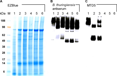
Anti-anthrose-trirhamnose tetrasaccharide MAbs and anti-anthrose-rhamnose disaccharide MAbs (14, 26–28) were tested for their reactivities with B. anthracis samples isolated from cattle carcasses in four African countries. In immunoblotting experiments with spore lysates, the anti-anthrose-rhamnose disaccharide MAb MTD5 reacted with strain Sterne (Fig. 1C, lane 1), with an internal control strain of B. anthracis (strain JF3783, belonging to the globally spread genotype cluster A4 [16]) (Fig. 1C, lane 2), and with an isolate from southern Africa (Zimbabwe) (Fig. 1C, lane 3). The observed staining pattern included both high-molecular-mass (∼180/150 kDa) and low-molecular-mass (∼60 kDa) bands, as is characteristic for the BclA glycoprotein (25, 27). In contrast, none of the B. anthracis isolates collected in Chad (13 isolates), Cameroon (6 isolates), and Mali (1 isolate) were recognized by the disaccharide-specific MAb MTD5 (Fig. 1C, lanes 4 to 6, and data not shown) or by the other antidisaccharide or antitetrasaccharide MAbs tested (data not shown). The BclA glycoprotein, found in B. anthracis, B. cereus, and B. thuringiensis, has highly conserved amino and carboxy termini and is also the immunodominant spore surface antigen in mice (24). Staining of endospore lysate proteins by immunoblotting with an anti-B. thuringiensis spore antiserum as the primary antibody yielded multiple-band staining patterns with a prominent high-molecular-mass band, suggesting the presence of the BclA protein moiety in all isolates (Fig. 1B). However, cross-reactivity of the B. thuringiensis mouse antiserum with other high-molecular-mass proteins cannot be excluded.
Fig. 1.
Immunodetection of anthrose-containing tetrasaccharide in B. anthracis strains isolated from cattle carcasses in African countries. (A) EZBlue (Sigma) protein staining of B. anthracis spore lysates (lane 1, Sterne strain; lane 2, internal reference strain JF3783 from Switzerland; lane 3, isolate from Zimbabwe; lane 4, isolate from Mali; lane 5, one representative isolate from Chad; lane 6, one representative isolate from Cameroon) separated on a 4 to 10% gradient SDS-polyacrylamide gel. (B) Western blot analysis of the reactivity of anti-B. thuringiensis antiserum diluted 1:1,000 (27). (C) Cross-reactivity of the anti-anthrose-rhamnose disaccharide MAb MTD5 with the blotted total spore lysates. Similar results were obtained with all other antidisaccharide (MTD1 to MTD4 and MTD6) and antitetrasaccharide (MTA1 to MTA3) MAbs (14, 26–28) tested. MAb MTD5 was used at a concentration of 0.01 μg/ml. The sizes of the molecular mass markers (lane M) are given in kDa.
Since the anthrose monosaccharide is an essential component of the epitopes of the tested MAbs (14, 27), the failure of the MAbs to bind to the isolates from Mali, Chad, and Cameroon suggested that these strains might be unable to produce anthrose. To test this hypothesis, the anthrose operons of the African isolates were sequenced. The sequence of the BAS3319 gene, encoding an acyltransferase, was the same for all African strains (data not shown) and identical to that of the sequenced B. anthracis strain Sterne (GenBank accession no. AE017225). Sequencing of the BAS3320 gene (encoding an aminotransferase) revealed the presence of three eight-base-pair (AAAAAAAG) tandem repeats at position 230 in all isolates from Chad, Cameroon, and Mali instead of two repeats in all other strains, including the Zimbabwe isolate (Table 2). The observed tandem repeat insertion causes a frameshift mutation, changing the protein sequence and creating a premature stop codon at position 306 (normal termination after base pair 1139). Comparison of the available aminotransferase gene (BAS3320) sequences revealed the presence of a point mutation at position 72 causing an Ile→Met amino acid sequence exchange in the sequenced B. anthracis strain A1055 but not in the African isolates. None of the currently published BAS3320 sequences contains three AAAAAAAG tandem repeats (Table 2). In all isolates from western Africa the BAS3321 (glycosyltransferase-encoding) gene carried a single base substitution from C to T at position 892, resulting in a premature a stop codon (normal termination at position 2556) (Table 3). This mutation is not present in any of the currently published BAS3321 gene sequences.
Table 2.
Sequence polymorphisms in the BAS3320 gene (1,139 bp, encoding aminotransferase) of the anthrose operon
| B. anthracis straina | Polymorphism (amino acid) |
|
|---|---|---|
| T→G | Tandem repeat | |
| Sterne | 70 ATT 72 (I) | 211 TTTAAAAAAAGAAAAAAAG--------GAA 232 |
| A1055 | 70 ATG 72 (M) | 211 TTTAAAAAAAGAAAAAAAG--------GAA 232 |
| KrugerB | 70 ATT 72 (I) | 211 TTTAAAAAAAGAAAAAAAG--------GAA 232 |
| A0442 | 70 ATT 72 (I) | 211 TTTAAAAAAAGAAAAAAAG--------GAA 232 |
| A0465 | 70 ATT 72 (I) | 211 TTTAAAAAAAGAAAAAAAG--------GAA 232 |
| CNEVA-9066 | 70 ATT 72 (I) | 211 TTTAAAAAAAGAAAAAAAG--------GAA 232 |
| Zimbabwe | 70 ATT 72 (I) | 211 TTTAAAAAAAGAAAAAAAG--------GAA 232 |
| Mali | 70 ATT 72 (I) | 211 TTTAAAAAAAGAAAAAAAGAAAAAAAGGAA 240 (frameshift → stop) |
| Chad (13x) | 70 ATT 72 (I) | 211 TTTAAAAAAAGAAAAAAAGAAAAAAAGGAA 240 (frameshift → stop) |
| Cameroon (6x) | 70 ATT 72 (I) | 211 TTTAAAAAAAGAAAAAAAGAAAAAAAGGAA 240 (frameshift → stop) |
The B. anthracis strains from Zimbabwe, Mali, Chad, and Cameroon were isolated from cattle carcasses. The B. anthracis strains Sterne, A1055, KrugerB, A0442, A0465, and CNEVA-9066 are representative hits from sequence comparisons using the BLAST program (http://blast.ncbi.nlm.nih.gov/Blast.cgi).
Table 3.
Sequence polymorphisms in the BAS3321 gene (2,256 bp, encoding glycosyltransferase) of the anthrose operon
|
B. anthracis straina |
Polymorphism (amino acid) |
|||
|---|---|---|---|---|
| C→T | C→T | C→T | A→T | |
| Sterne | 892 CAA 894 (Q) | 1150 ACG 1152 (T) | 1351 ACC 1353 (T) | 1933 AAA 1935 (K) |
| A1055 | 892 CAA 894 (Q) | 1150 ACG 1152 (T) | 1351 ACC 1353 (T) | 1933 AAA 1935 (K) |
| KrugerB | 892 CAA 894 (Q) | 1150 ATG 1152 (M) | 1351 ACC 1353 (T) | 1933 ATA 1935 (I) |
| A0442 | 892 CAA 894 (Q) | 1150 ATG 1152 (M) | 1351 ACC 1353 (T) | 1933 ATA 1935 (I) |
| A0465 | 892 CAA 894 (Q) | 1150 ATG 1152 (M) | 1351 ACC 1353 (T) | 1933 AAA 1935 (K) |
| CNEVA-9066 | 892 CAA 894 (Q) | 1150 ATG 1152 (M) | 1351 ACC 1353 (T) | 1933 AAA 1935 (K) |
| Zimbabwe | 892 CAA 894 (Q) | 1150 ATG 1152 (M) | 1351 ACC 1353 (T) | 1933 ATA 1935 (I) |
| Mali | 892 TAA 894 (stop) | 1150 ACG 1152 (T) | 1351 ATC 1353 (I) | 1933 AAA 1935 (K) |
| Chad (13x) | 892 TAA 894 (stop) | 1150 ACG 1152 (T) | 1351 ATC 1353 (I) | 1933 AAA 1935 (K) |
| Cameroon (6x) | 892 TAA 894 (stop) | 1150 ACG 1152 (T) | 1351 ATC 1353 (I) | 1933 AAA 1935 (K) |
The B. anthracis strains from Zimbabwe, Mali, Chad, and Cameroon were isolated from cattle carcasses. The B. anthracis strains Sterne, A1055, KrugerB, A0442, A0465, and CNEVA-9066 are representative hits from sequence comparisons using the BLAST program (http://blast.ncbi.nlm.nih.gov/Blast.cgi).
Additionally, in some B. anthracis strains three nonsynonymous single-nucleotide polymorphisms (SNPs) compared to the Sterne strain were identified in the BAS3321 gene at positions 1151, 1352, and 1934 (Table 3). Finally, an SNP at position 614 was found in the BAS3322 (enoyl-CoA hydratase-encoding) gene, which has also been reported in other sequenced B. anthracis strains (Table 4). At all four positions the strain from Zimbabwe diverged from the strains from Mali, Chad, and Cameroon.
Table 4.
Sequence polymorphisms in the BAS3322 gene (792 bp, encoding enoyl-CoA hydratase) of the anthrose operon
| B. anthracis straina | T→C polymorphism (amino acid) |
|---|---|
| Sterne | 613 TTT 615 (F) |
| A1055 | 613 TTT 615 (F) |
| KrugerB | 613 TCT 615 (S) |
| A0442 | 613 TCT 615 (S) |
| A0465 | 613 TCT 615 (S) |
| CNEVA-9066 | 613 TCT 615 (S) |
| Zimbabwe | 613 TCT 615 (S) |
| Mali | 613 TTT 615 (F) |
| Chad (13x) | 613 TTT 615 (F) |
| Cameroon (6x) | 613 TTT 615 (F) |
The B. anthracis strains from Zimbabwe, Mali, Chad, and Cameroon were isolated from cattle carcasses. The B. anthracis strains Sterne, A1055, KrugerB, A0442, A0465, and CNEVA-9066 are representative hits from sequence comparisons using the BLAST program (http://blast.ncbi.nlm.nih.gov/Blast.cgi).
Sequencing of the anthrose four-gene operon revealed the presence of premature stop codons in genes BAS3320 and BAS3321 in all isolates from Chad, Cameroon, and Mali, resulting in shortened open reading frames. It is most likely that these truncated proteins possess no or heavily impaired enzymatic activity. It has been shown previously that experimental inactivation of either gene BAS3321, BAS3320, or BAS3319 results in spores devoid of anthrose (6), indicating that their encoded enzymes are required for anthrose biosynthesis. In contrast, inactivation of gene BAS3322 resulted in spores with about half as much anthrose as found in wild-type spores (6). Furthermore it was reported that inactivation of gene BAS3321 resulted in BclA being substituted with trisaccharides only, suggesting that the enzyme is a dTDP-β-l-rhamnose α-1,3-l-rhamnosy transferase that attaches the fourth residue of the pentasaccharide side chain (7). Inactivation of genes BAS3320 and BAS3319 resulted in the disappearance of BclA pentasaccharides and the appearance of a tetrasaccharide lacking anthrose (7). Thus, the inactivation of enzymes BAS3320 and BAS3321 of the anthrose biosynthetic operon in the strains from Chad, Cameroon, and Mali represents a plausible explanation for the lack of binding of the anti-carbohydrate MAbs.
The observed deficiency in anthrose biosynthesis and the differences in the corresponding biosynthesis genes for the aminotransferase (BAS3320) and the glycosyltransferase (BAS3321), both resulting in premature stop codons, seem to be particular traits of strains from West Africa and are not found elsewhere. This is interesting in that B. anthracis strains from West Africa belong to a particular genetic lineage, the genotype cluster Aβ (12; P. Pilo [Bern], unpublished observations), as determined by variable-number tandem repeat (VNTR) analysis according the typing scheme of Keim et al. (9, 29). In this respect, the 8-nucleotide tandem repeat AAAAAAAG that is present in 2 copies in the anthrose biosynthesis aminotransferase gene (BAS3320) in most B. anthracis strains worldwide except those from West Africa, where the repeat is present in 3 copies, is proposed as a valuable new VNTR that allows an increase in the resolution of subtyping and is directly associated with the anthrose phenotype.
Antigenicity of the anthrose-containing pentasaccharide of a Chadian veterinary anthrax vaccine.
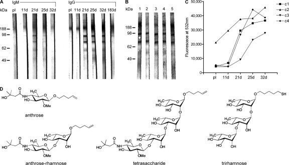
Spore preparations of the attenuated B. anthracis vaccine strain Sterne are used as a live veterinary anthrax vaccine. Since many countries manufacture veterinary vaccines on their own, quality control would be important to avoid a resurgence of the disease because of the usage of inadequate vaccines (32). Differences in vaccine quality have been observed but not systematically analyzed (32). In this study, a live animal anthrax vaccine manufactured in Chad from spores of strain Sterne (Weybridge no. 34F2) was used to immunize cattle. Serum antibody responses were analyzed in immunoblotting experiments with a B. anthracis spore lysate (Fig. 2A). These analyses provided evidence for the production of antibody responses against B. anthracis proteins. When using IgM-specific secondary antibodies, the preimmune serum revealed a weak and diffuse high-molecular-mass band (>188 kDa) which remained largely constant after immunization. The first traces of a vaccine-induced IgM response were observed at day 21, when at least two distinct protein bands of between 62 and 98 kDa appeared. Already at 25 days after vaccination staining of these bands decreased, and staining was no longer detectable at day 32. When using IgG-specific secondary antibodies, preimmune serum also stained a high-molecular-mass antigen. Starting at day 21 after immunization, the IgG signal strength of this band increased substantially. Simultaneously, a strong IgG response to two distinct protein bands of between 62 and 98 kDa also emerged. These analyses suggested that immunogenic B. anthracis antigens induced an initial production of anthrax antigen cross-reactive IgM and that activated B cells subsequently switched to the expression of Cγ heavy-chain genes. Six months after the single immunization, all animals showed a substantial drop of IgG levels reacting with spore lysates in Western blots, reflecting the relatively short duration of immunity of vaccinated animals. As recommended by WHO (33), this makes annual booster immunizations necessary.
Fig. 2.
Antigenicity of the BclA-associated pentasaccharide of the Sterne 34F2 veterinary vaccine in cattle. (A) Development of B. anthracis cross-reactive IgM and IgG in Western blotting with total spore lysate. Cattle serum samples taken preimmunity (pI) and at days 11, 21, 25, 32, and 183 after a single subcutaneous vaccination with the Sterne spore vaccine were tested at a dilution of 1:100. IgM and IgG responses of a representative animal are depicted. (B) Competition Western blot experiments with overlapping synthetic carbohydrates were used for epitope mapping. Immune serum of cattle no. 3 taken 32 days after vaccination was preincubated with synthetic competitors and afterwards added to cut strips. The different competitors used were as follows: lane 1, no competitor; lane 2, tetrasaccharide; lane 3, disaccharide; lane 4, anthrose; lane 5, trirhamnose. Immune sera were used at a dilution of 1:100 and synthetic competitors at a concentration of 100 μg/ml. (C) Glycan microarray analysis of the development of antitetrasaccharide IgG responses in the four vaccinated cattle (c1 to c4). Shown are results obtained with individual sera at a dilution of 1:100. (D) Synthetic structures derived from the BclA-associated pentasaccharide.
The specificity of the interaction of immune sera with the high-molecular-mass antigen was further investigated by competition Western blot analyses using a set of overlapping synthetic saccharides (Fig. 2D). Binding of serum IgG to the high-molecular-mass band, thought to be the BclA glycoprotein, was reduced greatly by the synthetic tetrasaccharide (Fig. 2B, lane 2) and partially by the anthrose-rhamnose disaccharide (Fig. 2B, lane 3) and anthrose (Fig. 2B, lane 4) but not by trirhamnose (Fig. 2B, lane 5). Competition with the glycan structures tested had no influence on the serological reactions with the lower-molecular-mass antigens that are unrelated to BclA. These results indicated that the spore vaccine induced a substantial IgG response against the tetrasaccharide, suggesting that the anthrose-containing carbohydrate constituent of the BclA protein is one of the immunodominant antigens in cattle. Thus, the anthrose-containing tetrasaccharide could be used as a target antigen to monitor vaccine-induced antibody responses as a quality control for the livestock vaccines.
Using a previously developed glycan microarray spotted with the tetrasaccharide (14), it was possible to monitor development of this antitetrasaccharide antibody response directly. A single immunization with spores of strain Sterne elicited high levels of specific antitetrasaccharide IgG titers (Fig. 2C) in all four cattle immunized. One of the animals had a substantial preimmunization antitetrasaccharide IgG titer, which was boosted by the vaccination. Since the animals were not reported to have been vaccinated with strain Sterne previously, the observed preimmunization antitetrasaccharide IgG titer in one animal suggests either exposure to an anthrose-expressing Bacillus species or the development of cross-reactive antibodies against structurally related antigenic saccharides found on other endemic microorganisms.
West Africa is a region where anthrax is highly endemic and where massive vaccination of livestock has taken place over long periods of time (22y). The geographic correlation of massive vaccination with the live vaccine strain Sterne, which produces an antigenic response to anthrose, with the lack of anthrose-positive B. anthracis field strains specifically in this area is interesting and leads to the hypothesis that the latter strains could represent escape mutants. This hypothesis could be tested in the future by comparing the protective efficacies of a Sterne vaccine and a similar vaccine derived from an anthrose-deficient B. anthracis strain toward anthrose-producing and anthrose-deficient strains.
Footnotes
Present address: Institute of Biochemistry and Molecular Medicine, Bühlstr. 28, CH 3011 Bern, Switzerland.
Present address: Department of Chemistry, Massachusetts Institute of Technology, Cambridge, MA 02139.
Passed away in 2008.
Published ahead of print on 13 May 2011.
REFERENCES
- 1. Adamo R., Saksena R., Kováč P. 2005. Synthesis of the β anomer of the spacer-equipped tetrasaccharide side chain of the major glycoprotein of the Bacillus anthracis exosporium. Carbohydr. Res. 340:2579–2582 [DOI] [PubMed] [Google Scholar]
- 2. Cohen S., et al. 2000. Attenuated nontoxinogenic and nonencapsulated recombinant Bacillus anthracis spore vaccines protect against anthrax. Infect. Immun. 8:4549–4558 [DOI] [PMC free article] [PubMed] [Google Scholar]
- 3. Crich D., Vinogradova O. 2007. Synthesis of the antigenic tetrasaccharide side chain from the major glycoprotein of Bacillus anthracis exosporium. J. Org. Chem. 72:6513–6520 [DOI] [PMC free article] [PubMed] [Google Scholar]
- 4. Daubenspeck J. M., et al. 2004. Novel oligosaccharide side chains of the collagen-like region of BclA, the major glycoprotein of the Bacillus anthracis exosporium. J. Biol. Chem. 30:30945–30953 [DOI] [PubMed] [Google Scholar]
- 5. Dhénin S. G., et al. 2008. Synthesis of an anthrose derivative and production of polyclonal antibodies for the detection of anthrax spores. Carbohydr. Res. 343:2101–2110 [DOI] [PubMed] [Google Scholar]
- 6. Dong S., McPherson S. A., Tan L., Chesnokova O. N., Turnbough C. L., Jr., Pritchard D. G. 2008. Anthrose biosynthetic operon of Bacillus anthracis. J. Bacteriol. 190:2350–2359 [DOI] [PMC free article] [PubMed] [Google Scholar]
- 7. Dong S., et al. 2010. Characterization of the enzymes encoded by the anthrose biosynthetic operon of Bacillus anthracis. J. Bacteriol. 192:5053–5062 [DOI] [PMC free article] [PubMed] [Google Scholar]
- 8. Guo H., O'Doherty G. A. 2007. De novo asymmetric synthesis of the anthrax tetrasaccharide by a palladium-catalyzed glycosylation reaction. Angew Chem. Int. 46:5206–5208 [DOI] [PubMed] [Google Scholar]
- 9. Keim P., et al. 2000. Multiple-locus variable-number tandem repeat analysis reveals genetic relationships within Bacillus anthracis. J. Bacteriol. 182:2928–2936 [DOI] [PMC free article] [PubMed] [Google Scholar]
- 10. Kubler-Kielb J., et al. 2008. Saccharides cross-reactive with Bacillus anthracis spore glycoprotein as an anthrax vaccine component. Proc. Natl. Acad. Sci. U. S. A. 25:8709–8712 [DOI] [PMC free article] [PubMed] [Google Scholar]
- 11. Kuehn A., et al. 2009. Development of antibodies against anthrose tetrasaccharide for specific detection of Bacillus anthracis spores. Clin. Vaccine Immunol. 12:1728–1737 [DOI] [PMC free article] [PubMed] [Google Scholar]
- 12. Maho A., et al. 2006. Antibiotic susceptibility and molecular diversity of Bacillus anthracis strains in Chad: detection of a new phylogenetic subgroup. J. Clin. Microbiol. 44:3422–3425 [DOI] [PMC free article] [PubMed] [Google Scholar]
- 13. Mehta A. S., et al. 2006. Synthesis and antigenic analysis of the BclA glycoprotein oligosaccharide from the Bacillus anthracis exosporium. Chem. Eur. J. 12:9136–9149 [DOI] [PubMed] [Google Scholar]
- 14. Oberli M. A., et al. 2010. Molecular analysis of carbohydrate-antibody interactions: case study using a Bacillus anthracis tetrasaccharide. J. Am. Chem. Soc. 132:10239–10241 [DOI] [PubMed] [Google Scholar]
- 15. Perreten V., et al. 2005. Microarray-based detection of 90 antibiotic resistance genes of gram-positive bacteria. J. Clin. Microbiol. 43:2291–2302 [DOI] [PMC free article] [PubMed] [Google Scholar]
- 16. Pilo P., Perreten. V., Frey J. 2008. Molecular epidemiology of Bacillus anthracis: getting the correct origin. Appl. Environ. Microbiol. 74:2928–2931 [DOI] [PMC free article] [PubMed] [Google Scholar]
- 17. Ryu C., et al. 2005. Molecular characterization of Korean Bacillus anthracis isolates by amplified fragment length polymorphism analysis and multilocus variable-number tandem repeat analysis. Appl. Environ. Microbiol. 71:4664–4671 [DOI] [PMC free article] [PubMed] [Google Scholar]
- 18. Saile E., et al. 2011. Antibody responses to a spore carbohydrate antigen as a marker of non-fatal inhalation anthrax in rhesus macaques. Clin. Vaccine Immunol. doi:10.1128/CVI.00475-10 [DOI] [PMC free article] [PubMed] [Google Scholar]
- 19. Saksena R., Adamo R., Kováč P. 2005. Studies toward a conjugate vaccine for anthrax. Synthesis and characterization of anthrose [4,6-dideoxy-4-(3-hydroxy-3-methylbutanamido)-2-O-methyl-d-glucopyranose] and its methyl glycosides. Carbohydr. Res. 340:1591–1600 [DOI] [PubMed] [Google Scholar]
- 20. Saksena R., Adamo R., Kováč P. 2006. Synthesis of the tetrasaccharide side chain of the major glycoprotein of the Bacillus anthracis exosporium. Bioorg. Med. Chem. Lett. 16:615–617 [DOI] [PubMed] [Google Scholar]
- 21. Saksena R., Adamo R., Kováč P. 2007. Immunogens related to the synthetic tetrasaccharide side chain of Bacillus anthracis exosporium. Bioorg. Med. Chem. Lett. 15:4283–4310 [DOI] [PMC free article] [PubMed] [Google Scholar]
- 22. Schelling E., Wyss K., Béchir M., Moto D. D., Zinsstag J. 2005. Synergy between public health and veterinary services to deliver human and animal health interventions in rural low income settings. BMJ 331:1264–1267 [DOI] [PMC free article] [PubMed] [Google Scholar]
- 23. Sterne M. 1939. The use of anthrax vaccines prepared from avirulent (uncapsulated) variants of Bacillus anthracis. Onderstepoort J. Vet. Sci. An. Ind. 13:307–312 [Google Scholar]
- 24. Swiecki M. K., Lisanby M. W., Shu F., Turnbough C. L., Jr., Kearney J. F. 2006. Monoclonal antibodies for Bacillus anthracis spore detection and functional analyses of spore germination and outgrowth. J. Immunol. 176:6076–6084 [DOI] [PubMed] [Google Scholar]
- 25. Sylvestre P., Couture-Tosi E., Mock M. 2003. Polymorphism in the collagen-like region of the Bacillus anthracis BclA protein leads to variation in exosporium filament length. J. Bacteriol. 185:1555–1563 [DOI] [PMC free article] [PubMed] [Google Scholar]
- 26. Tamborrini M., Holzer M., Seeberger P. H., Schürch N., Pluschke G. 2010. Anthrax spore detection by a Luminex assay based on monoclonal antibodies recognizing anthrose-containing oligosaccharides. Clin. Vaccine Immunol. 17:1446–1451 [DOI] [PMC free article] [PubMed] [Google Scholar]
- 27. Tamborrini M., et al. 2009. Immuno-detection of anthrose containing tetrasaccharide in the exosporium of Bacillus anthracis and Bacillus cereus strains. J. Appl. Microbiol. 5:1618–1628 [DOI] [PubMed] [Google Scholar]
- 28. Tamborrini M., Werz D. B., Frey J., Pluschke G., Seeberger P. H. 2006. Anti-carbohydrate antibodies for the de tection of anthrax spores. Angew Chem. Int. 45:6581–6582 [DOI] [PubMed] [Google Scholar]
- 29. Van Ert M. N., et al. 2007. Global genetic population structure of Bacillus anthracis. PLoS One 2:e461. [DOI] [PMC free article] [PubMed] [Google Scholar]
- 30. Welkos S. L., Friedlander A. M. 1988. Comparative safety and efficacy against Bacillus anthracis of protective antigen and live vaccines in mice. Microb. Pathog. 2:127–139 [DOI] [PubMed] [Google Scholar]
- 31. Werz D. B., Seeberger P. H. 2005. Total synthesis of antigen Bacillus anthracis tetrasaccharide-creation of an anthrax vaccine candidate. Angew Chem. Int. 44:6315–6318 [DOI] [PubMed] [Google Scholar]
- 32. World Health Organization 1998. Guidelines for the surveillance and control of anthrax in humans and animals, 3rd ed World Health Organization, Geneva, Switzerland [Google Scholar]
- 33. World Organisation for Animal Health 2009. Manual of diagnostic tests and vaccines for terrestrial animals. World Organisation for Animal Health, Paris, France [Google Scholar]


