Abstract
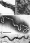
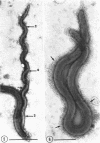
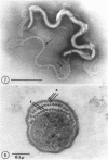
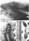
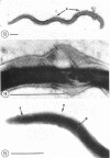
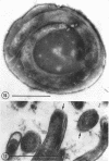
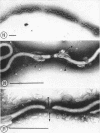
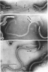
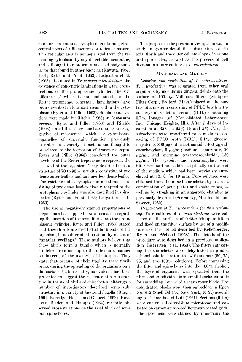
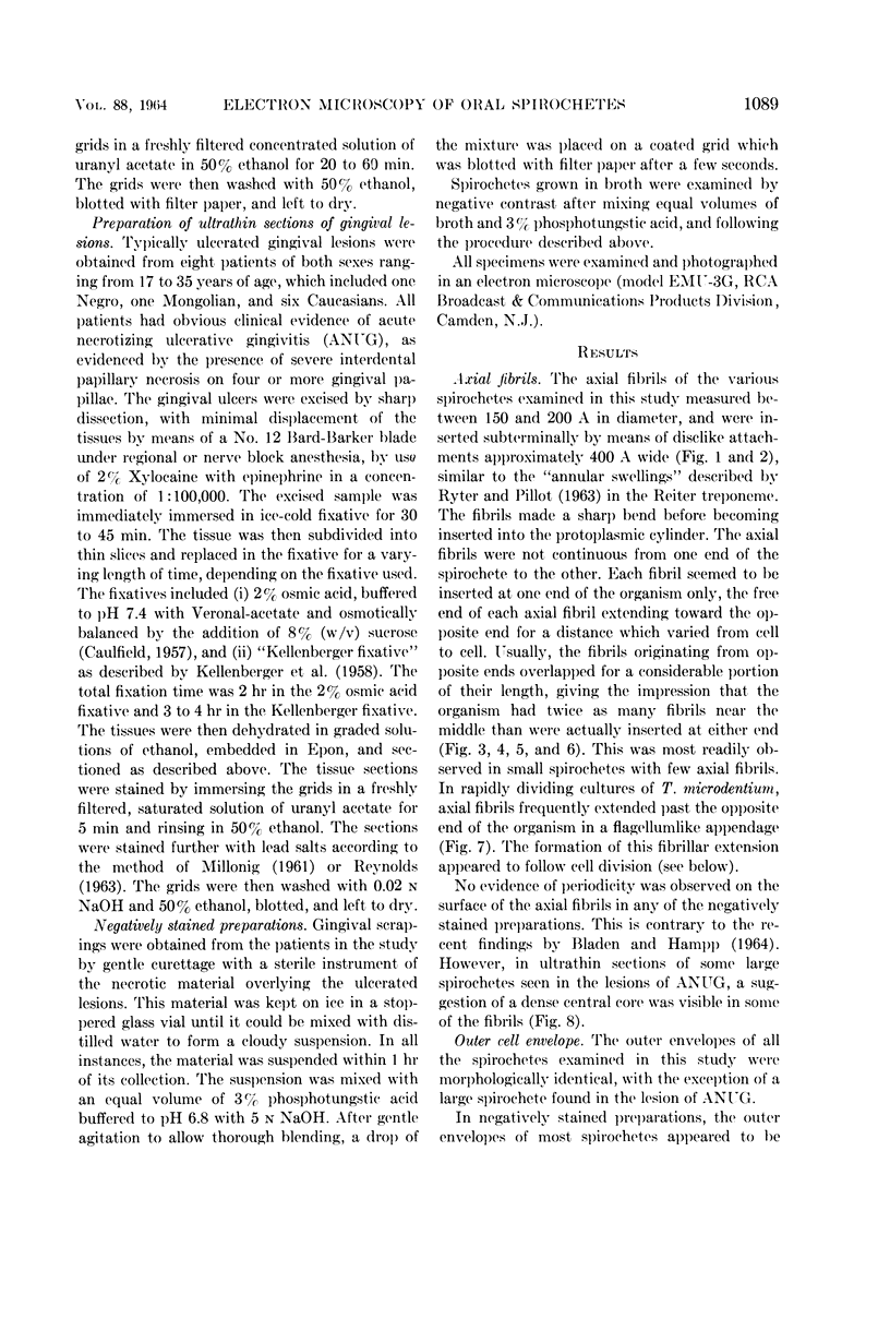
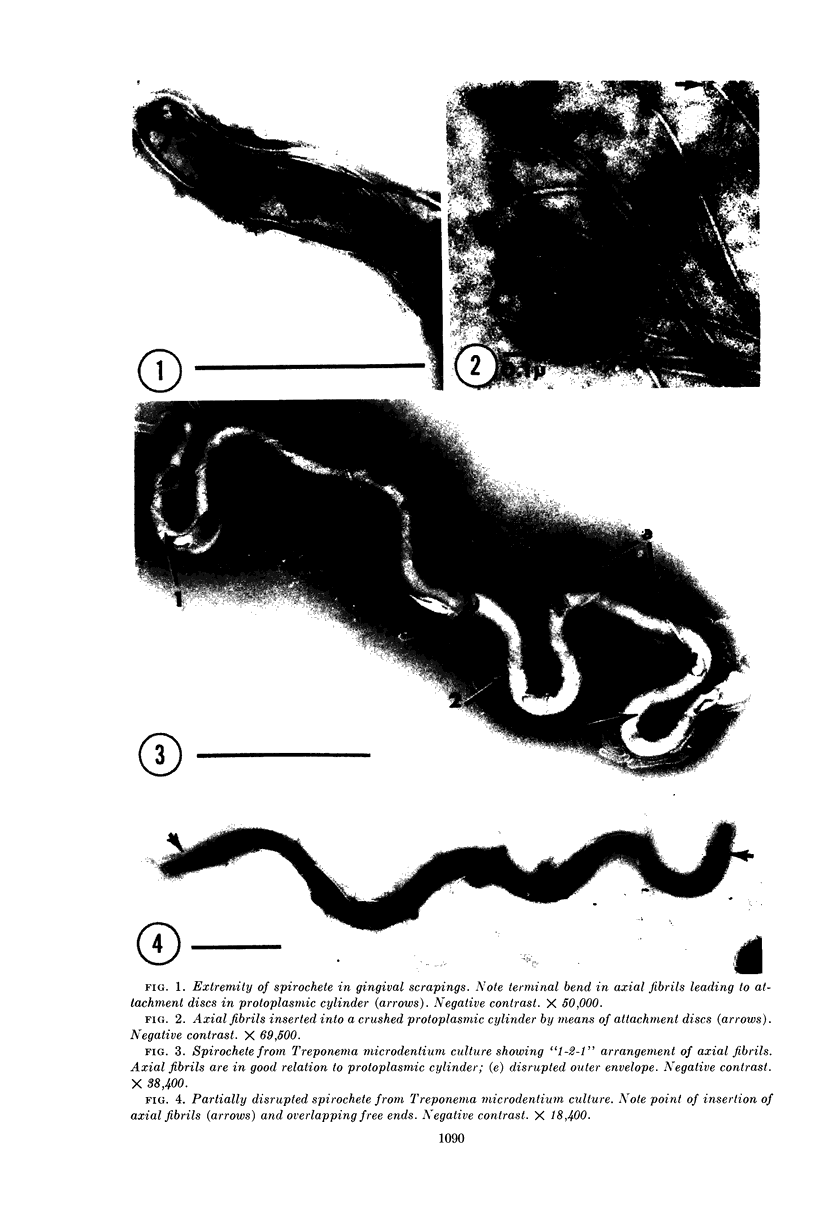
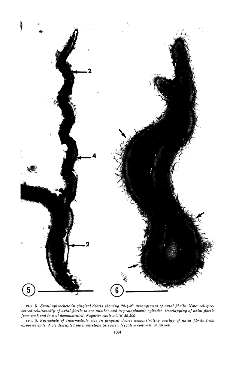
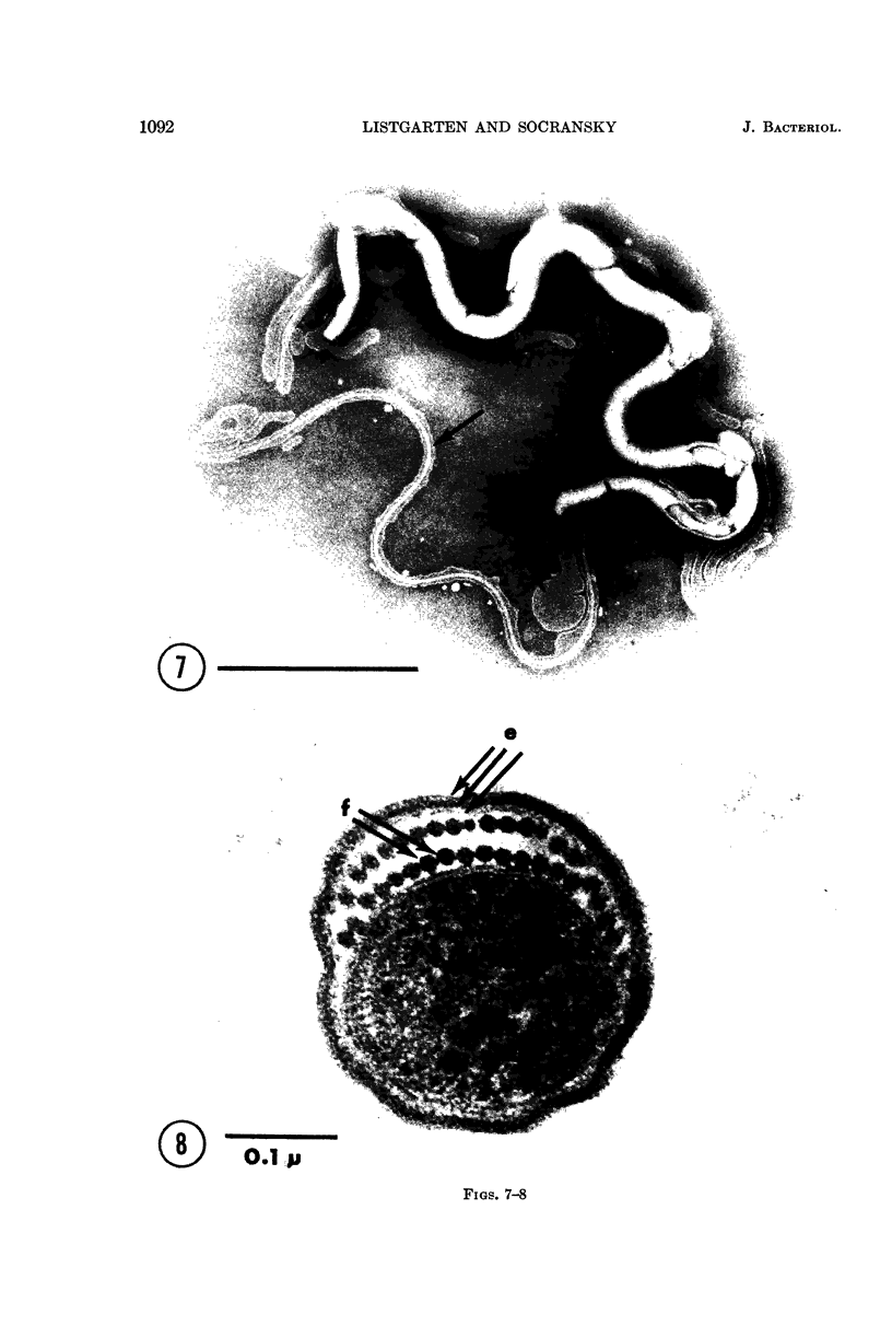
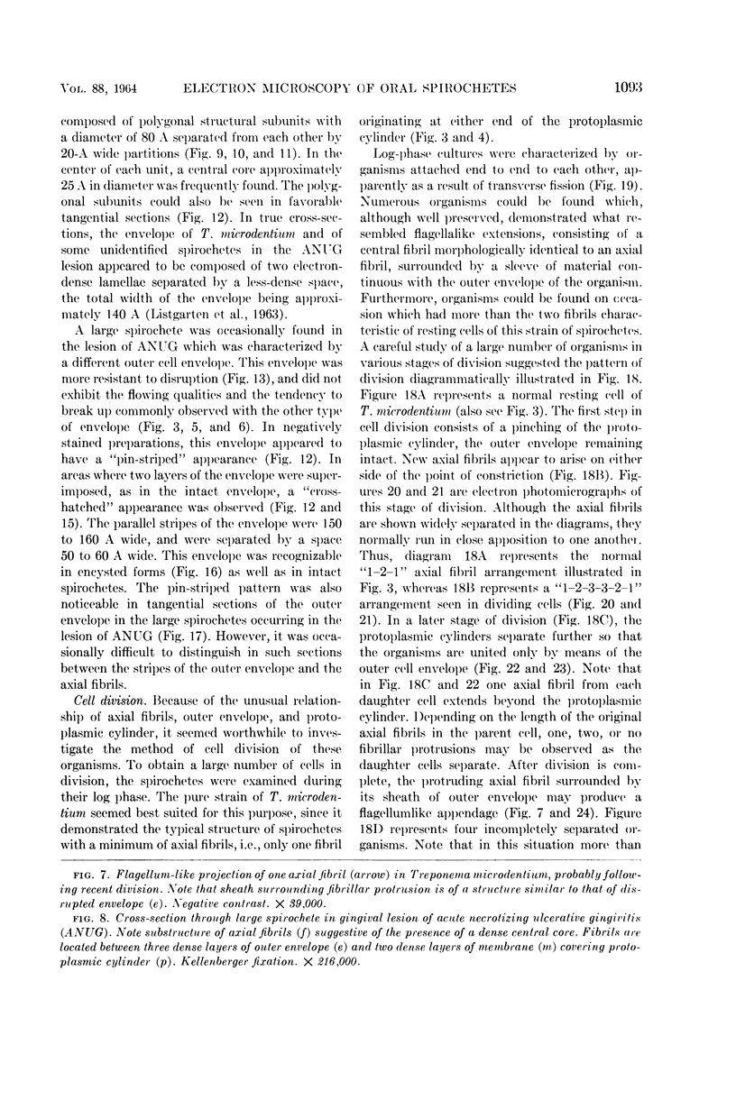
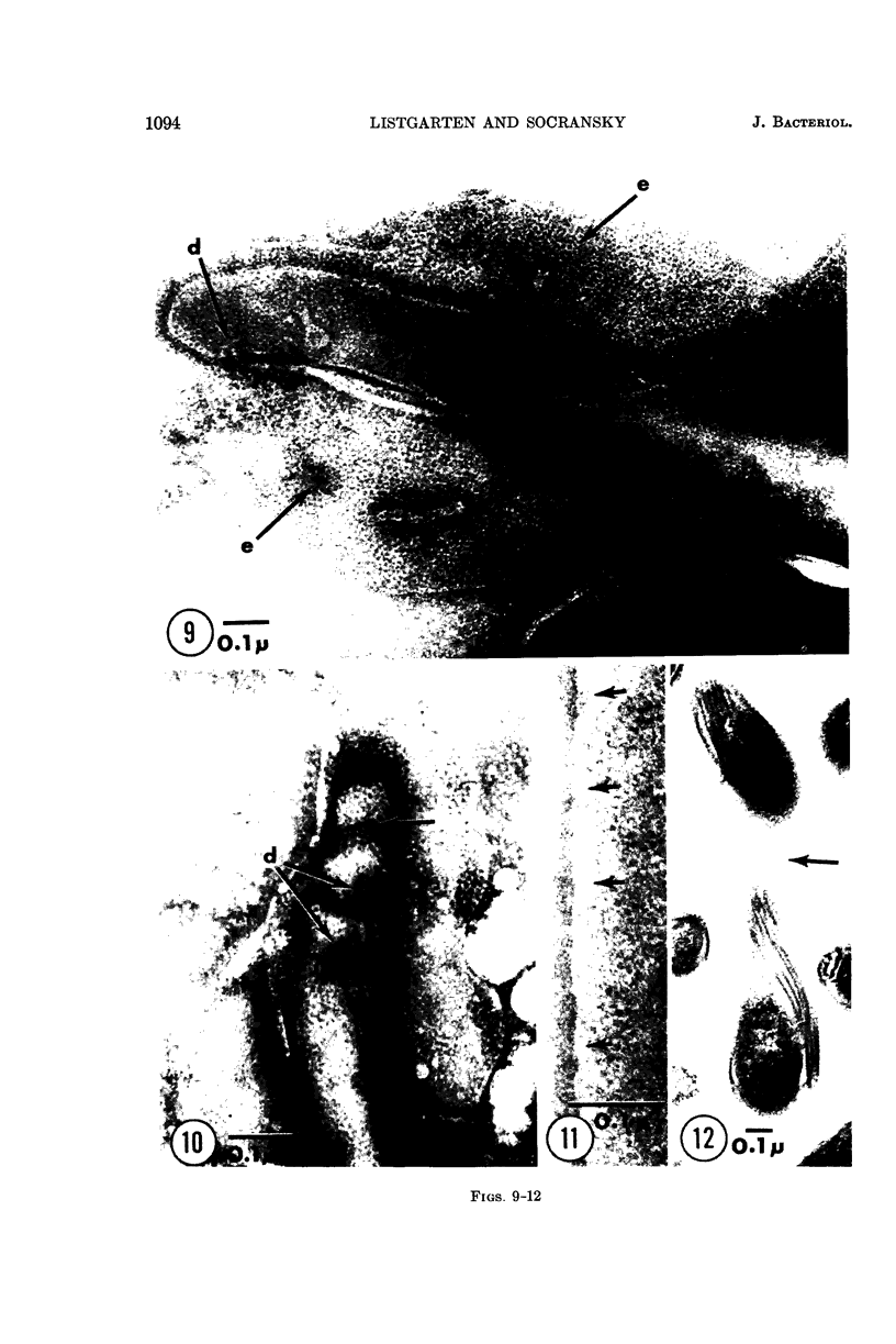
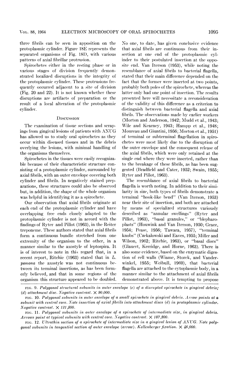
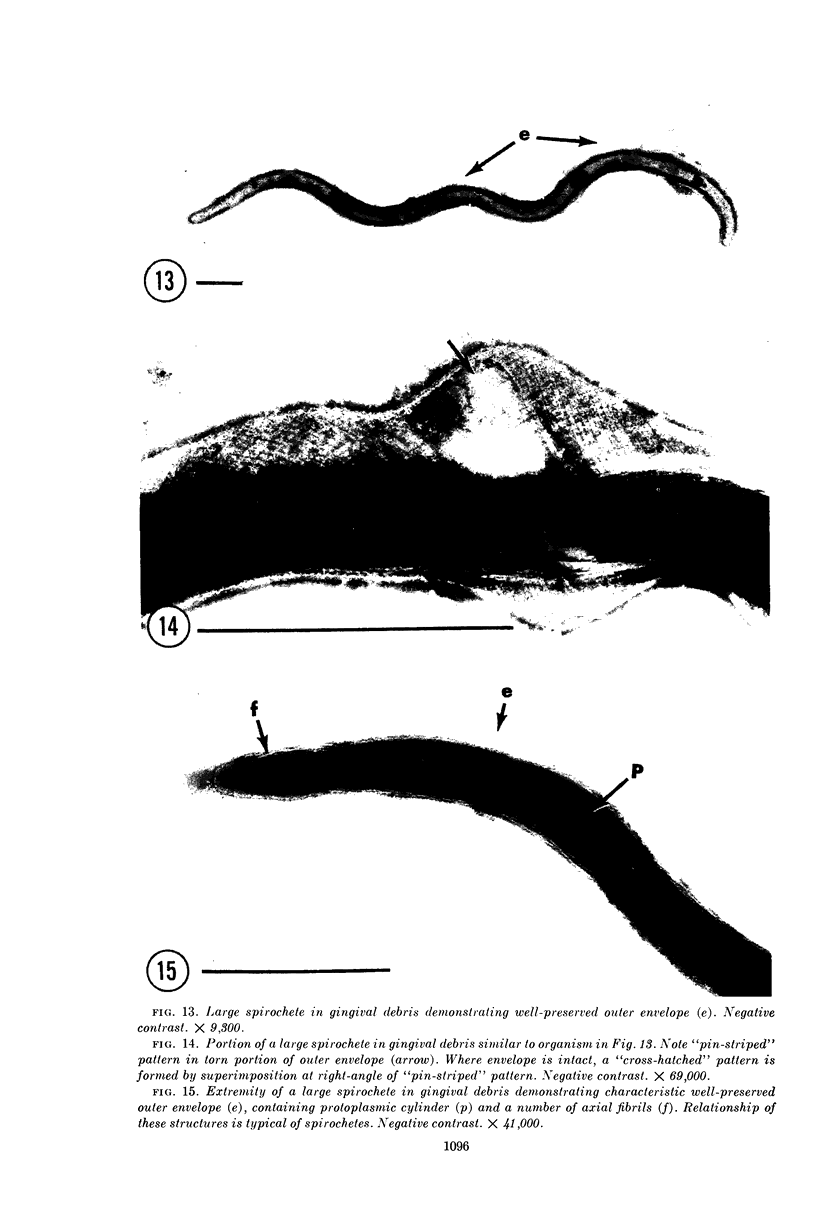
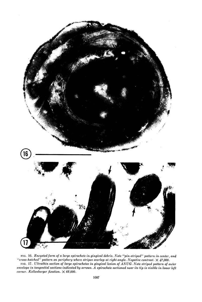
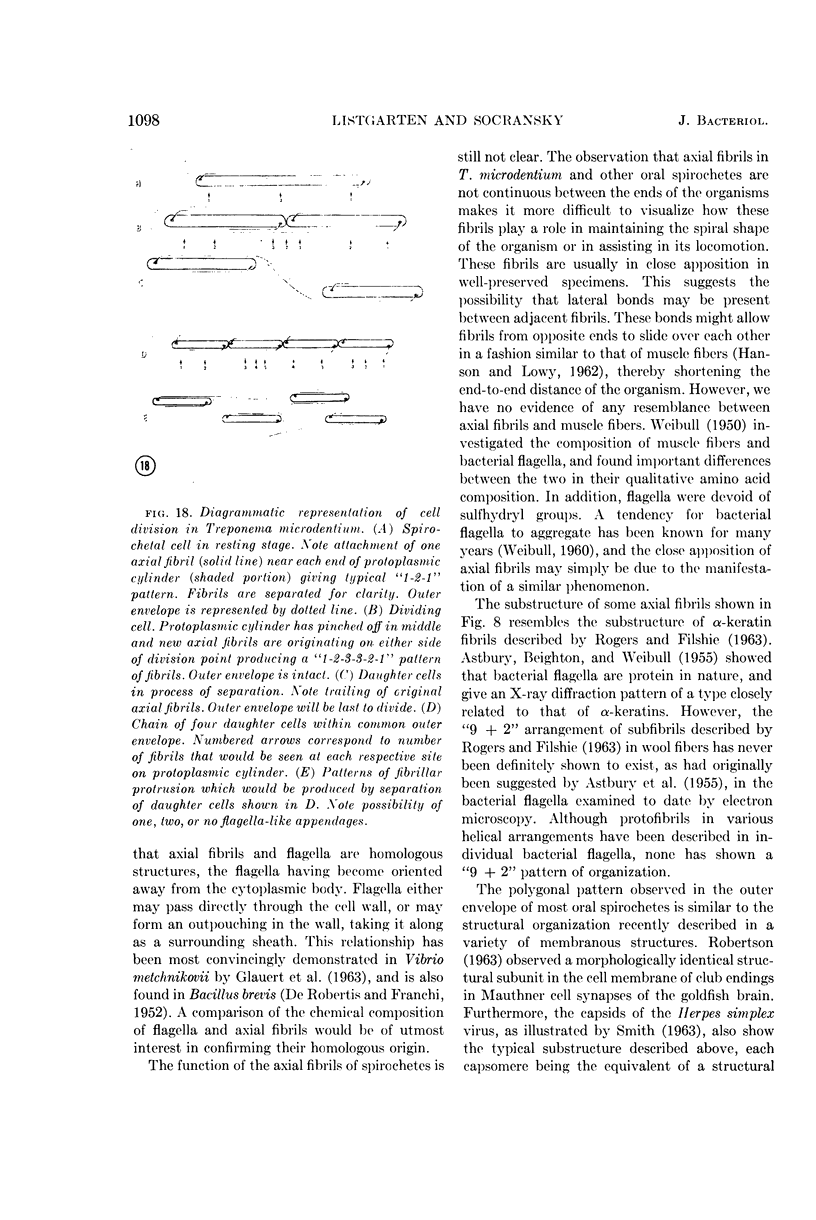
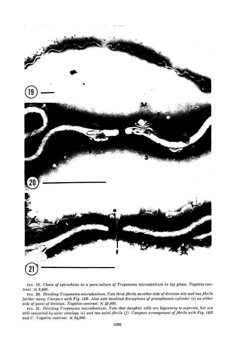
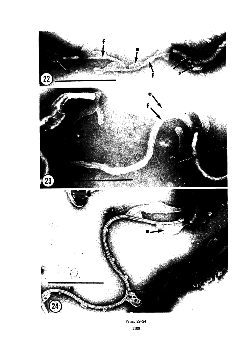
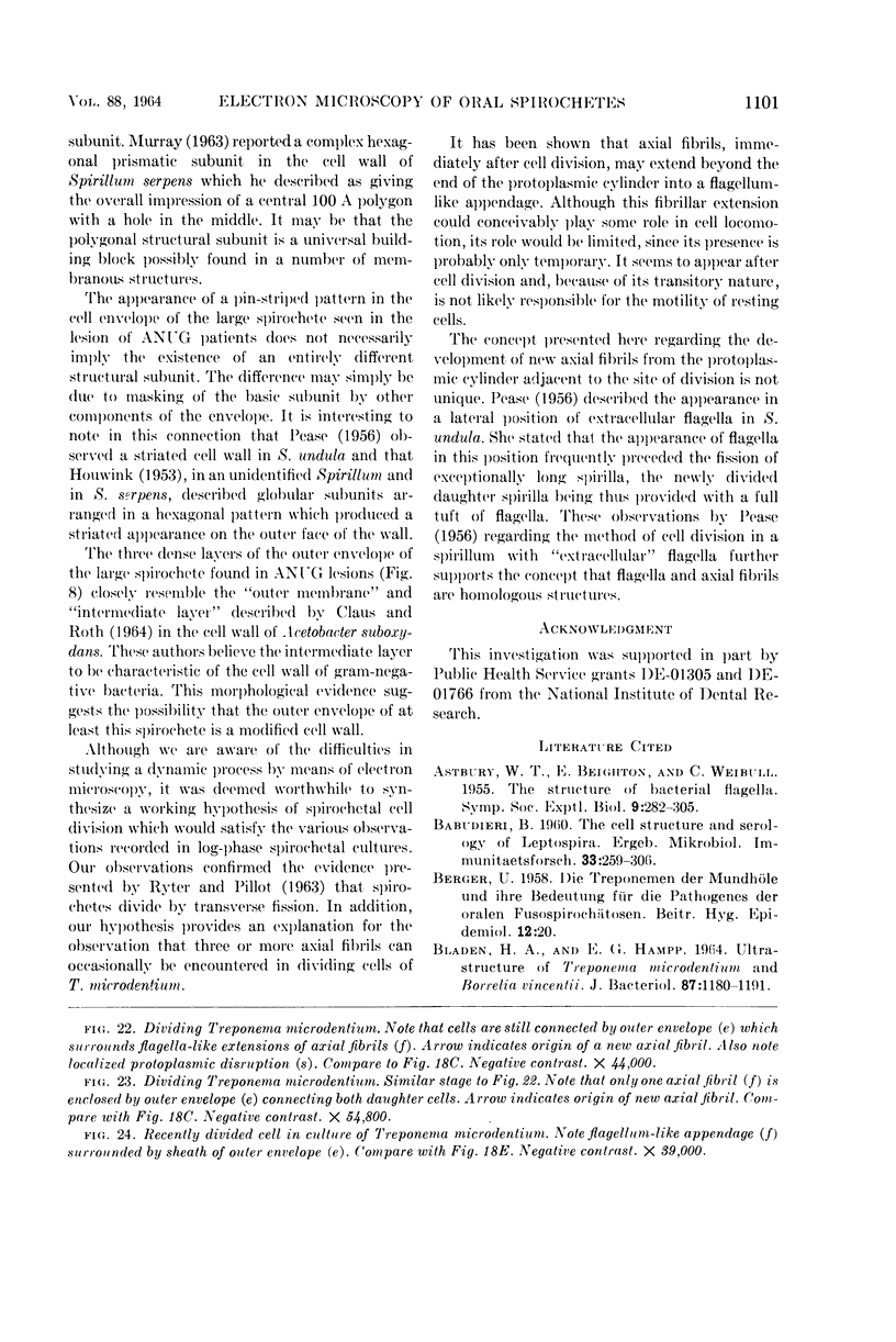
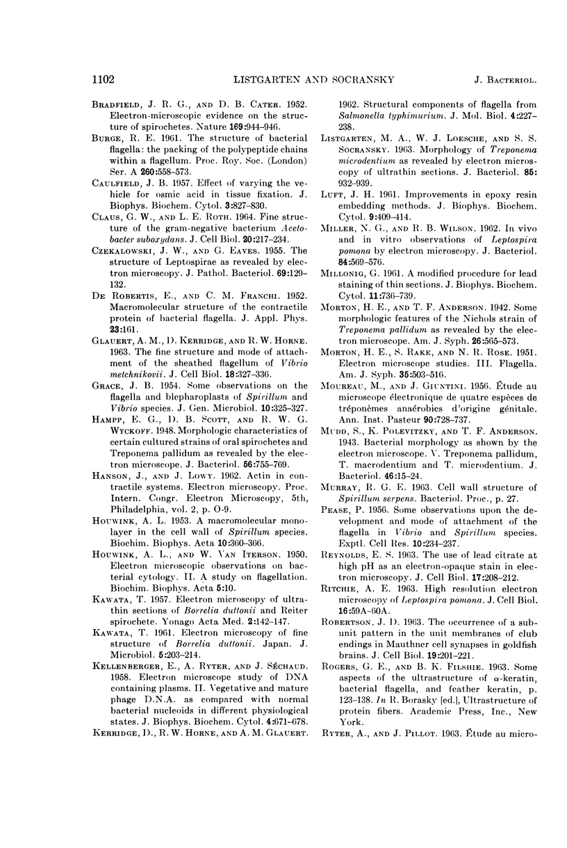
Listgarten, M. A. (Harvard School of Dental Medicine and Forsyth Dental Center, Boston, Mass.), and S. S. Socransky. Electron microscopy of axial fibrils, outer envelope, and cell division of certain oral spirochetes. J. Bacteriol. 88:1087–1103. 1964.—The ultrastructure of axial fibrils and outer envelopes of a number of oral spirochetes was studied in thin sections and by negative contrast. The axial fibrils measured 150 to 200 A in diameter. Only one end of each fibril was inserted subterminally into the protoplasmic cylinder by means of a 400 A wide disc. The free ends of fibrils inserted near one end of the cylinder extended toward, and overlapped in close apposition, the free ends of fibrils inserted at the other end. In thin sections, some axial fibrils showed a substructure, suggestive of a dense central core. The outer envelopes of most spirochetes appeared to consist of 80 A wide polygonal structural subunits. However, in one large spirochete, the outer envelope demonstrated a “pin-striped” pattern. Cell division in a pure culture of Treponema microdentium was studied by negative contrast. Results suggested that this organism divides by transverse fission, the outer envelope being last to divide. During the course of division, new axial fibrils appeared to originate on either side of the point of constriction of the protoplasmic cylinder. Flagellalike extensions which were found in rapidly dividing organisms were due to protruding axial fibrils, and appeared to be the result of cell division. Some evidence is presented to support the concept of a homologous origin for axial fibrils and flagella.
Full text
PDF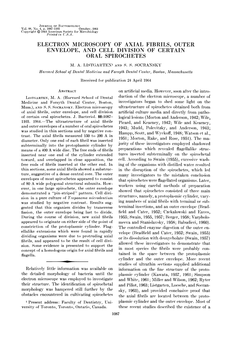
Images in this article
Selected References
These references are in PubMed. This may not be the complete list of references from this article.
- BABUDIERI B. [The cell structure and serology of Leptospira]. Ergeb Mikrobiol Immunitatsforsch Exp Ther. 1960;33:259–306. [PubMed] [Google Scholar]
- BRADFIELD J. R. G., CATER D. B. Electron-microscopic evidence on the structure of spirochaetes. Nature. 1952 Jun 7;169(4310):944–946. doi: 10.1038/169944a0. [DOI] [PubMed] [Google Scholar]
- CAULFIELD J. B. Effects of varying the vehicle for OsO4 in tissue fixation. J Biophys Biochem Cytol. 1957 Sep 25;3(5):827–830. doi: 10.1083/jcb.3.5.827. [DOI] [PMC free article] [PubMed] [Google Scholar]
- CLAUS G. W., ROTH L. E. FINE STRUCTURE OF THE GRAM-NEGATIVE BACTERIUM ACETOBACTER SUBOXYDANS. J Cell Biol. 1964 Feb;20:217–233. doi: 10.1083/jcb.20.2.217. [DOI] [PMC free article] [PubMed] [Google Scholar]
- CZEKALOWSKI J. W., EAVES G. The structure of leptospirae as revealed by electron microscopy. J Pathol Bacteriol. 1955 Jan-Apr;69(1-2):129–132. doi: 10.1002/path.1700690118. [DOI] [PubMed] [Google Scholar]
- GLAUERT A. M., KERRIDGE D., HORNE R. W. THE FINE STRUCTURE AND MODE OF ATTACHMENT OF THE SHEATHED FLAGELLUM OF VIBRIO METCHNIKOVII. J Cell Biol. 1963 Aug;18:327–336. doi: 10.1083/jcb.18.2.327. [DOI] [PMC free article] [PubMed] [Google Scholar]
- GRACE J. B. Some observations on the flagella and blepharoplasts of Spirillum and Vibrio spp. J Gen Microbiol. 1954 Apr;10(2):325–327. doi: 10.1099/00221287-10-2-325. [DOI] [PubMed] [Google Scholar]
- HOUWINK A. L. A macromolecular mono-layer in the cell wall of Spirillum spec. Biochim Biophys Acta. 1953 Mar;10(3):360–366. doi: 10.1016/0006-3002(53)90266-2. [DOI] [PubMed] [Google Scholar]
- HOUWINK A. L., van ITERSON W. Electron microscopical observations on bacterial cytology; a study on flagellation. Biochim Biophys Acta. 1950 Mar;5(1):10–44. doi: 10.1016/0006-3002(50)90144-2. [DOI] [PubMed] [Google Scholar]
- Hampp E. G., Scott D. B., Wyckoff R. W. Morphologic Characteristics of Certain Cultured Strains of Oral Spirochetes and Treponema pallidum as Revealed by the Electron Microscope. J Bacteriol. 1948 Dec;56(6):755–769. doi: 10.1128/jb.56.6.755-769.1948. [DOI] [PMC free article] [PubMed] [Google Scholar]
- KAWATA T. Electron microscopy of fine structure of Borrelia duttonii. Jpn J Microbiol. 1961 Apr;5:203–214. doi: 10.1111/j.1348-0421.1961.tb00201.x. [DOI] [PubMed] [Google Scholar]
- KELLENBERGER E., RYTER A., SECHAUD J. Electron microscope study of DNA-containing plasms. II. Vegetative and mature phage DNA as compared with normal bacterial nucleoids in different physiological states. J Biophys Biochem Cytol. 1958 Nov 25;4(6):671–678. doi: 10.1083/jcb.4.6.671. [DOI] [PMC free article] [PubMed] [Google Scholar]
- KERRIDGE D., HORNE R. W., GLAUERT A. M. Structural components of flagella from Salmonella typhimurium. J Mol Biol. 1962 Apr;4:227–238. doi: 10.1016/s0022-2836(62)80001-1. [DOI] [PubMed] [Google Scholar]
- LISTGARTEN M. A., LOESCHE W. J., SOCRANSKY S. S. MORPHOLOGY OF TREPONEMA MICRODENTIUM AS REVEALED BY ELECTRON MICROSCOPY OF ULTRATHIN SECTIONS. J Bacteriol. 1963 Apr;85:932–939. doi: 10.1128/jb.85.4.932-939.1963. [DOI] [PMC free article] [PubMed] [Google Scholar]
- LUFT J. H. Improvements in epoxy resin embedding methods. J Biophys Biochem Cytol. 1961 Feb;9:409–414. doi: 10.1083/jcb.9.2.409. [DOI] [PMC free article] [PubMed] [Google Scholar]
- MILLONIG G. A modified procedure for lead staining of thin sections. J Biophys Biochem Cytol. 1961 Dec;11:736–739. doi: 10.1083/jcb.11.3.736. [DOI] [PMC free article] [PubMed] [Google Scholar]
- MORTON H. E., RAKE G., ROSE N. R. Electron microscope studies of treponemes. III. Flagella. Am J Syph Gonorrhea Vener Dis. 1951 Nov;35(6):503–516. [PubMed] [Google Scholar]
- MOUREAU M., GIUNTINI J. Etude au microscope électronique de quatre espèces de tréponèmes anaérobies d'origine génitale. Ann Inst Pasteur (Paris) 1956 Jun;90(6):728–737. [PubMed] [Google Scholar]
- Miller N. G., Wilson R. B. IN VIVO AND IN VITRO OBSERVATIONS OF LEPTOSPIRA POMONA BY ELECTRON MICROSCOPY. J Bacteriol. 1962 Sep;84(3):569–576. doi: 10.1128/jb.84.3.569-576.1962. [DOI] [PMC free article] [PubMed] [Google Scholar]
- Mudd S., Polevitzky K., Anderson T. F. Bacterial Morphology as shown by the Electron Microscope: V. Treponema pallidum, T. macrodentium and T. microdentium. J Bacteriol. 1943 Jul;46(1):15–24. doi: 10.1128/jb.46.1.15-24.1943. [DOI] [PMC free article] [PubMed] [Google Scholar]
- PEASE P. Some observations upon the development and mode of attachment of the flagella in Vibrio and Spirillum species. Exp Cell Res. 1956 Feb;10(1):234–237. doi: 10.1016/0014-4827(56)90092-1. [DOI] [PubMed] [Google Scholar]
- REYNOLDS E. S. The use of lead citrate at high pH as an electron-opaque stain in electron microscopy. J Cell Biol. 1963 Apr;17:208–212. doi: 10.1083/jcb.17.1.208. [DOI] [PMC free article] [PubMed] [Google Scholar]
- ROBERTSON J. D. THE OCCURRENCE OF A SUBUNIT PATTERN IN THE UNIT MEMBRANES OF CLUB ENDINGS IN MAUTHNER CELL SYNAPSES IN GOLDFISH BRAINS. J Cell Biol. 1963 Oct;19:201–221. doi: 10.1083/jcb.19.1.201. [DOI] [PMC free article] [PubMed] [Google Scholar]
- RYTER A., PILLOT J. [Electron microscope study of the external and internal structure of the Reiter treponema]. Ann Inst Pasteur (Paris) 1963 Apr;104:496–501. [PubMed] [Google Scholar]
- SIMPSON C. F., WHITE F. H. Electron microscope studies and staining reactions of leptospires. J Infect Dis. 1961 Nov-Dec;109:243–250. doi: 10.1093/infdis/109.3.243. [DOI] [PubMed] [Google Scholar]
- SMITH K. O. PHYSICAL AND BIOLOGICAL OBSERVATIONS ON HERPESVIRUS. J Bacteriol. 1963 Nov;86:999–1009. doi: 10.1128/jb.86.5.999-1009.1963. [DOI] [PMC free article] [PubMed] [Google Scholar]
- TAWARA J. Electron-microscopic study on the flagella of Vibrio comma. J Bacteriol. 1957 Jan;73(1):89–90. doi: 10.1128/jb.73.1.89-90.1957. [DOI] [PMC free article] [PubMed] [Google Scholar]
- VARPHOLOMEEVA A. A., STANISLAVSKY E. S. Recherches sur la morphologie des Leptospires à l'aide du microscope électronique. Ann Inst Pasteur (Paris) 1958 Mar;94(3):361–366. [PubMed] [Google Scholar]
- WATSON J. H. L., ANGULO J. J., LEON-BLANCO F., VARELA G., WEDDERBURN C. C. Electron microscopic observations of flagellation in some species of the genus Treponema Schaudinn. J Bacteriol. 1951 Apr;61(4):455–461. doi: 10.1128/jb.61.4.455-461.1951. [DOI] [PMC free article] [PubMed] [Google Scholar]
- WIAME J. M., STORCK R., VANDERWINKEL E. Biosynthèse induite d'arabokinase dans les protoplastes de Bacillus subtilis. Biochim Biophys Acta. 1955 Nov;18(3):353–357. doi: 10.1016/0006-3002(55)90097-4. [DOI] [PubMed] [Google Scholar]


