Abstract
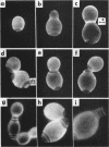
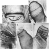
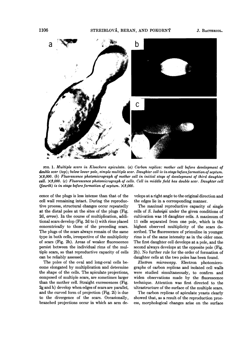
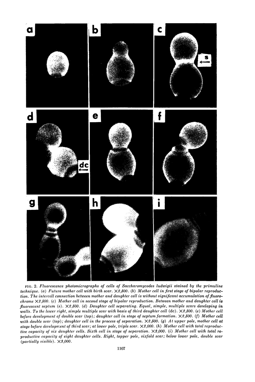
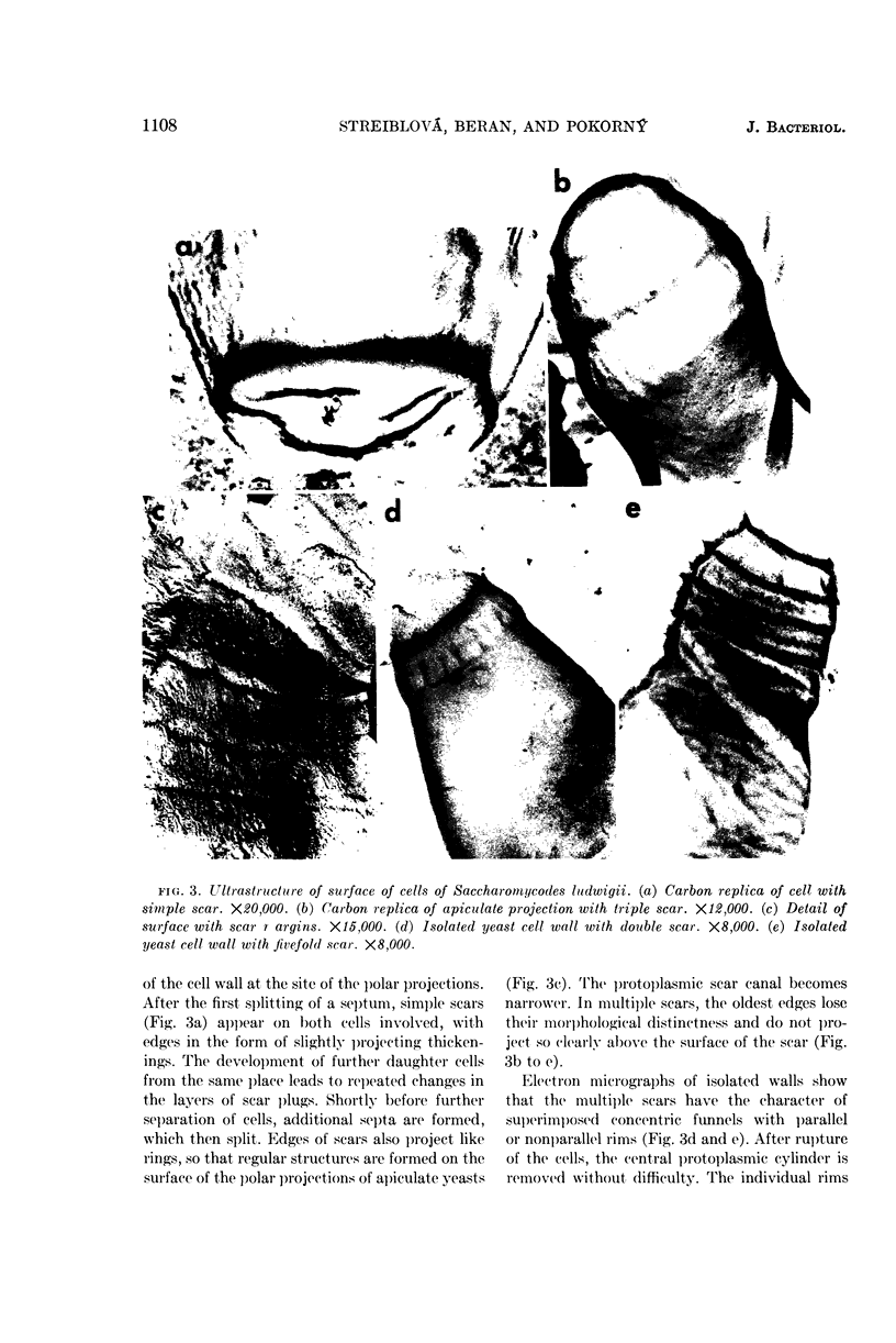
Streiblová, Eva (Czechoslovak Academy of Sciences, Prague, Czechoslovakia), K. Beran, and V. Pokorný. Multiple scars, a new type of yeast scar in apiculate yeasts. J. Bacteriol. 88:1104–1111. 1964.—A new type of yeast scar is described in apiculate yeasts: Saccharomycodes, Nadsonia, Hanseniaspora, and Kloeckera. These scars are formed on the distal poles of the cell walls in the course of vegetative reproduction, and are the cause of the formation of the apiculate form of the cells. The structure of multiple scars was studied by fluorescence microscopy and by electron microscopy on carbon replicas and isolated cell walls. The discussion deals with the importance of described cytological structures for the morphogenesis of cells and for determining individual reproductive capacity of cells, and considers some questions related to the interpretation of the development of multiple scars.
Full text
PDF
Images in this article
Selected References
These references are in PubMed. This may not be the complete list of references from this article.
- AGAR H. D., DOUGLAS H. C. Studies of budding and cell wall structure of yeast; electron microscopy of the sections. J Bacteriol. 1955 Oct;70(4):427–434. doi: 10.1128/jb.70.4.427-434.1955. [DOI] [PMC free article] [PubMed] [Google Scholar]
- BARTHOLOMEW J. W., LEVIN R. The structure of Saccharomyces carlsbergensis and S. cerevisiae as determined by ultra-thin sectioning methods and electron microscopy. J Gen Microbiol. 1955 Jun;12(3):473–477. doi: 10.1099/00221287-12-3-473. [DOI] [PubMed] [Google Scholar]
- BARTHOLOMEW J. W., MITTWER T. Demonstration of yeast bud scars with the electron microscope. J Bacteriol. 1953 Mar;65(3):272–275. doi: 10.1128/jb.65.3.272-275.1953. [DOI] [PMC free article] [PubMed] [Google Scholar]
- BARTON A. A. Some aspects of cell division in saccharomyces cerevisiae. J Gen Microbiol. 1950 Jan;4(1):84–86. doi: 10.1099/00221287-4-1-84. [DOI] [PubMed] [Google Scholar]
- CONTI S. F., NAYLOR H. B. Electron microscopy of ultrathin sections of Schizosaccharomyces octosporus. I. Cell division. J Bacteriol. 1959 Dec;78:868–877. doi: 10.1128/jb.78.6.868-877.1959. [DOI] [PMC free article] [PubMed] [Google Scholar]
- DE BECZE G. I. A microbiological process report; yeasts. I. Morphology. Appl Microbiol. 1956 Jan;4(1):1–12. doi: 10.1128/am.4.1.1-12.1956. [DOI] [PMC free article] [PubMed] [Google Scholar]
- HOUWINK A. L., KREGER D. R. Observations on the cell wall of yeasts; an electron microscope and x-ray diffraction study. Antonie Van Leeuwenhoek. 1953;19(1):1–24. doi: 10.1007/BF02594830. [DOI] [PubMed] [Google Scholar]
- MUNDKUR B. ELECTRON MICROSCOPICAL STUDIES OF FROZEN-DRIED YEAST. IV. SCHIZOSACCHAROMYCES, NADSONIA AND SACCHAROMYCODES. Z Naturforsch B. 1963 Dec;18:1073–1082. doi: 10.1515/znb-1963-1215. [DOI] [PubMed] [Google Scholar]
- MUNDKUR B. Electron microscopical studies of frozen-dried yeast. I. Localization of polysaccharides. Exp Cell Res. 1960 Jun;20:28–42. doi: 10.1016/0014-4827(60)90219-6. [DOI] [PubMed] [Google Scholar]
- NICKERSON W. J. SYMPOSIUM ON BIOCHEMICAL BASES OF MORPHOGENESIS IN FUNGI. IV. MOLECULAR BASES OF FORM IN YEASTS. Bacteriol Rev. 1963 Sep;27:305–324. doi: 10.1128/br.27.3.305-324.1963. [DOI] [PMC free article] [PubMed] [Google Scholar]
- NORTHCOTE D. H., HORNE R. W. The chemical composition and structure of the yeast cell wall. Biochem J. 1952 May;51(2):232–236. doi: 10.1042/bj0510232. [DOI] [PMC free article] [PubMed] [Google Scholar]
- Olson B. H., Johnson M. J. FACTORS PRODUCING HIGH YEAST YIELDS IN SYNTHETIC MEDIA. J Bacteriol. 1949 Feb;57(2):235–246. doi: 10.1128/jb.57.2.235-246.1949. [DOI] [PMC free article] [PubMed] [Google Scholar]
- SENTHESHANMUGATHAN S., NICKERSON W. J. Composition of cells and cell walls of triangular and ellipsoidal forms of Trigonopsis variabilis. J Gen Microbiol. 1962 Mar;27:451–464. doi: 10.1099/00221287-27-3-451. [DOI] [PubMed] [Google Scholar]
- STREIBLOVA E., BERAN K. Types of multiplication scars in yeasts, demonstrated by fluorescence microscopy. Folia Microbiol (Praha) 1963 Jul;8:221–227. doi: 10.1007/BF02872585. [DOI] [PubMed] [Google Scholar]


