Abstract
Background
Bone changes are common in sickle cell disease, but the pathogenesis is not fully understood. Tartrate-resistant acid phosphatase (TRACP) type 5b is produced by bone-resorbing osteoclasts. In other forms of hemolytic anemia, increased iron stores are associated with osteoporosis. We hypothesized that transfusional iron overload would be associated with increased osteoclast activity in patients with sickle cell disease.
Design and Methods
We examined tartrate-resistant acid phosphatase 5b concentrations in patients with sickle cell disease and normal controls of similar age and sex distribution at steady state. Serum tartrate-resistant acid phosphatase 5b concentration was measured using an immunocapture enzyme assay and plasma concentrations of other cytokines were assayed using the Bio-Plex suspension array system. Tricuspid regurgitation velocity, an indirect measure of systolic pulmonary artery pressure, was determined by echocardiography.
Results
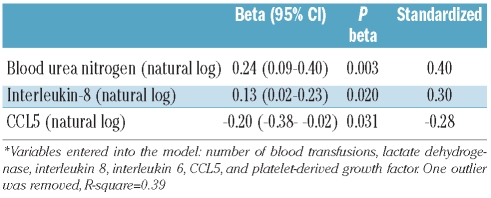
Tartrate-resistant acid phosphatase 5b concentrations were higher in 58 adults with sickle cell disease than in 22 controls (medians of 4.4 versus 2.4 U/L, respectively; P=0.0001). Among the patients with sickle cell disease, tartrate-resistant acid phosphatase 5b independently correlated with blood urea nitrogen (standardized beta=0.40, P=0.003), interleukin-8 (standardized beta=0.30, P=0.020), and chemokine C-C motif ligand 5 (standardized beta=−0.28, P=0.031) concentrations, but not with serum ferritin concentration. Frequent blood transfusions (>10 units in life time) were not associated with higher tartrate-resistant acid phosphatase 5b levels in multivariate analysis. There were strong correlations among tartrate-resistant acid phosphatase 5b, alkaline phosphatase and tricuspid regurgitation velocity (r>0.35, P<0.001).
Conclusions
Patients with sickle cell disease have increased osteoclast activity as reflected by serum tartrate-resistant acid phosphatase 5b concentrations. Our results may support a potential role of inflammation rather than increased iron stores in stimulating osteoclast activity in sickle cell disease. The positive relationships among tartrate-resistant acid phosphatase 5b, alkaline phosphatase and tricuspid regurgitation velocity raise the possibility of a common pathway in the pulmonary and bone complications of sickle cell disease.
Keywords: sickle cell disease, osteoclast activity, bone turnover, inflammation, TRACP 5b, pulmonary complications
Introduction
Markers of bone turnover are increased in sickle cell disease and in β-thalassemia major and intermedia.1,2 Osteoporosis and osteopenia are common complications of these conditions,3–5 with altered bone metabolism beginning in early childhood.4,5 Multiple blood transfusions, bone marrow expansion, iron overload and inflammation are recognized risk factors for osteoporosis in patients with beta-thalassemia.3 Both in vitro and animal experiments indicate that iron may play a role in bone formation and/or destruction.6–8 An increased amount of iron has been associated with reduced in vitro osteoblast proliferation9 and extracellular calcium deposition.10 In a mouse model, iron dextran injection increased the osteoclast population in the bone structure and reduced the number and thickness of trabeculae.11 Another study in mice indicated that osteoclast differentiation and mitochondria biogenesis are regulated by iron: increased iron stimulated osteoclast activity while iron chelation both inhibited osteoclast-mediated bone resorption and protected against bone loss.8
Tartrate-resistant acid phosphatase (TRACP) type 5 is an enzyme that is produced by bone-resorbing osteoclasts, inflammatory macrophages and dendritic cells. TRACP is encoded by a single gene, ACP5, on chromosome 19p13.3–13.2.12 TRACP 5a is derived from macrophages and dendritic cells and TRACP 5b is a bone resorption marker which is produced specifically by activated osteoclasts.13,14 The protein structure of these two isoforms is similar and the only difference is in their sugar chain.15 Normal bone metabolism requires TRACP 5b expression.16 There is a negative correlation between TRACP 5b and bone mass density.17 TRACP 5b also correlates positively with other markers of bone turnover.17
In this study, we hypothesized that transfusional iron overload would be associated with increased osteoclast activity in patients with sickle cell disease. To test this hypothesis, we examined serum TRACP 5b concentrations in adults with sickle cell disease.
Design and Methods
Study participants
Participants with sickle cell disease were recruited from the out-patient sickle cell disease clinic at Howard University Hospital as a part of the Pulmonary Hypertension and the Hypoxic Response in SCD (PUSH) study (ClinicalTrials.gov identifier NCT00495638). PUSH studies two different populations of patients: children and adults. Subjects for this analysis were selected from the adults with sickle cell disease who were in steady-state, which is defined as having no history of hospital admissions, emergency room visits or blood transfusions in the preceding 3 weeks. Cases were selected from two different strata: patients with normal tricuspid regurgitation velocity determined by echocardiography and patients with elevated tricuspid regurgitation velocity. A tricuspid regurgitation velocity of 2.70 m/sec or above was considered to be elevated, consistent with the definition of the Walk-PHaSST study of adults and adolescents.18 Control subjects were free of sickle cell disease, were of similar age, gender and ethnicity as the patients, and had not been admitted to hospital or visited the emergency room for an acute illness in the preceding 3 weeks. Controls were selected from the community, from hospital staff, or from relatives or friends of the cases. Clinical and laboratory data were collected at the time of recruitment. The study protocol was approved by the Howard University Institutional Review Board (IRB-05-SCD-01). All participants gave written informed consent.
Plasma and serum samples for biomarkers
Venous blood was drawn from non-fasting subjects. EDTA plasma and serum were prepared and stored at −80°C for future analysis.
Analysis of biological markers
The concentration of TRACP 5b in serum was measured by an enzyme-linked immunosorbent assay (ELISA) format immunocapture enzyme assay kit (Cat #8033, Quidel Corporation, San Diego, CA, USA). Plasma concentrations of seven biological markers were assayed in patients and controls using the Bio-Plex suspension array system (Bio-Rad, Hercules, CA): interleukins-6, 8 and 10, basic fibroblastic growth factor, platelet-derived growth factor (PDGF)-BB, vascular endothelial growth factor (VEGF), and chemokine (C-C motif) ligand 5 (CCL5), also known as RANTES. This system allows for simultaneous identification of cytokines in a 96-well filter plate, as described fully by Niu et al).19 Plasma concentrations of endothelin-1 were measured with a commercially available ELISA kit (R&D Systems, Minneapolis, MN, USA).
Statistical analysis
We used the best transformation of continuous variables to normal distribution. Student’s t-test was applied to compare continuous variables between patients with sickle cell disease and controls. For categorical variables, we used either the χ2 test or Fisher’s exact test as appropriate. Spearman’s or Pearson’s correlation was applied to study the correlation between different variables. Because of the small sample size, we considered correlation coefficients of 0.25 or greater as potentially important instead of using a cut-off P value for selecting significant correlations. Multiple linear regression analysis was used to identify independent predictors of serum TRACP 5b concentration. To build the final model, we used a combined biological and statistical approach in which potential predictors of TRACP 5b having a P value of 0.2 or less in univariate analysis were entered into the model. The final variables were selected using a P value less than 0.05. The models were checked for distribution of residuals and co-linearity. Statistical analysis was performed with Stata 10.1 software (StataCorp, College Station, TX, USA).
Results
Comparison of patients with sickle cell disease and controls
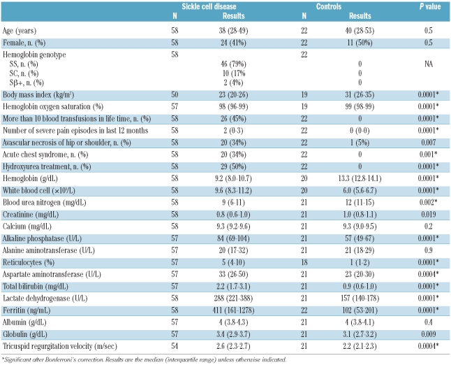
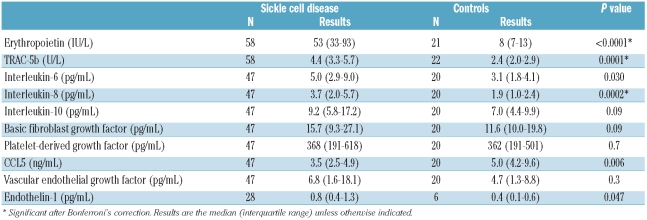
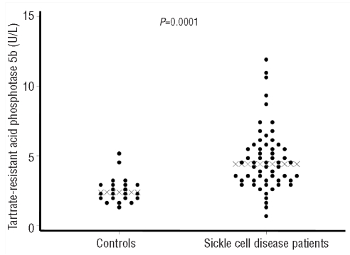
Fifty-eight adults with sickle cell disease (comprising 46 with hemoglobin SS, 10 with hemoglobin SC and 2 with hemoglobin S beta-thalassemia) and 22 normal controls (comprising 19 with hemoglobin AA, 2 with hemoglobin AC, and 1 with hemoglobin AS) were studied. The demographic and clinical characteristics of these subjects are summarized in Table 1. Serum TRACP 5b concentrations were significantly higher in the sickle cell disease patients than in the controls (medians of 4.4 versus 2.4 U/L; P=0.0001). Sickle cell disease patients also had lower body mass index (BMI) values and lower concentrations of hemoglobin, blood urea nitrogen (BUN) and creatinine than controls. They had higher values for markers of hemolysis, mean corpuscular value, white blood cell count, and serum concentrations of globulin and ferritin. Of the inflammatory and angiogenic factors studied, interleukin-6 and −8 and endothelin-1 concentrations were higher in the sickle cell disease patients while the level of CCL5 was lower (Table 2).
Table 1.
Distribution of demographic and clinical data in sickle cell patients and normal controls.
Table 2.
Distribution of biomarkers in sickle cell patients and normal controls.
Predictors of TRACP 5b in patients with sickle cell disease
Clinical and hematologic variables
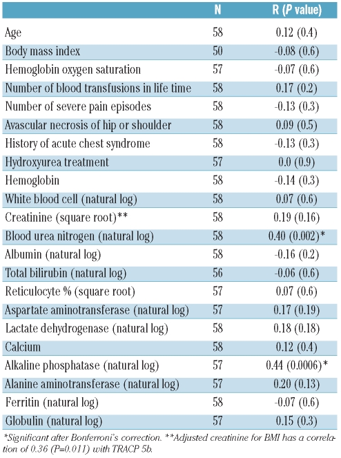
In bivariate analysis, serum TRACP 5b concentration was positively correlated with BUN concentration (Table 3). There was also a trend to higher TRACP 5b levels among the patients who had had more than ten blood transfusions (median 4.8 U/L versus 3.1 U/L in patients with ≤10 transfusions, P=0.025). However, TRACP 5b levels did not differ according to serum ferritin concentration quartiles (median of 4.3 U/L in the lowest quartile versus 4.3 U/L in the highest quartile, P for trend = 0.9). Furthermore, TRACP 5b concentration was not significantly higher in the group of 20 patients who had both a ferritin level greater than 500 ng/mL and had received more than ten blood transfusions when compared to patients who had either normal ferritin or had received ten or fewer blood transfusions (medians of 4.8 U/L versus 4.1 U/L, P=0.17). In addition, TRACP 5b concentration did not correlate with age, gender, BMI, sickle cell genotype, history of severe pain episodes, history of acute chest syndrome, history of avascular necrosis, hemoglobin concentration, or markers of hemolysis or calcium. TRACP 5b concentration did correlate with the levels of BUN and creatinine (this latter adjusted for BMI).
Table 3.
Potential clinical predictors of TRACP 5b in patients with sickle cell disease.
Biological markers
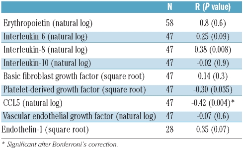
In bivariate analysis among sickle cell disease patients, serum TRACP 5b concentration correlated negatively with plasma CCL5 concentration (Table 4). There were also trends to positive correlations with plasma endothelin-1, interleukin-8 and interleukin-6 concentrations and a negative correlation with PDGF. In a separate analysis, the median (interquartile range) concentration of interleukin-8 was 5.2 (3.6–9.8) pg/mL in patients who had had more than ten blood transfusions compared to 2.9 (1.4–4.7) pg/mL in patients who had received ten or fewer units of blood (P=0.004). Other biomarkers were not significantly different between these two groups (data not shown).
Table 4.
Potential biological predictors of TRACP 5b in patients with sickle cell disease.
Independent predictors of serum TRACP b5 concentration
In multivariate analysis, the concentrations of BUN (beta=0.24, P=0.003), interleukin-8 (beta=0.13, P=0.020) and CCL5 (beta=−0.20, P=0.031) independently correlated with serum TRACP 5b concentration. This model predicted 39% of TRACP 5b variation. The number of units of blood transfused and serum ferritin concentration were not independently correlated with TRACP 5b concentration (Table 5).
Table 5.
Independent relationship of clinical variables and biomarkers with serum TRACP 5b (natural log) concentration in multiple linear regression analysis.
Clinical correlates of TRACP b5
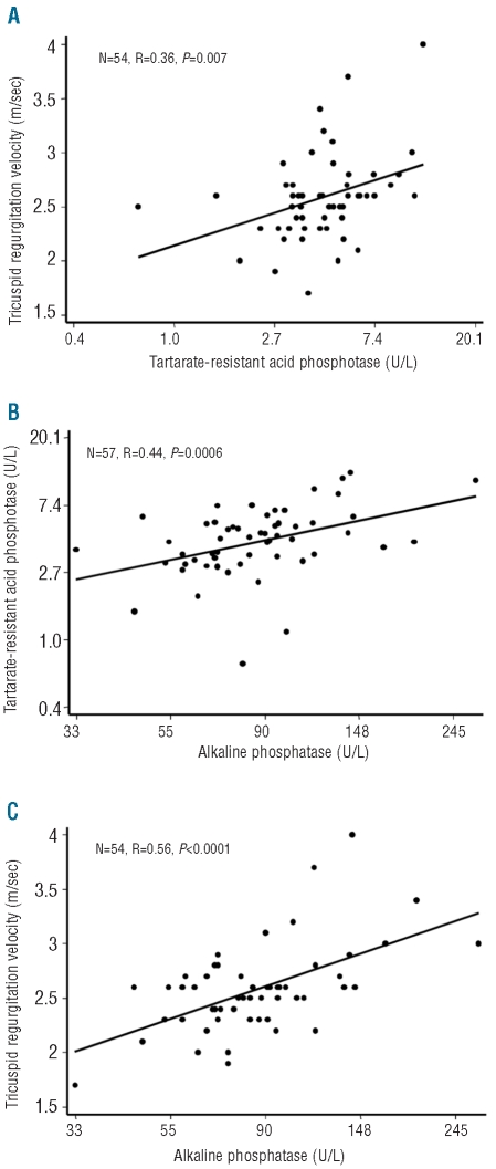
Alkaline phosphatase concentration correlated strongly with TRACP 5b concentration (r=0.44, P=0.0006). Furthermore, this relationship was more robust than the relationship of alkaline phosphatase with markers of liver function, alanine aminotransferase (r=0.29, P=0.03) and total bilirubin (r=−0.03, P=0.08). The concentrations of both TRACP 5b (r=0.56, P<0.0001) and alkaline phosphatase (r=0.56, P<0.0001) were significantly correlated with tricuspid regurgitation velocity (Figure 2).
Figure 2.
Linear relationship between (A) tricuspid regurgitation velocity and TRACP 5b, (B) TRACP 5b and alkaline phosphatase concentrations, (C) tricuspid regurgitation velocity and alkaline phosphatase.
Discussion
In this study, we found that osteoclast activity, as measured by serum TRACP 5b concentration, is elevated in patients with sickle cell disease. Renal function and inflammatory markers were the most important independent predictors of TRACP 5b concentration in patients with sickle cell disease. There were strong correlations among TRACP 5b, tricuspid regurgitation velocity and alkaline phosphatase.
Independent studies indicate that more than 65% of adult patients with sickle cell disease suffer from low bone mineral density20,21 Bone density abnormalities have been reported in other forms of hemolytic anemia as well.3,22 For example, more than 40% of patients with well-managed beta-thalassemia major are affected by osteopenia or osteoporosis.3
The pathogenesis of osteoporosis in hemolytic anemia is multifactorial. In beta-thalassemia, iron overload, decreased physical activity, vitamin C and D deficiencies, and endocrine complications such as hypogonadism and hypothyroidism may increase the susceptibility to osteoporosis.3,23 Iron overload is associated with osteoporosis in hereditary hemochromatosis as well.24 Iron could potentially trigger osteoporosis through several mechanisms, including, but not limited to, lower osteoblast activity, hypogonadism, hypothyroidism and reactive oxidative stress.11 The TRACP 5 gene (Acp5) is highly regulated by iron.16 Iron chelation inhibits osteoclastic bone resorption in vitro.8
Figure 1.
Distribution of serum tartrate-resistance acid phosphatase 5b concentrations among sickle cell disease patients and normal controls (crosses indicate the median in each group).
In sickle cell disease, iron overload was reported to be associated with a lower bone mass index in one study.25 However, current information is inconclusive for a possible correlation between bone mineral density and iron stores as reflected by serum ferritin concentration in patients with sickle cell disease.20,26–29 In our study of sickle cell disease patients, BUN, interleukin-8 and interleukin-6 concentrations were associated with higher osteoclast activity as reflected by TRACP 5b concentration while PDGF and CCL5 were associated with lower osteoclast activity. Higher osteoclast activity, as reflected by serum TRACP 5b concentration, was also associated with higher tricuspid regurgitation velocity. However, in our patients, serum ferritin concentration was not correlated with TRACP 5b. Furthermore, in multivariate analysis, TRACP 5b concentrations did not correlate with the number of units of blood transfused. In our study, increased interleukin-8 concentrations correlated significantly with both frequent blood transfusions and serum TRACP 5b concentrations. This finding, in addition to the lack of a relationship between ferritin and TRACP 5b concentrations, may support a potential role of inflammation, rather than increased iron stores, in stimulating osteoclast activity in patients with sickle cell disease.
We found a positive correlation between renal dysfunction (as reflected by increased BUN concentration) and TRACP 5b concentration. Studies in renal osteopathy have produced inconclusive results with regard to the effect of renal function on serum TRACP 5b concentrations. While some studies found that TRACP 5b was not increased with renal dysfunction,30,31 other studies showed that secondary hyperparathyroidism in chronic renal failure was associated with increased TRACP 5b32 and that TRACP 5b concentration correlated inversely with glomerular filtration rate in pre-dialysis patients with chronic kidney disease.33 The lack of a correlation between serum creatinine and TRACP 5b concentrations in this study may reflect the effect of body mass on creatinine. The BMI was lower in patients with sickle cell disease and it had a low but negative correlation with TRACP 5b, which could obscure the correlation with creatinine. In fact, creatinine values adjusted for BMI correlated positively with TRACP 5b, confirming an association of TRACP 5b with renal function in the present study.
Inflammation plays an important role in causing osteoporosis.34 Osteoclasts are the only source of TRACP 5b production.16 Macrophages and dendritic cells derived from the same myeloid progenitors as osteoclasts express high levels of TRACP 5a.35 Interleukin-6 has been linked to osteoporosis in cancer36 as well as in chronic inflammatory conditions, such as inflammatory bowel disease and rheumatoid arthritis.37,38 It is thought that interleukin-6 induces receptor activator for NF-κB ligand (RANKL), which leads to osteoclast activation and, subsequently, to bone resorption.39 Interleukin-8 is released by normal human osteoclasts.40 High serum interleukin-8 levels have been related to osteoclast activity, osteoporosis in patients with cancer41 and patients with orthopedic implants.42 In this study, the correlation of osteoclast activity with interleukin-8 and −6 supports a potential role of inflammation in causing the osteoporosis of sickle cell disease patients. In multivariate analysis, CCL5 was associated with lower TRACP 5b concentrations. CCL5 induces osteoclast migration and differentiation.43 However, in mouse models, it reportedly had no effect on bone resorption.44 Osteoclasts are one of the most important sources of CCL5 secretion. In response to high local calcium concentration, osteoclasts secrete more CCL5, which, in turn induces osteoblast migration and may increase bone formation.32 Mice that are CCR55−/− (CCL5 receptor) have higher alveolar bone resorption during orthodontic movement.45 A negative relationship of CCL5 with osteoclast activity was recently observed in patients with chronic obstructive pulmonary disease who suffer from low bone mineral density.46
Elevated tricuspid regurgitation velocity is a non-invasive marker that can predict pulmonary hypertension and is associated with higher mortality rates among adult patients with sickle cell disease.47 Higher osteoclast activity, as reflected by TRACP 5b levels, was correlated with higher tricuspid regurgitation velocity among our subjects. Alkaline phosphatase also correlated strongly with tricuspid regurgitation velocity in our study, as in another study of adults with sickle cell disease.47 In our study, alkaline phosphatase showed a stronger correlation with bone markers than with liver markers, so it could be a marker of bone turnover, similar to TRACP 5b. Osteoporosis and pulmonary hypertension coexist in other clinical situations as well, such as chronic obstructive pulmonary disease48 and advanced parenchymal lung disease.49 Furthermore, severe World Health Organization group I pulmonary hypertension has emerged as a risk factor for osteoporosis.50 In pulmonary disorders, this relationship could partially be explained by the effect of confounders such as low BMI or long-term corticosteroid medication. In our study, neither BMI nor medications could explain this relationship; rather, there may be common pathways underlying elevated pulmonary artery pressure and increased osteoclast activity in sickle cell disease. Correlations of both tricuspid regurgitation velocity and osteoclast activity with inteleukin-619 and endothelin-151 levels in sickle cell disease could support the possibility that inflammatory and endothelial responses are common pathways in pulmonary hypertension and osteoporosis in this disorder. Furthermore, there is a growing body of literature supporting the belief that macrophage activation may trigger pulmonary hypertension.52
In conclusion, our data suggest that osteoclast activity is increased in patients with sickle cell disease. This could reflect the effect of inflammation in such patients. The relationship between osteoclast activity and higher tricuspid regurgitation velocity could also reveal important mechanistic pathways for further studies. These future studies could specifically focus on the role of macrophagemonocyte activation in the pulmonary and bone complications of sickle cell disease.
Supplementary Material
Footnotes
Funding: supported in part by grant numbers 2 R25 HL003679-08 and 1 R01 HL079912-02 from NHLBI, by Howard University GCRC grant number 2MOI RR10284-10 from NCRR, NIH, Bethesda, MD, and by the National Institutes of Health intramural research program grant number 1ZIAHL006016.
Authorship and Disclosures
The information provided by the authors about contributions from persons listed as authors and in acknowledgments is available with the full text of this paper at www.haematologica.org.
Financial and other disclosures provided by the authors using the ICMJE (www.icmje.org) Uniform Format for Disclosure of Competing Interests are also available at www.haematologica.org.
References
- 1.Buchowski MS, de la Fuente FA, Flakoll PJ, Chen KY, Turner EA. Increased bone turnover is associated with protein and energy metabolism in adolescents with sickle cell anemia. Am J Physiol Endocrinol Metab. 2001;280(3):E518–27. doi: 10.1152/ajpendo.2001.280.3.E518. [DOI] [PubMed] [Google Scholar]
- 2.Dresner Pollack R, Rachmilewitz E, Blumenfeld A, Idelson M, Goldfarb AW. Bone mineral metabolism in adults with beta-thalassaemia major and intermedia. Br J Haematol. 2000;111(3):902–7. [PubMed] [Google Scholar]
- 3.Voskaridou E, Terpos E. New insights into the pathophysiology and management of osteoporosis in patients with beta thalassaemia. Br J Haematol. 2004;127(2):127–39. doi: 10.1111/j.1365-2141.2004.05143.x. [DOI] [PubMed] [Google Scholar]
- 4.Fung EB, Kawchak DA, Zemel BS, Rovner AJ, Ohene-Frempong K, Stallings VA. Markers of bone turnover are associated with growth and development in young subjects with sickle cell anemia. Pediatr Blood Cancer. 2008;50(3):620–3. doi: 10.1002/pbc.21147. [DOI] [PMC free article] [PubMed] [Google Scholar]
- 5.Serarslan Y, Kalaci A, Ozkan C, Dogramaci Y, Cokluk C, Yanat AN. Morphometry of the thoracolumbar vertebrae in sickle cell disease. J Clin Neurosci. 2010;17(2):182–6. doi: 10.1016/j.jocn.2009.05.010. [DOI] [PubMed] [Google Scholar]
- 6.Isomura H, Fujie K, Shibata K, Inoue N, Iizuka T, Takebe G, et al. Bone metabolism and oxidative stress in postmenopausal rats with iron overload. Toxicology. 2004;197(2):93–100. doi: 10.1016/j.tox.2003.12.006. [DOI] [PubMed] [Google Scholar]
- 7.Kudo H, Suzuki S, Watanabe A, Kikuchi H, Sassa S, Sakamoto S. Effects of colloidal iron overload on renal and hepatic siderosis and the femur in male rats. Toxicology. 2008;246(2–3):143–7. doi: 10.1016/j.tox.2008.01.004. [DOI] [PubMed] [Google Scholar]
- 8.Ishii KA, Fumoto T, Iwai K, Takeshita S, Ito M, Shimohata N, et al. Coordination of PGC-1beta and iron uptake in mitochondrial biogenesis and osteoclast activation. Nat Med. 2009;15(3):259–66. doi: 10.1038/nm.1910. [DOI] [PubMed] [Google Scholar]
- 9.Yamasaki K, Hagiwara H. Excess iron inhibits osteoblast metabolism. Toxicol Lett. 2009;191(2–3):211–5. doi: 10.1016/j.toxlet.2009.08.023. [DOI] [PubMed] [Google Scholar]
- 10.Zarjou A, Jeney V, Arosio P, Poli M, Zavaczki E, Balla G, Balla J. Ferritin ferroxidase activity: a potent inhibitor of osteogenesis. J Bone Miner Res. 2010;25(1):164–72. doi: 10.1359/jbmr.091002. [DOI] [PubMed] [Google Scholar]
- 11.Tsay J, Yang Z, Ross FP, Cunningham- Rundles S, Lin H, Coleman R, et al. Bone loss due to iron overload in a murine model: importance of oxidative stress. Blood. 2010;116(14):2582–9. doi: 10.1182/blood-2009-12-260083. [DOI] [PMC free article] [PubMed] [Google Scholar]
- 12.Lord DK, Cross NC, Bevilacqua MA, Rider SH, Gorman PA, Groves AV, et al. Type 5 acid phosphatase. Sequence, expression and chromosomal localization of a differentiation-associated protein of the human macrophage. Eur J Biochem. 1990;189(2):287–93. doi: 10.1111/j.1432-1033.1990.tb15488.x. [DOI] [PubMed] [Google Scholar]
- 13.Halleen JM, Tiitinen SL, Ylipahkala H, Fagerlund KM, Vaananen HK. Tartrateresistant acid phosphatase 5b (TRACP 5b) as a marker of bone resorption. Clin Lab. 2006;52(9–10):499–509. [PubMed] [Google Scholar]
- 14.Janckila AJ, Takahashi K, Sun SZ, Yam LT. Tartrate-resistant acid phosphatase isoform 5b as serum marker for osteoclastic activity. Clin Chem. 2001;47(1):74–80. [PubMed] [Google Scholar]
- 15.Kawaguchi T, Nakano T, Sasagawa K, Ohashi T, Miura T, Komoda T. Tartrateresistant acid phosphatase 5a and 5b contain distinct sugar moieties. Clin Biochem. 2008;41(14–15):1245–9. doi: 10.1016/j.clinbiochem.2008.07.010. [DOI] [PubMed] [Google Scholar]
- 16.Janckila AJ, Yam LT. Biology and clinical significance of tartrate-resistant acid phosphatases: new perspectives on an old enzyme. Calcif Tissue Int. 2009;85(6):465–83. doi: 10.1007/s00223-009-9309-8. [DOI] [PubMed] [Google Scholar]
- 17.Voskaridou E, Stoupa E, Antoniadou L, Premetis E, Konstantopoulos K, Papassotiriou I, Terpos E. Osteoporosis and osteosclerosis in sickle cell/beta-thalassemia: the role of the RANKL/osteoprotegerin axis. Haematologica. 2006;91(6):813–6. [PubMed] [Google Scholar]
- 18.Machado RF, Barst RJ, Yovetich NA, Hassell KL, Goldsmith JC, Woolson R, et al. Safety and efficacy of sildenafil therapy for Doppler-defined pulmonary hypertension in patients with sickle cell disease: preliminary results of the walk-PHaSST clinical trial. ASH Annual Meeting. Blood. 2009;114:571. [Google Scholar]
- 19.Niu X, Nouraie M, Campbell A, Rana S, Minniti CP, Sable C, et al. Angiogenic and inflammatory markers of cardiopulmonary changes in children and adolescents with sickle cell disease. PLoS One. 2009;4(11):e7956. doi: 10.1371/journal.pone.0007956. [DOI] [PMC free article] [PubMed] [Google Scholar]
- 20.Sarrai M, Duroseau H, D’Augustine J, Moktan S, Bellevue R. Bone mass density in adults with sickle cell disease. Br J Haematol. 2007;136(4):666–72. doi: 10.1111/j.1365-2141.2006.06487.x. [DOI] [PubMed] [Google Scholar]
- 21.Miller RG, Segal JB, Ashar BH, Leung S, Ahmed S, Siddique S, et al. High prevalence and correlates of low bone mineral density in young adults with sickle cell disease. Am J Hematol. 2006;81(4):236–41. doi: 10.1002/ajh.20541. [DOI] [PubMed] [Google Scholar]
- 22.Gurevitch O, Slavin S. The hematological etiology of osteoporosis. Med Hypotheses. 2006;67(4):729–35. doi: 10.1016/j.mehy.2006.03.051. [DOI] [PubMed] [Google Scholar]
- 23.Haidar R, Musallam KM, Taher AT. Bone disease and skeletal complications in patients with beta thalassemia major. Bone. 2011;48(3):425–32. doi: 10.1016/j.bone.2010.10.173. [DOI] [PubMed] [Google Scholar]
- 24.Guggenbuhl P, Deugnier Y, Boisdet JF, Rolland Y, Perdriger A, Pawlotsky Y, Chales G. Bone mineral density in men with genetic hemochromatosis and HFE gene mutation. Osteoporos Int. 2005;16(12):1809–14. doi: 10.1007/s00198-005-1934-0. [DOI] [PubMed] [Google Scholar]
- 25.Sadat-Ali M, Sultan O, Al-Turki H, Alelq A. Does high serum iron level induce low bone mass in sickle cell anemia ? Biometals. 2011;24(1):19–22. doi: 10.1007/s10534-010-9391-4. [DOI] [PubMed] [Google Scholar]
- 26.Karimi M, Ghiam AF, Hashemi A, Alinejad S, Soweid M, Kashef S. Bone mineral density in beta-thalassemia major and intermedia. Indian Pediatr. 2007;44(1):29–32. [PubMed] [Google Scholar]
- 27.Kyriakou A, Savva SC, Savvides I, Pangalou E, Ioannou YS, Christou S, Skordis N. Gender differences in the prevalence and severity of bone disease in thalassaemia. Pediatr Endocrinol Rev. 2008;6(Suppl 1):116–22. [PubMed] [Google Scholar]
- 28.Lal A, Fung EB, Pakbaz Z, HackneyStephens E, Vichinsky EP. Bone mineral density in children with sickle cell anemia. Pediatr Blood Cancer. 2006;47(7):901–6. doi: 10.1002/pbc.20681. [DOI] [PubMed] [Google Scholar]
- 29.Vogiatzi MG, Autio KA, Schneider R, Giardina PJ. Low bone mass in prepubertal children with thalassemia major: insights into the pathogenesis of low bone mass in thalassemia. J Pediatr Endocrinol Metab. 2004;17(10):1415–21. doi: 10.1515/jpem.2004.17.10.1415. [DOI] [PubMed] [Google Scholar]
- 30.Fahrleitner-Pammer A, Herberth J, Browning SR, Obermayer-Pietsch B, Wirnsberger G, Holzer H, et al. Bone markers predict cardiovascular events in chronic kidney disease. J Bone Miner Res. 2008;23(11):1850–8. doi: 10.1359/jbmr.080610. [DOI] [PubMed] [Google Scholar]
- 31.Hamano T, Tomida K, Mikami S, Matsui I, Fujii N, Imai E, et al. Usefulness of bone resorption markers in hemodialysis patients. Bone. 2009;45(Suppl 1):S19–25. doi: 10.1016/j.bone.2009.03.663. [DOI] [PubMed] [Google Scholar]
- 32.Yano S, Suzuki K, Sumi M, Tokumoto A, Shigeno K, Himeno Y, Sugimoto T. Bone metabolism after cinacalcet administration in patients with secondary hyperparathyroidism. J Bone Miner Metab. 2010;28(1):49–54. doi: 10.1007/s00774-009-0102-6. [DOI] [PubMed] [Google Scholar]
- 33.Yamada S, Inaba M, Kurajoh M, Shidara K, Imanishi Y, Ishimura E, Nishizawa Y. Utility of serum tartrate-resistant acid phosphatase (TRACP5b) as a bone resorption marker in patients with chronic kidney disease: independence from renal dysfunction. Clin Endocrinol (Oxf) 2008;69(2):189–96. doi: 10.1111/j.1365-2265.2008.03187.x. [DOI] [PubMed] [Google Scholar]
- 34.Mundy GR. Osteoporosis and inflammation. Nutr Rev. 2007;65(12 Pt 2):S147–51. doi: 10.1111/j.1753-4887.2007.tb00353.x. [DOI] [PubMed] [Google Scholar]
- 35.Moss DW. Changes in enzyme expression related to differentiation and regulatory factors: the acid phosphatase of osteoclasts and other macrophages. Clin Chim Acta. 1992;209(1–2):131–8. doi: 10.1016/0009-8981(92)90344-p. [DOI] [PubMed] [Google Scholar]
- 36.Ara T, Declerck YA. Interleukin-6 in bone metastasis and cancer progression. Eur J Cancer. 2010;46(7):1223–31. doi: 10.1016/j.ejca.2010.02.026. [DOI] [PMC free article] [PubMed] [Google Scholar]
- 37.Edwards CJ, Williams E. The role of interleukin-6 in rheumatoid arthritis-associated osteoporosis. Osteoporos Int. 2010;21(8):1287–93. doi: 10.1007/s00198-010-1192-7. [DOI] [PubMed] [Google Scholar]
- 38.Pollak RD, Karmeli F, Eliakim R, Ackerman Z, Tabb K, Rachmilewitz D. Femoral neck osteopenia in patients with inflammatory bowel disease. Am J Gastroenterol. 1998;93(9):1483–90. doi: 10.1111/j.1572-0241.1998.468_q.x. [DOI] [PubMed] [Google Scholar]
- 39.Kwan Tat S, Padrines M, Theoleyre S, Heymann D, Fortun Y. IL-6, RANKL, TNF-alpha/IL-1: interrelations in bone resorption pathophysiology. Cytokine Growth Factor Rev. 2004;15(1):49–60. doi: 10.1016/j.cytogfr.2003.10.005. [DOI] [PubMed] [Google Scholar]
- 40.Rothe L, Collin-Osdoby P, Chen Y, Sunyer T, Chaudhary L, Tsay A, et al. Human osteoclasts and osteoclast-like cells synthesize and release high basal and inflammatory stimulated levels of the potent chemokine interleukin-8. Endocrinology. 1998;139(10):4353–63. doi: 10.1210/endo.139.10.6247. [DOI] [PubMed] [Google Scholar]
- 41.Bendre MS, Montague DC, Peery T, Akel NS, Gaddy D, Suva LJ. Interleukin-8 stimulation of osteoclastogenesis and bone resorption is a mechanism for the increased osteolysis of metastatic bone disease. Bone. 2003;33(1):28–37. doi: 10.1016/s8756-3282(03)00086-3. [DOI] [PubMed] [Google Scholar]
- 42.Tuan RS, Lee FY, Konttinen YT, Wilkinson JM, Smith RL. What are the local and systemic biologic reactions and mediators to wear debris, and what host factors deter-mine or modulate the biologic response to wear particles? J Am Acad Orthop Surg. 2008;16(Suppl 1):S42–8. doi: 10.5435/00124635-200800001-00010. [DOI] [PMC free article] [PubMed] [Google Scholar]
- 43.Yano S, Mentaverri R, Kanuparthi D, Bandyopadhyay S, Rivera A, Brown EM, Chattopadhyay N. Functional expression of beta-chemokine receptors in osteoblasts: role of regulated upon activation, normal T cell expressed and secreted (RANTES) in osteoblasts and regulation of its secretion by osteoblasts and osteoclasts. Endocrinology. 2005;146(5):2324–35. doi: 10.1210/en.2005-0065. [DOI] [PubMed] [Google Scholar]
- 44.Yu X, Huang Y, Collin-Osdoby P, Osdoby P. CCR1 chemokines promote the chemotactic recruitment, RANKL development, and motility of osteoclasts and are induced by inflammatory cytokines in osteoblasts. J Bone Miner Res. 2004;19(12):2065–77. doi: 10.1359/JBMR.040910. [DOI] [PubMed] [Google Scholar]
- 45.Andrade I, Jr, Taddei SR, Garlet GP, Garlet TP, Teixeira AL, Silva TA, Teixeira MM. CCR5 down-regulates osteoclast function in orthodontic tooth movement. J Dent Res. 2009;88(11):1037–41. doi: 10.1177/0022034509346230. [DOI] [PubMed] [Google Scholar]
- 46.Bon JM, Zhang Y, Duncan SR, Pilewski JM, Zaldonis D, Zeevi A, et al. Plasma inflammatory mediators associated with bone metabolism in COPD. COPD. 2010;7(3):186–91. doi: 10.3109/15412555.2010.482114. [DOI] [PMC free article] [PubMed] [Google Scholar]
- 47.Gladwin MT, Sachdev V, Jison ML, Shizukuda Y, Plehn JF, Minter K, et al. Pulmonary hypertension as a risk factor for death in patients with sickle cell disease. N Engl J Med. 2004;350(9):886–95. doi: 10.1056/NEJMoa035477. [DOI] [PubMed] [Google Scholar]
- 48.Anderson D, Macnee W. Targeted treatment in COPD: a multi-system approach for a multi-system disease. Int J Chron Obstruct Pulmon Dis. 2009;4(2):321–35. doi: 10.2147/copd.s2999. [DOI] [PMC free article] [PubMed] [Google Scholar]
- 49.Caplan-Shaw CE, Arcasoy SM, Shane E, Lederer DJ, Wilt JS, O’Shea MK, et al. Osteoporosis in diffuse parenchymal lung disease. Chest. 2006;129(1):140–6. doi: 10.1378/chest.129.1.140. [DOI] [PubMed] [Google Scholar]
- 50.Tschopp O, Schmid C, Speich R, Seifert B, Russi EW, Boehler A. Pretransplantation bone disease in patients with primary pulmonary hypertension. Chest. 2006;129(4):1002–8. doi: 10.1378/chest.129.4.1002. [DOI] [PubMed] [Google Scholar]
- 51.Sundaram N, Tailor A, Mendelsohn L, Wansapura J, Wang X, Higashimoto T, et al. High levels of placenta growth factor in sickle cell disease promote pulmonary hypertension. Blood. 2010;116(1):109–12. doi: 10.1182/blood-2009-09-244830. [DOI] [PMC free article] [PubMed] [Google Scholar]
- 52.Robbins IM, Barst RJ, Rubin LJ, Gaine SP, Price PV, Morrow JD, Christman BW. Increased levels of prostaglandin D(2) suggest macrophage activation in patients with primary pulmonary hypertension. Chest. 2001;120(5):1639–44. doi: 10.1378/chest.120.5.1639. [DOI] [PubMed] [Google Scholar]
Associated Data
This section collects any data citations, data availability statements, or supplementary materials included in this article.


