Abstract
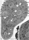
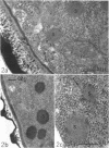
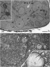
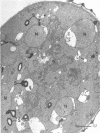
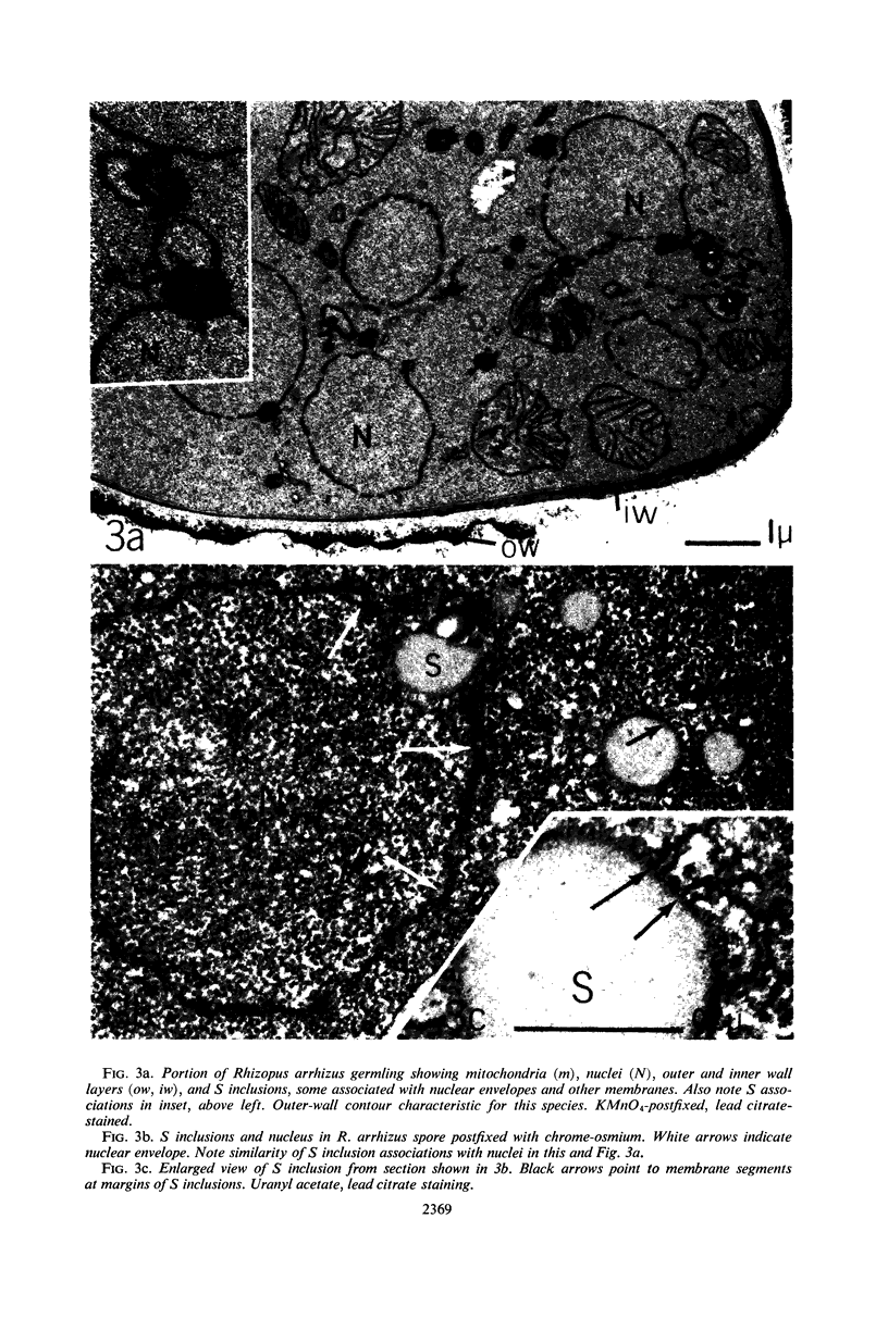
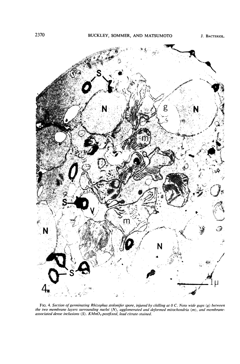
Electron microscope examination of sporangiospore sections from Rhizopus stolonifer (Ehrenb. ex Fr.) Lind. and R. arrhizus Fischer revealed details on intracellular organization not previously reported. Aldehyde fixation followed by chromeosmium postfixation permitted clear depiction of ribosomes hitherto unrevealed in these cells. Mitochondria were diversiform. Spore wall structures in the two species were generally similar, but outer contours differed sufficiently to permit easy species identification in examination of sections. The spores of both species abounded in cytosomes, corresponding in size, shape, and heavy-metal “stain” affinities to spherosomes in cells of higher plants. The osmiophilic response of these spherosome-like inclusions was intensified by treatment of sections with thiocarbohydrazide solution and subsequent application of aqueous osmium tetroxide, which strengthens an assumption that they are lipid-rich. The margins of the spherosome-like inclusions in lead citrate-stained sections included dense particles, about 60 A across, whose crystalline-like arrangements suggested that protein as well as lipid was present. Frequent and close associations between the spherosome-like inclusions and various cell membranes suggested that such bodies participate in membrane elaboration during germination.
Full text
PDF
Images in this article
Selected References
These references are in PubMed. This may not be the complete list of references from this article.
- Buckley P. M., Sjaholm V. E., Sommer N. F. Electron microscopy of Botrytis cinerea conidia. J Bacteriol. 1966 May;91(5):2037–2044. doi: 10.1128/jb.91.5.2037-2044.1966. [DOI] [PMC free article] [PubMed] [Google Scholar]
- CARASSO N., FAVARD P. L origine des plaquettes vitellines de l'oeuf de Planorbe. C R Hebd Seances Acad Sci. 1958 Mar 10;246(10):1594–1597. [PubMed] [Google Scholar]
- EKUNDAYO J. A., CARLILE M. J. THE GERMINATION OF SPORANGIOSPORES OF RHIZOPUS ARRHIZUS; SPORE SWELLING AND GERM-TUBE EMERGENCE. J Gen Microbiol. 1964 May;35:261–269. doi: 10.1099/00221287-35-2-261. [DOI] [PubMed] [Google Scholar]
- ERICSSON J. L., TRUMP B. F. ELECTRON MICROSCOPIC STUDIES OF THE EPITHELIUM OF THE PROXIMAL TUBULE OF THE RAT KIDNEY. I. THE INTRACELLULAR LOCALIZATION OF ACID PHOSPHATASE. Lab Invest. 1964 Nov;13:1427–1456. [PubMed] [Google Scholar]
- HAWKER L. E. FINE STRUCTURE OF FUNGI AS REVEALED BY ELECTRON MICROSCOPY. Biol Rev Camb Philos Soc. 1965 Feb;40:52–92. doi: 10.1111/j.1469-185x.1965.tb00795.x. [DOI] [PubMed] [Google Scholar]
- Hyde J. M., Walkinshaw C. H. Ultrastructure of basidiospores and mycelium of Lenzites saepiaria. J Bacteriol. 1966 Oct;92(4):1218–1227. doi: 10.1128/jb.92.4.1218-1227.1966. [DOI] [PMC free article] [PubMed] [Google Scholar]
- NECAS O., HAVELKOVA M., SOUDEK D. SUBMICROSCOPIC MORPHOLOGY OF RHIZOPUS NIGRICANS. Folia Microbiol (Praha) 1963 Sep;40:290–292. doi: 10.1007/BF02868772. [DOI] [PubMed] [Google Scholar]
- REYNOLDS E. S. The use of lead citrate at high pH as an electron-opaque stain in electron microscopy. J Cell Biol. 1963 Apr;17:208–212. doi: 10.1083/jcb.17.1.208. [DOI] [PMC free article] [PubMed] [Google Scholar]
- SOMMER N. F., CREASY M., ROMANI R. J., MAXIE E. C. AN OXYGEN-DEPENDENT POSTIRRADIATION RESTORATION OF RHIZOPUS STOLONIFER SPORANGIOSPORES. Radiat Res. 1964 May;22:21–28. [PubMed] [Google Scholar]
- Seligman A. M., Wasserkrug H. L., Hanker J. S. A new staining method (OTO) for enhancing contrast of lipid--containing membranes and droplets in osmium tetroxide--fixed tissue with osmiophilic thiocarbohydrazide(TCH). J Cell Biol. 1966 Aug;30(2):424–432. doi: 10.1083/jcb.30.2.424. [DOI] [PMC free article] [PubMed] [Google Scholar]
- VOELZ H., NIEDERPRUEM D. J. FINE STRUCTURE OF BASIDIOSPORES OF SCHIZOPHYLLUM COMMUNE. J Bacteriol. 1964 Nov;88:1497–1502. doi: 10.1128/jb.88.5.1497-1502.1964. [DOI] [PMC free article] [PubMed] [Google Scholar]


