Abstract
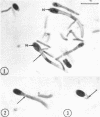
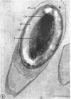
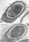
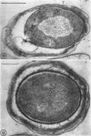
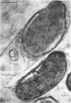
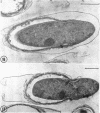
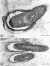
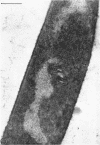
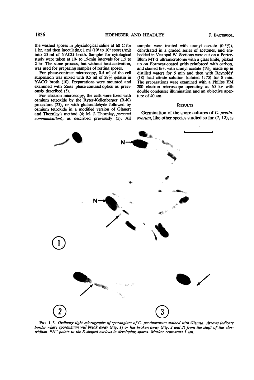
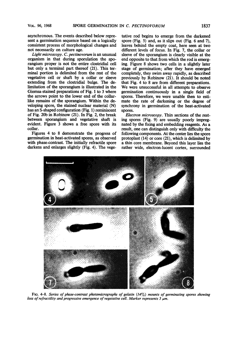
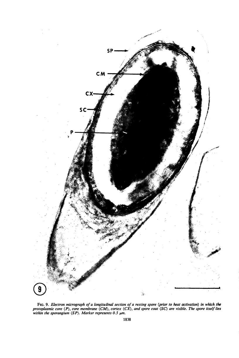
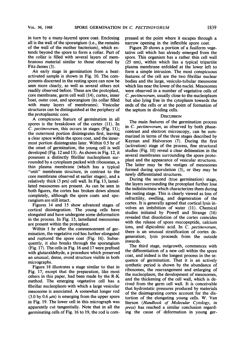
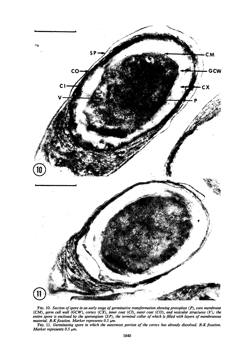
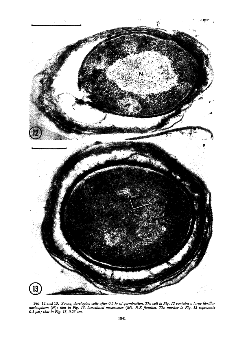
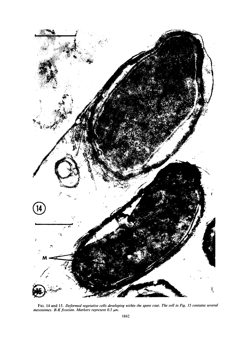
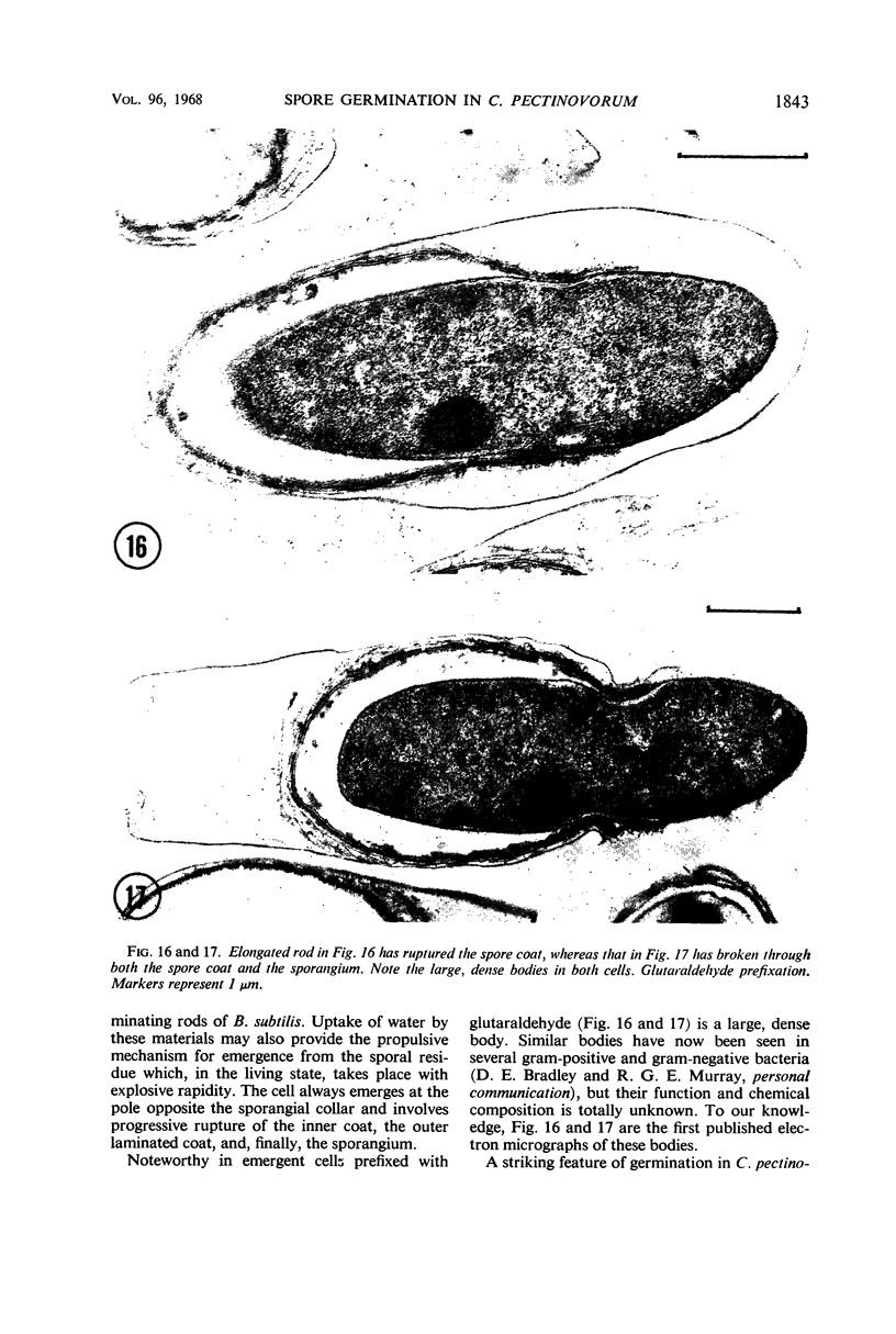
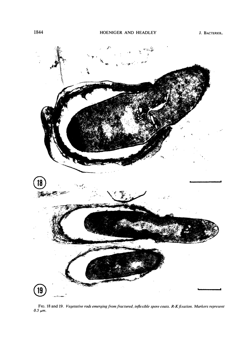
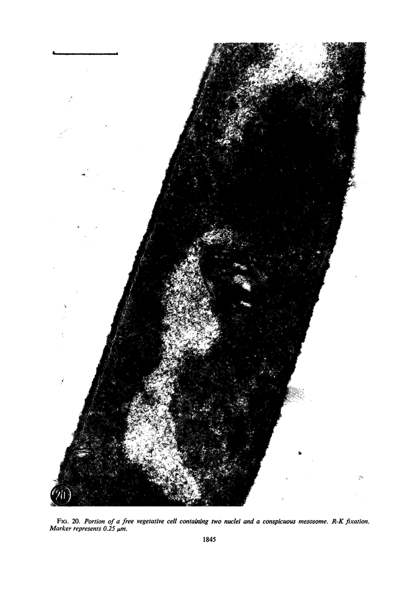
The process of spore germination in Clostridium pectinovorum has been followed by phase-contrast and electron microscopy. Unlike most other Bacillaceae, germination of this species takes place within the sporangium. Under phase-contrast, the spore darkens and swells slightly, and then the vegetative rod slips out through the end opposite the collar-like extension of the sporangium. In thin sections, a spore from an early stage in germination consists of a central protoplast, core membrane, germ cell wall, cortex, and two coats. Within a short period, the cortex disintegrates and the young cell develops. It possesses a large fibrillar nucleoplasm and several mesosomes. Subsequently, the young cell elongates, becomes somewhat deformed, and then emerges through a narrow aperture in the inflexible coats of the spore, finally rupturing the sporangium. Free vegetative cells of C. pectinovorum resemble in their structure other gram-positive rods.
Full text
PDF
Images in this article
Selected References
These references are in PubMed. This may not be the complete list of references from this article.
- CHAPMAN G. B., ZWORYKIN K. A. Study of germinating Bacillus cereus spores employing television microscopy of living cells and electron microscopy of ultrathin sections. J Bacteriol. 1957 Aug;74(2):126–132. doi: 10.1128/jb.74.2.126-132.1957. [DOI] [PMC free article] [PubMed] [Google Scholar]
- Fitz-James P. C. MORPHOLOGY OF SPORE DEVELOPMENT IN CLOSTRIDIUM PECTINOVORUM. J Bacteriol. 1962 Jul;84(1):104–114. doi: 10.1128/jb.84.1.104-114.1962. [DOI] [PMC free article] [PubMed] [Google Scholar]
- Hoeniger J. F., Stuart P. F., Holt S. C. Cytology of spore formation in Clostridium perfringens. J Bacteriol. 1968 Nov;96(5):1818–1834. doi: 10.1128/jb.96.5.1818-1834.1968. [DOI] [PMC free article] [PubMed] [Google Scholar]
- Knaysi G., Baker R. F., Hillier J. A Study, with the High-Voltage Electron Microscope, of the Endospore and Life Cycle of Bacillus mycoides. J Bacteriol. 1947 May;53(5):525–537. doi: 10.1128/jb.53.5.525-537.1947. [DOI] [PMC free article] [PubMed] [Google Scholar]
- Knaysi G., Hillier J. PRELIMINARY OBSERVATIONS ON THE GERMINATION OF THE ENDOSPORE IN BACILLUS MEGATHERIUM AND THE STRUCTURE OF THE SPORE COAT. J Bacteriol. 1949 Jan;57(1):23–29. doi: 10.1128/jb.57.1.23-29.1949. [DOI] [PMC free article] [PubMed] [Google Scholar]
- MASON D. J., POWELSON D. M. Nuclear division as observed in live bacteria by a new technique. J Bacteriol. 1956 Apr;71(4):474–479. doi: 10.1128/jb.71.4.474-479.1956. [DOI] [PMC free article] [PubMed] [Google Scholar]
- Moberly B. J., Shafa F., Gerhardt P. Structural details of anthrax spores during stages of transformation into vegetative cells. J Bacteriol. 1966 Jul;92(1):220–228. doi: 10.1128/jb.92.1.220-228.1966. [DOI] [PMC free article] [PubMed] [Google Scholar]
- POWELL J. F., STRANGE R. E. Biochemical changes occurring during the germination of bacterial spores. Biochem J. 1953 May;54(2):205–209. doi: 10.1042/bj0540205. [DOI] [PMC free article] [PubMed] [Google Scholar]
- Propst A., Möse J. R. Sporulation und Germination beim Clostridium butyricum M 55. Elektronenmikroskopische Untersuchung. Zentralbl Bakteriol Orig. 1966 Nov;201(3):373–395. [PubMed] [Google Scholar]
- REYNOLDS E. S. The use of lead citrate at high pH as an electron-opaque stain in electron microscopy. J Cell Biol. 1963 Apr;17:208–212. doi: 10.1083/jcb.17.1.208. [DOI] [PMC free article] [PubMed] [Google Scholar]
- Rousseau M., Fléchon J., Hermier J. Etude au microscope électronique de la germination de la spore chex Bacillus subtilis. Ann Inst Pasteur (Paris) 1966 Aug;111(2):149–160. [PubMed] [Google Scholar]
- TAKAGI A., KAWATA T., YAMAMOTO S. Electron microscope studies on ultrathin sections of spores of the Clostridium group, with special reference to the sporulation and germination process. J Bacteriol. 1960 Jul;80:37–46. doi: 10.1128/jb.80.1.37-46.1960. [DOI] [PMC free article] [PubMed] [Google Scholar]
- TAKAGI A., KAWATA T., YAMAMOTO S., KUBO T., OKITA S. Electron microscopic studies on ultrathin sections of spores of Clostridium tetani and Clostridium histolyticum, with special reference to sporulation and spore germination process. Jpn J Microbiol. 1960 Apr;4:137–155. doi: 10.1111/j.1348-0421.1960.tb00162.x. [DOI] [PubMed] [Google Scholar]


