Abstract
Using affinity chromatography and surface plasmon resonance analysis, we have identified cubilin, a 460-kDa receptor heavily expressed in kidney proximal tubule epithelial cells, as an albumin binding protein. Dogs with a functional defect in cubilin excrete large amounts of albumin in combination with virtually abolished proximal tubule reabsorption, showing the critical role for cubilin in the uptake of albumin by the proximal tubule. Also, by immunoblotting and immunocytochemistry we show that previously identified low–molecular-weight renal albumin binding proteins are fragments of cubilin. In addition, we find that mice lacking the endocytic receptor megalin show altered urinary excretion, and reduced tubular reabsorption, of albumin. Because cubilin has been shown to colocalize and interact with megalin, we propose a mechanism of albumin reabsorption mediated by both of these proteins. This process may prove important for understanding interstitial renal inflammation and fibrosis caused by proximal tubule uptake of an increased load of filtered albumin.
Introduction
Proteinuria, in particular albuminuria, has long been considered a marker of progression not only in renal, but also in selected forms of cardiovascular disease. Several studies in recent years have suggested that the presence of proteins in the ultrafiltrate may be an independent factor causing deterioration in kidney function. Hyperfiltration of albumin is followed by increased reabsorption in the kidney proximal tubule (1). Both animal and clinical studies have suggested that protein and albumin itself can be toxic to the proximal tubule epithelium causing interstitial inflammation and fibrosis (2–7). This implicates that tubular uptake of albumin may be an important factor for the development and progression of chronic renal disease.
Cubilin is a 460-kDa glycoprotein with no transmembrane domain (8). The receptor is heavily expressed in the kidney proximal tubule brush border and intracellular endocytic compartments (9). Cubilin binds to the transmembrane, endocytic receptor megalin, and it has been suggested that megalin is a coreceptor involved in the endocytosis and intracellular trafficking of cubilin (8). Cubilin is also the established intrinsic factor receptor involved in the intestinal uptake of the intrinsic factor vitamin B12 complex (10, 11). Little or no intrinsic factor (IF) is present in plasma, and although cubilin recently was identified as a receptor for HDL and apolipoprotein A-I (12, 13), the physiological significance of cubilin within the kidney is not fully elucidated. Recently, mutations in the cubilin gene have been identified in patients suffering from Imerslund-Gräsbeck syndrome, an inherited vitamin B12 deficiency disease characterized by vitamin B12 malabsorption (14). These patients have varying degrees of proteinuria resistant to vitamin B12 supplementation (15, 16). Furthermore, an inherited vitamin B12 malabsorption syndrome in dogs characterized by abnormal processing and the absence of apical cubilin in the intestinal and renal epithelia cells is associated with proteinuria (17, 18). Our finding of heavy albuminuria in these animals prompted us to study the significance of cubilin to normal tubular albumin reabsorption.
Using different biochemical and morphological approaches, including affinity chromatography, surface plasmon resonance (SPR) analysis, and immunocytochemistry to analyze renal tissues and urine from animal models of cubilin and megalin deficiency, we have identified cubilin as an albumin binding protein important to normal tubular albumin reabsorption. On the basis of these data, we suggest a new and complex mechanism of albumin reabsorption involving the two high–molecular-weight receptors, possibly through inter-receptor interactions.
Methods
Renal tissues and urine.
Rat kidney cortical membranes were obtained from Wistar rats. The kidney cortex was dissected from individual kidneys, minced, and homogenized in 0.31 M sucrose, 15 mM HEPES, 8.5 μM leupeptin, 0.4 mM Pefabloc (pH 7.5) by five strokes of a Potter-Elvehjem homogenizer at 1,250 rpm. The homogenate was centrifuged at 4,000 g for 15 minutes, rehomogenized, and the supernatants were pooled and centrifuged at 200,000 g for 1 hour. The pellet representing crude membranes was solubilized overnight in 0.14 M NaCl, 2 mM CaCl2, 10 mM HEPES, 0.4 mM Pefabloc, and 1% Triton X-100 (pH 7.4). The solubilized membranes were recovered as the supernatant after centrifugation at 30,000 g for 1 hour. All procedures were carried out on ice or at 4°C.
A family of mixed-breed dogs originating from giant schnauzer dogs exhibiting simple autosomal recessive inheritance of selective cobalamin malabsorption has previously been thoroughly characterized (17, 18). Biochemical analyses revealed a decrease in IF-B12 binding activity in both kidney homogenate (tenfold) and isolated kidney cortical brush border membranes (180-fold) supported by the absence of immunodetectable cubilin in the brush border membrane fraction by immunoblotting. Furthermore, the cubilin from affected dog kidney was shown to be largely endo-H sensitive in contrast to cubilin from normal dogs (18). Thus, these dogs exhibit abnormal processing and defective insertion of cubilin into the luminal plasma membranes of cubilin expressing cells, causing failure of intestinal absorption of IF-B12 and selective proteinuria resembling the Imerslund-Gräsbeck disease in humans (17). All affected dogs were treated regularly with parenteral vitamin B12 and were in hematologic and metabolic remission. Urine samples were collected from six affected and six normal dogs of various strains, age, and sex. For immunocytochemistry, kidneys from three affected and three normal dogs were analyzed. Megalin-deficient mice were produced by gene targeting as described elsewhere (19), and littermates were used as controls. Urine was collected during a 6- to 24-hour period. Mouse kidneys were fixed by perfusion through the left ventricle of the heart; rat kidneys were fixed by retrograde perfusion through the abdominal aorta; and dog kidneys were fixed by perfusion through the renal arteries. The fixative used was 1–4% paraformaldehyde in 0.1 M sodium cacodylate buffer (pH 7.4).
Albumin affinity purification of cubilin.
Rat serum albumin (RSA; 10 mg/mL; Sigma Chemical Co., St. Louis, Missouri, USA) was coupled to CNBr-activated Sepharose (Amersham Pharmacia Biotech AB, Uppsala, Sweden) using 0.1 M NaHCO3 and 0.5 M NaCl (pH 8.3) coupling buffer according to the protocol provided by the manufacturer. Unreacted active sites were blocked with 0.2 M glycine buffer (pH 8.0) followed by repeated alternating washes with 0.1 M NaC2H3O2 and 0.5 M NaCl (pH 4.0) or the coupling buffer. A final volume of 5 mL gel was prepared. The solubilized rat kidney cortical membranes were recirculated over the column overnight at 0.2 mL/min. The column was extensively washed in 20 mM Tris and 0.15 M NaCl (pH 7.4) followed by elution of cubilin with 20 mM Tris, 0.15 M NaCl, and 6 M urea (pH 7.4). Fractions (1 mL) were collected, subjected to SDS-PAGE, and stained by Coomassie blue. Additional gels were blotted onto nitrocellulose membranes for further protein identification by immunoblotting.
SPR analysis.
For the SPR analyses, the BIAcore sensor chips (type CM5; Biacore AB, Uppsala, Sweden) were activated with a 1:1 mixture of 0.2 M N-ethyl-N′-(3-dimethylaminopropyl) carbodiimide and 0.05 M N-hydroxysuccinimide in water according to the manufacturer’s directions. Rat cubilin purified by IF-B12 affinity chromatography was immobilized as described elsewhere (11). The SPR signal from immobilized cubilin generated BIAcore response units (RUs) equivalent to 25–35 fmol/mm2. The flow cells were regenerated with 20 μL and 1.5 M glycine-HCl (pH 3.0). The flow buffer was 10 mM HEPES, 0.15 M NaCl, 1.5 mM CaCl2, and 1 mM EGTA (pH 7.4). The Kd for binding was estimated using a BIAevaluation program (Biacore AB).
Receptors, ligands, and antibodies.
Cubilin was purified from rat kidney cortical membranes by IF-B12 affinity chromatography of renal cortical membranes according to the procedure described elsewhere (11). Rat albumin was obtained commercially as fraction V powder and as globulin and/or fatty acid free, lyophilized powder (Sigma Chemical Co.). Glycated or palmitylated albumin was prepared from globulin and fatty acid free rat albumin according to protocols described by others previously (20, 21). In short, for glycation, 2 mg of rat albumin was dissolved in 60 μL of 0.5 M glucose, 33 mM PBS, and 15 mM NaN3, and was incubated for 24 hours at 37°C giving an approximately 3:1 ratio of glucose/albumin. This was followed by dialysis against 10 mM HEPES, 0.14 M NaCl, and 2 mM CaCl2 (pH 7.4) for 24 hours at 5°C. For palmitylation, 2.8 μL of a 90 mM solution of palmitic acid in ethanol was added to 138 μL of rat albumin solution (300 μM) under stirring. The solution was lyophilized and dissolved in 10 mM HEPES, 0.14 M NaCl, and 2 mM CaCl2 (pH 7.4) before use. Samples of possible low–molecular-weight albumin binding proteins was generated by Cessac-Guillemet et al. as described previously (22).
The following polyclonal antibodies were used for RIA, immunoblotting, and immunocytochemistry: rabbit anti-rat cubilin antibodies raised against immunopurified protein (23); rabbit anti-dog cubilin (18); rabbit anti-albumin binding protein raised against affinity purified albumin binding proteins as described elsewhere (22); rabbit anti-dog albumin (Organon Teknika Corp., Boxtel, The Netherlands); rabbit anti-rat albumin (Nordic Immunological, Tilbury, The Netherlands); sheep anti-rat albumin (Biogenesis, Poole, United Kingdom); and sheep anti-mouse albumin (The Binding Site, Birmingham, United Kingdom). A mouse monoclonal anti-rat cubilin antibody (75; ref. 24) and a sheep anti-megalin (25) was used for triple labeling immunocytochemistry. The secondary antibodies used were horseradish peroxidase–conjugated (HRP-conjugated) goat anti-rabbit, goat anti-mouse, and rabbit anti-sheep immunoglobulins (DAKO A/S, Glostrup, Denmark), and colloidal gold coupled goat anti-rabbit, goat anti-mouse, and donkey anti-sheep immunoglobulins (British BioCell International, Cardiff, United Kingdom).
Determination of urinary albumin excretion by RIA.
Dog or mouse urine samples were incubated with polyclonal antibodies against dog (1:10–1:500) or rat albumin (1:10, cross-reacting fully with mouse serum albumin). After 48 hours’ incubation, polyethylene glycol was used for separation of free and antibody-bound iodinated albumin as described previously (26). Dog or rat albumin (Sigma Chemical Co.) was used for iodination and calibration. Albumin excretion both in dogs and mice was related to creatinine excretion measured by standard procedure. Data was compared using the Student’s t test.
SDS-PAGE and immunoblotting.
Samples of membrane fractions or urine proteins were subjected to SDS-PAGE using polyacrylamide minigels (Bio-Rad Mini Protean II; Bio-Rad Laboratories Inc., Hercules, California, USA), and transferred to nitrocellulose membranes. Blots were blocked with 5% nonfat milk in PBS-T (80 mM Na2HPO4, 20 mM NaH2PO4, 0.1 M NaCl, and 0.1% Tween 20 [pH 7.5]) for 1 hour, and incubated overnight at 4°C with primary antibody in PBS-T with 1% BSA. After washing in PBS-T, the blots were incubated for 1 hour with HRP-conjugated secondary antibody diluted 1:3,000. After final wash, antibody binding was visualized using the ECL enhanced chemiluminescence system (Amersham Pharmacia Biotech AB). Controls involving incubation without primary antibody and incubation with nonspecific serum revealed no significant labeling.
Immunocytochemistry.
For light microscope immunocytochemistry, semithin cryosections were cut on a Reichert Ultracut S (Reichert-Jung, Vienna, Austria) and placed on gelatine-coated glass slides. Sections were incubated 1 hour with the primary antibody diluted 1:3,000 to 1:200,000 in 10 mM PBS, 0.15 M NaCl, 0.1% nonfat milk, and 20 mM NaN3, followed by incubation for 1 hour with HRP-conjugated secondary antibody and visualization by incubation with diaminobenzidine and 0.03% H2O2 for 10 minutes. Preincubation experiments were performed by mixing anti-rat albumin binding protein antibody with excess rat cubilin purified by IF-B12 affinity chromatography before the incubation with sections. All incubations were performed at room temperature, and sections were counterstained with Mayers hematoxylin stain before examination in a Leica DMR microscope equipped with a Sony SCCD color video camera and a Sony Digital Still recorder (Sony Corp., Tokyo, Japan). Electron microscope immunocytochemistry was performed on ultrathin (90 nm) cryosections incubated overnight at 5°C with primary antibodies diluted 1:2,500–1:200,000 followed by incubation with gold conjugated secondary antibodies in 50 mM Tris buffer, 0.15 M NaCl, 0.1% BSA, 0.06% PEG, 1% fish gelatine, and 20 mM NaN3 for 2 hours at room temperature. Sections were photographed in a Phillips CM100 or EM208 electron microscope (Phillips, Eindhoven, The Netherlands). Controls involving incubation without primary antibody or incubation with nonspecific mouse immunoglobulins or rabbit or sheep serum revealed no significant labeling.
Results
Cubilin is an albumin binding protein.
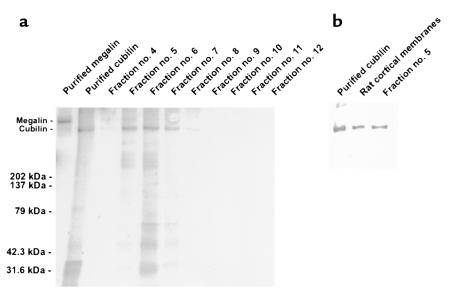
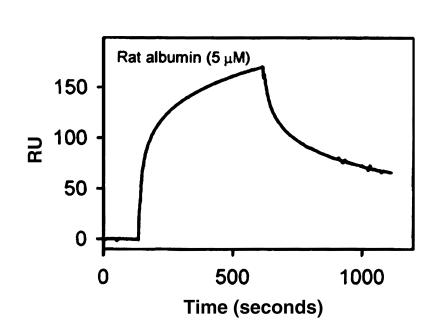
Cubilin was identified as an albumin binding protein by affinity chromatography. When rat cortical membranes were applied to a column of rat albumin, significant amounts of an approximately 460-kDa protein were recovered. This protein could be identified as cubilin by immunoblotting using antibodies raised against immunopurified rat cubilin (Figure 1). SPR analysis confirmed binding of albumin to purified and immobilized cubilin (Figure 2). Using the BIAevaluation program, Kd was estimated to 0.63 μM. A slightly lower binding affinity was found for palmitylated albumin, whereas no difference was observed with fatty acid free or glycated albumin (data not shown).
Figure 1.
Albumin affinity purification of cubilin. (a) Eluted fractions were analyzed by SDS-PAGE and Coomassie stained. Lane 1: 0.1 μg of RAP affinity purified rabbit megalin; lane 2: IF-B12 affinity purified cubilin. Lanes 3–11: fractions 4–12 (12 μL/lane). The positions of standard molecular weight markers are indicated. A strong band corresponding to the size of cubilin is clearly identified in fractions 5–8. Faint low–molecular-weight bands are seen with both the IF-B12 purified and albumin purified cubilin. (b) The eluted protein was further identified as cubilin by immunoblotting using anti-rat cubilin antibodies.
Figure 2.
SPR sensorgram of the binding of rat albumin to purified rat cubilin immobilized to a BIAcore sensor chip. The BIAevaluation software estimated Kd = 0.63 μM when fitting the curves to a one-site model. The on and off rates for binding of rat albumin were recorded by a flow of 5 μM albumin followed by flow of buffer alone. The SPR signal is displayed as the mass-equivalent RUs.
Cubilin is important to normal albumin reabsorption.
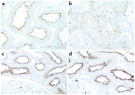
To establish the physiological significance of cubilin to renal tubular albumin reabsorption, we examined dogs exhibiting an inherited defect in cubilin expression. Biochemical studies have shown abnormal processing and defective insertion of cubilin into the luminal plasma membranes of affected dogs (18). We confirmed this by immunocytochemistry. In contrast to the luminal localization in normal dog proximal tubules, the affected dogs revealed an intracellular, vesicular labeling with no detectable receptor in the luminal plasma membrane (Figure 3, a and b). No difference was observed in the labeling intensity or in the distribution of megalin (Figure 3, c and d), showing that this receptor is normally expressed in the affected dogs.
Figure 3.
Localization of cubilin (a and b) and megalin (c and d) in kidney cortex of normal (a and c) and affected (b and d) dogs. Sections were incubated with anti-dog cubilin (1:4,000). In normal dogs, cubilin is localized in luminal plasma membranes of kidney proximal tubules (a), whereas the labeling in affected dogs is intracellular and vesicular with no detectable receptor in the luminal plasma membrane (b), suggesting retention in an intracellular compartment. No difference in the labeling intensity or in the distribution of megalin is observed between normal (c) and affected (d) dogs when evaluated by immunocytochemistry using a sheep anti-rat megalin (1:10,000). ×600 (a and b); ×350 (c and d).
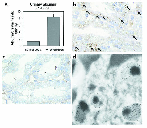
A significant albuminuria reflected by an approximately sevenfold increase in urinary albumin/creatinine excretion ratio (Figure 4a) was observed in six dogs with defective cubilin expression compared with six normal control dogs (P = 0.00003). To evaluate the origin of this albuminuria, we analyzed tubular albumin reabsorption by immunohistochemistry. In normal dogs, efficient tubular reabsorption was reflected by intense intracellular labeling for albumin in kidney proximal tubule cells (Figure 4b). Labeling appeared vesicular, and electron microscope immunocytochemistry revealed labeling of the endocytic apparatus, in particular lysosomes reflecting the accumulation of endocytosed albumin in this compartment (Figure 4d). In affected dogs, no intracellular labeling was observed in the renal tubules (Figure 4c), suggesting defective internalization. Thus, the functional defect in luminal cubilin expression is associated with decreased proximal tubule albumin endocytosis and increased urinary albumin excretion.
Figure 4.
Increased urinary excretion and decreased tubular reabsorption of albumin in affected dogs. (a) Dogs with a functional defect in cubilin expression (n = 6) reveals a significant, approximately sevenfold increase (P = 0.00003) in urinary albumin/creatinine excretion compared with normal control dogs (n = 6). (b and c) Immunohistochemistry using an anti-dog albumin antibody (1:100,000) to identify albumin in the kidney cortex of normal (b) and affected (c) dogs. In normal dogs (b), evidence of reabsorbed and endocytosed albumin in proximal tubules is clear (arrows), whereas no albumin can be identified inside the proximal tubules of affected dogs (c). In all dogs, a faint labeling was observed in basement membranes due to a basal deposition of albumin. ×600. Three affected and three normal dogs were examined using two different anti-albumin antibodies, all showing the same labeling pattern. (d) Electron microscopic immunocytochemistry using anti-dog albumin antibody (1:5,000) on a section of normal dog proximal tubule cells. Intense labeling of four lysosomes and part of a fifth is observed corresponding to the vesicular labeling observed with light microscopic immunohistochemistry. ×33,000.
Immunological similarity between cubilin and previously identified low–molecular-weight albumin binding proteins.
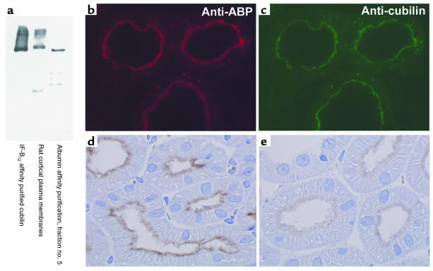
Antibodies raised against previously described albumin binding proteins of 31 and 55 kDa (22) reacted against cubilin purified on both IF-B12 and albumin affinity columns (Figure 5a). The apical expression of these proteins in the kidney proximal tubule cells as revealed by immunohistochemistry using an antibody against the low–molecular-weight albumin binding proteins was very similar to that of cubilin identified by anti-cubilin antibodies (Figure 5, b and c). Furthermore, the labeling by the antibody against the low–molecular-weight albumin binding proteins could be strongly inhibited by preincubation with IF-B12 affinity purified cubilin (Figure 5, d and e). Finally, immunoblotting using the anti-cubilin antibody on the albumin binding protein samples purified by Cessac-Guillemet et al. (22) revealed the same low–molecular-weight bands as identified by the antibody against the low–molecular-weight albumin binding proteins, suggesting that these protein bands represent fragments of cubilin (data not shown).
Figure 5.
Immunological cross-reaction and colocalization of antibodies raised against cubilin and antibodies against previously identified albumin binding proteins (ABP). (a) Immunoblotting using polyclonal anti-ABP antibody on affinity purified rat cubilin and rat kidney cortex. Both the IF-B12 and albumin affinity purified cubilin, as well as a similar band in rat kidney cortex, were recognized by the anti-ABP. (b and c) Fluorescent double labeling reveals identical labeling using rabbit anti-ABP (1:3,000) and monoclonal mouse anti-cubilin (1:5,000) on the same section of rat cortex followed by secondary TRITC labeled anti-rabbit IgG (red fluorescent in b) and FITC-labeled anti-mouse IgG (green fluorescent in c). The antibodies colocalize along the brush border and apical cytoplasm, as well as in vesicular structures in the proximal tubules. (d and e) Inhibition of labeling with anti-ABP by preincubation with IF-B12 affinity purified cubilin. Immunocytochemical labeling using anti-ABP (1:8,000) on cryosections of kidney cortex reveals proximal tubule apical labeling (d) that is clearly inhibited when the anti-ABP is preincubated with affinity purified cubilin (e). ×700.
Megalin is involved in albumin reabsorption.
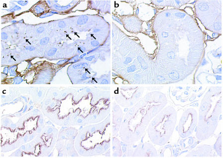
An approximately threefold increase in albumin/creatinine excretion ratio was observed in five megalin-deficient mice compared with five wild-type mice (31 ± 12 vs. 11 ± 6 μg/mg). Although not statistically significant (P = 0.17), the observation is in accordance with decreased tubular albumin reabsorption. Owing to the poor viability of megalin-deficient mice (19), we were unable to collect urine from more than five megalin-knockout mice. Tubular albumin reabsorption in megalin-deficient and control mice was further evaluated by immunohistochemistry identifying internalized albumin in kidney tubules. In normal mice, albumin could be identified as a vesicular labeling in proximal tubules, whereas no albumin was detected in proximal tubules from megalin-deficient mice (Figure 6). These findings were consistent when analyzing four different knockout and five corresponding wild-type or heterozygous, normal mice. Thus, albumin reabsorption was strongly inhibited in megalin-knockout mice.
Figure 6.
Decreased tubular reabsorption of albumin in megalin-deficient mice. (a and b) Immunohistochemistry using an anti-mouse albumin antibody (1:3,000) to identify albumin in the kidney cortex of normal (a) and megalin-deficient (b) mice. In normal mice (a), evidence of reabsorbed and endocytosed albumin in proximal tubules is clear (arrows), whereas no albumin can be identified inside the proximal tubules of megalin-deficient mice (b). ×1,000. Four megalin-deficient and five normal mice were examined, showing the same difference in labeling pattern. (c and d) Immunocytochemical localization of cubilin using an anti-dog cubilin antibody (1:200) in normal (c) and megalin-deficient (d) mice. Cubilin is expressed and normally distributed in the megalin-deficient mice (d) when compared with controls (c); however, the labeling intensity is reduced. ×600.
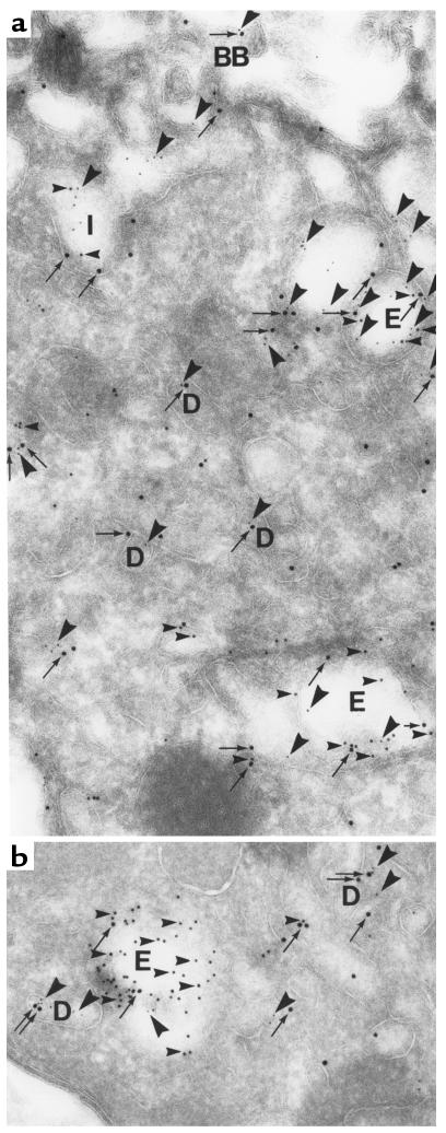
The involvement of both cubilin and megalin in albumin reabsorption was supported by the colocalization of endogenous albumin, megalin, and cubilin in endocytic compartments of proximal tubule cells as demonstrated by electron microscope immunocytochemistry (Figure 7). Cubilin and megalin colocalized on the luminal membrane, in endocytic compartments, and in the recycling compartment dense apical tubules. Albumin was found in similar compartments, however concentrating in late endosomes and lysosomes, suggesting that most of the endocytosed ligand is internalized for degradation, whereas the receptors recycle to the luminal membrane.
Figure 7.
Colocalization of endogenous albumin, cubilin, and megalin in endocytic compartments of normal rat proximal tubules. Section of rat kidney proximal tubule incubated with rabbit anti-rat albumin (1:10,000), sheep anti-rat megalin (1:50,000), and monoclonal mouse anti-rat cubilin (1:2,500) followed by incubation with gold conjugated goat anti-rabbit immunoglobulins (10 nm), donkey anti-sheep immunoglobulins (15 nm), and goat anti-mouse immunoglobulins (5 nm). Megalin (15-nm gold particles; arrows) and cubilin (5-nm gold particles; large arrowheads) colocalize (a) at the brush border (BB) and in endocytic vesicles (E) as well as in the recycling compartment dense apical tubules (D). Albumin (10-nm gold particles; small arrowheads) colocalize with the receptors in endocytic invaginations (I) and vesicles (E) and is concentrated in later endocytic compartments and lysosomes containing only little labeling for the receptors (b). Notice that most tubular structures (D) contain only labeling for the receptors supporting efficient recycling of these in contrast to albumin, which is degraded (b). ×58,000.
Discussion
The present data demonstrate the involvement of cubilin in kidney proximal tubule albumin reabsorption. Cubilin binds to albumin during affinity chromatography and binding is confirmed by SPR analysis. Furthermore, dogs with an abnormal processing and defective insertion of cubilin into the luminal membranes (Figure 1) causing a defect in intestinal absorption of IF-B12 and proteinuria display significant albuminuria and decreased tubular uptake of albumin. The clinically related Imerslund-Gräsbeck disease is also associated with various degrees of albuminuria (approximately two thirds of total urinary protein) (27). Recently, two independent mutations in the cubilin gene was identified in 17 Finnish families suffering from this disease (14), indicating a heterogeneity of mutations that is likely to explain some of the differences in protein and albuminuria observed in Imerslund-Gräsbeck patients (15, 27).
The Kd of albumin binding to cubilin estimated by SPR analysis is 0.63 μM. This is in accordance with previously described affinities for albumin binding to isolated proximal tubule segments and cultured opossum kidney cells (28–31). Most studies identified both high- and low-affinity binding sites with affinities ranging 0.3–2.0 and 18–240 μM, respectively (30, 31). Whereas the affinity for albumin binding to cubilin demonstrated here compares to the previously described high-affinity albumin binding sites, the affinity for albumin binding is low compared with the other known ligands for cubilin, i.e., IF (11) and apolipoprotein A-1 (13). A relatively low affinity of albumin reabsorption explains why increases in the load of filtered albumin lead to increases in urinary albumin excretion rates (32), and why the uptake of native albumin is significantly reduced when microinjected into late parts of the proximal convoluted tubule (33).
Cubilin is also heavily expressed the yolk sac epithelia (34). In early gestation of rodents, the yolk sac serves an important function for embryonic development as the major source of amino acids after the uptake and degradation of maternal protein (35). Because albumin constitutes the major plasma protein, it can be speculated that cubilin is important for the delivery of amino acids in the early embryonic period of rodents.
Several putative albumin binding proteins have been described in endothelial cells including 18-, 30-, and 60-kDa glycoproteins (36–38). Receptors of similar molecular weight were purified from kidney cortex by albumin affinity chromatography and identified by immunoblotting and immunocytochemistry (22). The present study shows strong cross reactivity between antibodies raised against these low–molecular-weight binding proteins and cubilin in addition to a very similar cellular distribution in the proximal tubule cells. Furthermore, the anti-cubilin antibody identified the same low–molecular-weight protein bands as the antibody raised by Cessac-Guillemet et al. (22), strongly indicating that these low–molecular-weight proteins are in fact fragments of cubilin. When performing albumin affinity chromatography similar to the protocol described by Cessac-Guillemet et al. (22), we were able to purify significant amounts of cubilin, which however, were not identified in their study. This may be explained by their use of 12% polyacrylamide separating gels to identify purified proteins, not allowing the migration of high–molecular-weight proteins such as cubilin. Electrophoresis of both IF-B12 and albumin purified cubilin also revealed very faint low–molecular-weight protein bands. Most likely these are fragments of cubilin. The bands are of varying molecular weight and have been observed previously when purifying cubilin by IF-B12 or RAP affinity chromatography (11). The present study does not exclude the presence of other albumin binding proteins, particularly in other cell types and possibly serving different physiological functions. A different affinity for modified albumin has been described for the 18- and 30-kDa binding proteins, suggesting that these are involved in the clearance of conformationally modified albumin (36). Because there is no evidence that only modified albumin is filtered in the glomeruli, it appears reasonable that receptors involved in tubular uptake would bind different forms of albumin with similar affinity, as is the case with binding of palmitylated or glycated albumin to cubilin.
The present study also supports a role for the multiligand, endocytic receptor megalin (9, 39) in tubular albumin uptake by showing decreased tubular albumin uptake and possibly increased urinary albumin excretion in megalin-deficient mice. These mice express cubilin apically as shown by immunocytochemistry. The difference in urinary albumin excretion between megalin-deficient and wild-type mice was less than the difference observed between affected and normal dogs. The average albumin/creatinine excretion ratio was 11 ± 6 μg/mg in normal mice compared with 1.2 ± 0.3 μg/mg in normal dogs. Assuming that the normal amount of albumin filtered is proportional to the creatinine excretion, this may suggest that the reabsorption of albumin in the mice is less efficient than in the dogs. Thus, a defective albumin reabsorption in the mice may result in a smaller relative increase in urinary albumin excretion when compared with the dogs. Megalin may be involved in tubular albumin reabsorption in at least two different ways. First, a previous study suggested that megalin was able to bind and mediate the endocytosis of albumin in kidney proximal tubules in vivo (40). Second, it was recently shown by megalin affinity chromatography and SPR analysis that cubilin binds with high affinity to megalin, and it was hypothesized that megalin mediates the endocytosis and intracellular trafficking of cubilin (8). This suggests an additional, indirect role of megalin in the reabsorption of albumin involving the binding of albumin to cubilin followed by megalin mediated endocytosis of the cubilin-albumin complex. The present finding of colocalization between endogenous albumin, cubilin, and megalin in endocytic compartments of rat proximal tubule cells supports this notion.
In conclusion, the present study shows that cubilin constitutes an albumin binding protein important for normal proximal tubule reabsorption of albumin. However, both cubilin and megalin deficiency is associated with decreased albumin uptake in the proximal tubule. Thus, a mechanism for reabsorption involving both receptors is suggested. Because excessive proximal tubule albumin uptake, under conditions of increased glomerular filtration of proteins, may contribute to the progression of renal disease, this mechanism may prove important to the understanding of renal pathophysiology and for establishing new strategies to prevent renal deterioration.
Acknowledgments
The work was supported in part by the Danish Medical Research Council, the University of Aarhus, the NOVO-Nordisk Foundation, the Leo Nielsen Foundation, the Ruth König-Petersen Foundation, Fonden til Lœgevidenskabens Fremme, the Beckett Foundation, the Biomembrane Research Center, Association pour la Recherche sur le Cancer grant number 9914 (to P.J. Verroust), and National Institutes of Health grant DK-45341 (to J.C. Fyfe). The skillful technical assistance by A.V. Steffensen, H. Sidelmann, I. Kristoffersen, K. Nyborg, and A. Meier is fully appreciated. The authors thank K. Ljungmann and S. Gregersen for providing rat kidneys.
Footnotes
This work was presented in part at the American Society of Nephrology 32nd annual meeting in Miami, Florida, USA, November 5–8, 1999. Portions of this work have appeared in abstract form (1999. J. Am. Soc. Nephrol. 10:A0316).
References
- 1.Exaire E, Pollak VE, Pesce AJ, Ooi BS. Albumin and -globulin in the nephron of the normal rat and following the injection of aminonucleoside. Nephron. 1972;9:42–54. doi: 10.1159/000180132. [DOI] [PubMed] [Google Scholar]
- 2.Remuzzi G, Bertani T. Pathophysiology of progressive nephropathies. N Engl J Med. 1998;339:1448–1456. doi: 10.1056/NEJM199811123392007. [DOI] [PubMed] [Google Scholar]
- 3.Thomas ME, et al. Proteinuria induces tubular cell turnover: a potential mechanism for tubular atrophy. Kidney Int. 1999;55:890–898. doi: 10.1046/j.1523-1755.1999.055003890.x. [DOI] [PubMed] [Google Scholar]
- 4.Remuzzi G. Abnormal protein traffic through the glomerular barrier induces proximal tubular cell dysfunction and causes renal injury. Curr Opin Nephrol Hypertens. 1995;4:339–342. doi: 10.1097/00041552-199507000-00009. [DOI] [PubMed] [Google Scholar]
- 5.Schreiner GF. Renal toxicity of albumin and other lipoproteins. Curr Opin Nephrol Hypertens. 1995;4:369–373. doi: 10.1097/00041552-199507000-00015. [DOI] [PubMed] [Google Scholar]
- 6.Gansevoort RT, Navis GJ, Wapstra FH, de Jong PE, de Zeeuw D. Proteinuria and progression of renal disease: therapeutic implications. Curr Opin Nephrol Hypertens. 1997;6:133–140. doi: 10.1097/00041552-199703000-00005. [DOI] [PubMed] [Google Scholar]
- 7.Jerums G, et al. Why is proteinuria such an important risk factor for progression in clinical trials? Kidney Int Suppl. 1997;63:S87–S92. [PubMed] [Google Scholar]
- 8.Moestrup SK, et al. The intrinsic factor-vitamin B12 receptor and target of teratogenic antibodies is a megalin-binding peripheral membrane protein with homology to developmental proteins. J Biol Chem. 1998;273:5235–5242. doi: 10.1074/jbc.273.9.5235. [DOI] [PubMed] [Google Scholar]
- 9.Christensen EI, Birn H, Verroust P, Moestrup SK. Membrane receptors for endocytosis in the renal proximal tubule. Int Rev Cytol. 1998;180:237–284. doi: 10.1016/s0074-7696(08)61772-6. [DOI] [PubMed] [Google Scholar]
- 10.Seetharam B, Christensen EI, Moestrup SK, Hammond TG, Verroust PJ. Identification of rat yolk sac target protein of teratogenic antibodies, gp280, as intrinsic factor-cobalamin receptor. J Clin Invest. 1997;99:2317–2322. doi: 10.1172/JCI119411. [DOI] [PMC free article] [PubMed] [Google Scholar]
- 11.Birn H, et al. Characterization of an epithelial 460 kDa protein that facilitates endocytosis of intrinsic factor-vitamin B12 and binds receptor-associated protein. J Biol Chem. 1997;272:26497–26504. doi: 10.1074/jbc.272.42.26497. [DOI] [PubMed] [Google Scholar]
- 12.Hammad SM, et al. Cubilin, the endocytic receptor for intrinsic factor-vitamin B12 complex, mediates high-density lipoprotein holoparticle endocytosis. Proc Natl Acad Sci USA. 1999;96:10158–10163. doi: 10.1073/pnas.96.18.10158. [DOI] [PMC free article] [PubMed] [Google Scholar]
- 13.Kozyraki R, et al. The intrinsic factor-vitamin B12 receptor, cubilin, is a high-affinity apolipoprotein A-I receptor facilitating endocytosis of high-density lipoprotein. Nat Med. 1999;5:656–661. doi: 10.1038/9504. [DOI] [PubMed] [Google Scholar]
- 14.Aminoff M, et al. Mutations in CUBN, encoding the intrinsic factor-vitamin B12 receptor, cubilin, cause hereditary megaloblastic anaemia 1. Nat Genet. 1999;21:309–313. doi: 10.1038/6831. [DOI] [PubMed] [Google Scholar]
- 15.Imerslund O. Idiopathic chronic megaloblastic anemia in children. Acta Paediatr Scand. 1960;49:1–115. [PubMed] [Google Scholar]
- 16.Grasbeck R, Gordon R, Kantero I, Kuhlback B. Selective vitamin B12 malabsorption and proteinuria in young people. Acta Med Scand. 1960;167:289–296. doi: 10.1111/j.0954-6820.1960.tb03549.x. [DOI] [PubMed] [Google Scholar]
- 17.Fyfe JC, et al. Inherited selective intestinal cobalamin malabsorption and cobalamin deficiency in dogs. Pediatr Res. 1991;29:24–31. doi: 10.1203/00006450-199101000-00006. [DOI] [PubMed] [Google Scholar]
- 18.Fyfe JC, Ramanujam KS, Ramaswamy K, Patterson DF, Seetharam B. Defective brush-border expression of intrinsic factor-cobalamin receptor in canine inherited intestinal cobalamin malabsorption. J Biol Chem. 1991;266:4489–4494. [PubMed] [Google Scholar]
- 19.Willnow TE, et al. Defective forebrain development in mice lacking gp330/megalin. Proc Natl Acad Sci USA. 1996;93:8460–8464. doi: 10.1073/pnas.93.16.8460. [DOI] [PMC free article] [PubMed] [Google Scholar]
- 20.Vorum H, Honore B. Influence of fatty acids on the binding of warfarin and phenprocoumon to human serum albumin with relation to anticoagulant therapy. J Pharm Pharmacol. 1996;48:870–875. doi: 10.1111/j.2042-7158.1996.tb03990.x. [DOI] [PubMed] [Google Scholar]
- 21.Vorum H, Fisker K, Otagiri M, Pedersen AO, Kragh-Hansen U. Calcium ion binding to clinically relevant chemical modifications of human serum albumin. Clin Chem. 1995;41:1654–1661. [PubMed] [Google Scholar]
- 22.Cessac-Guillemet AL, et al. Characterization and distribution of albumin binding protein in normal rat kidney. Am J Physiol. 1996;271:F101–F107. doi: 10.1152/ajprenal.1996.271.1.F101. [DOI] [PubMed] [Google Scholar]
- 23.Sahali D, et al. Comparative immunochemistry and ontogeny of two closely related coated pit proteins. The 280-kD target of teratogenic antibodies and the 330-kD target of nephritogenic antibodies. Am J Pathol. 1993;142:1654–1667. [PMC free article] [PubMed] [Google Scholar]
- 24.Ronco P, et al. Production and characterization of monoclonal antibodies against rat brush border antigens of the proximal convoluted tubule. Immunology. 1984;53:87–95. [PMC free article] [PubMed] [Google Scholar]
- 25.Moestrup SK, et al. Alpha 2-macroglobulin-proteinase complexes, plasminogen activator inhibitor type-1-plasminogen activator complexes, and receptor-associated protein bind to a region of the alpha 2-macroglobulin receptor containing a cluster of eight complement-type repeats. J Biol Chem. 1993;268:13691–13696. [PubMed] [Google Scholar]
- 26.Christensen CK, Ørskov C. Rapid screening PEG radioimmunoassay for quantification of pathological microalbuminuria. Diabetic Nephropathy. 1984;3:92–94. [Google Scholar]
- 27.Broch H, Imerslund O, Monn E, Hovig T, Seip M. Imerslund-Grasbeck anemia. A long-term follow-up study. Acta Paediatr Scand. 1984;73:248–253. doi: 10.1111/j.1651-2227.1984.tb09937.x. [DOI] [PubMed] [Google Scholar]
- 28.Gekle M, Mildenberger S, Freudinger R, Silbernagl S. Functional characterization of albumin binding to the apical membrane of OK cells. Am J Physiol. 1996;271:F286–F291. doi: 10.1152/ajprenal.1996.271.2.F286. [DOI] [PubMed] [Google Scholar]
- 29.Schwegler JS, Heppelmann B, Mildenberger S, Silbernagl S. Receptor-mediated endocytosis of albumin in cultured opossum kidney cells: a model for proximal tubular protein reabsorption. Pflugers Arch. 1991;418:383–392. doi: 10.1007/BF00550876. [DOI] [PubMed] [Google Scholar]
- 30.Brunskill NJ, Nahorski S, Walls J. Characteristics of albumin binding to opossum kidney cells and identification of potential receptors. Pflugers Arch. 1997;433:497–504. doi: 10.1007/s004240050305. [DOI] [PubMed] [Google Scholar]
- 31.Park CH, Maack T. Albumin absorption and catabolism by isolated perfused proximal convoluted tubules of the rabbit. J Clin Invest. 1984;73:767–777. doi: 10.1172/JCI111270. [DOI] [PMC free article] [PubMed] [Google Scholar]
- 32.Maack, T., Park, C.H., and Camargo, J.F. 1992. Renal filtration, transport and metabolism of proteins. In The kidney. Physiology and pathophysiology. D.W. Seldin and G. Giebisch, editors. Raven Press. New York, New York, USA. 3005–3038.
- 33.Christensen EI, Rennke HG, Carone FA. Renal tubular uptake of protein: effect of molecular charge. Am J Physiol. 1983;244:F436–F441. doi: 10.1152/ajprenal.1983.244.4.F436. [DOI] [PubMed] [Google Scholar]
- 34.Sahali D, et al. Characterization of a 280-kD protein restricted to the coated pits of the renal brush border and the epithelial cells of the yolk sac. Teratogenic effect of the specific monoclonal antibodies. J Exp Med. 1988;167:213–218. doi: 10.1084/jem.167.1.213. [DOI] [PMC free article] [PubMed] [Google Scholar]
- 35.Lloyd JB. Cell physiology of the rat visceral yolk sac: a study of pinocytosis and lysosome function. Teratology. 1990;41:383–393. doi: 10.1002/tera.1420410404. [DOI] [PubMed] [Google Scholar]
- 36.Schnitzer JE, Oh P. Albondin-mediated capillary permeability to albumin. Differential role of receptors in endothelial transcytosis and endocytosis of native and modified albumins. J Biol Chem. 1994;269:6072–6082. [PubMed] [Google Scholar]
- 37.Schnitzer JE, Carley WW, Palade GE. Albumin interacts specifically with a 60-kDa microvascular endothelial glycoprotein. Proc Natl Acad Sci USA. 1988;85:6773–6777. doi: 10.1073/pnas.85.18.6773. [DOI] [PMC free article] [PubMed] [Google Scholar]
- 38.Ghinea N, et al. Identification of albumin-binding proteins in capillary endothelial cells. J Cell Biol. 1988;107:231–239. doi: 10.1083/jcb.107.1.231. [DOI] [PMC free article] [PubMed] [Google Scholar]
- 39.Saito A, Pietromonaco S, Loo AK, Farquhar MG. Complete cloning and sequencing of rat gp330/“megalin,” a distinctive member of the low density lipoprotein receptor gene family. Proc Natl Acad Sci USA. 1994;91:9725–9729. doi: 10.1073/pnas.91.21.9725. [DOI] [PMC free article] [PubMed] [Google Scholar]
- 40.Cui S, Verroust PJ, Moestrup SK, Christensen EI. Megalin/gp330 mediates uptake of albumin in renal proximal tubule. Am J Physiol. 1996;271:F900–F907. doi: 10.1152/ajprenal.1996.271.4.F900. [DOI] [PubMed] [Google Scholar]


