Abstract
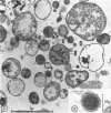
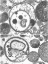
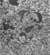
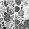
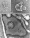
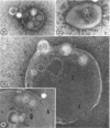
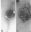
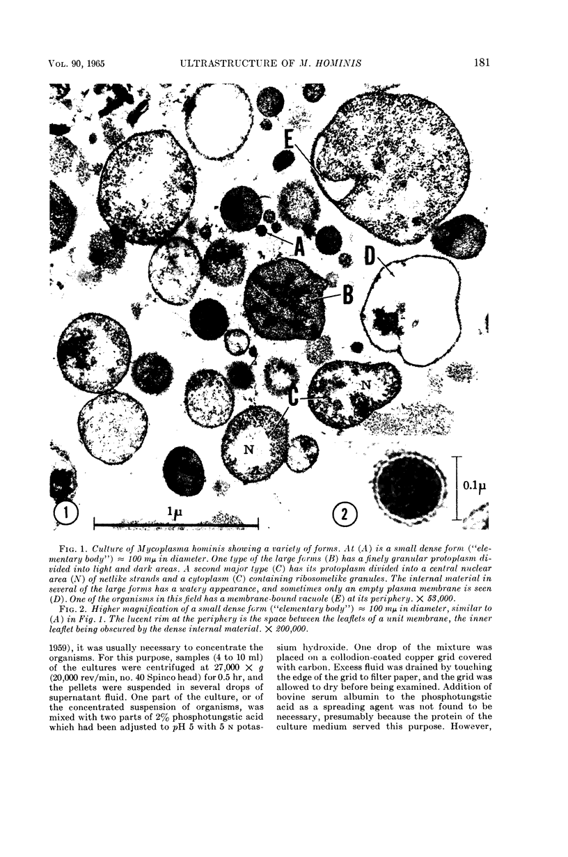
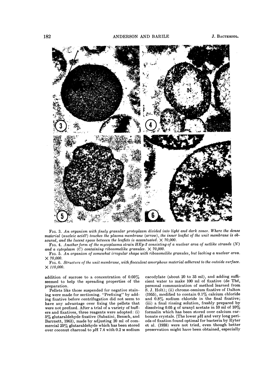
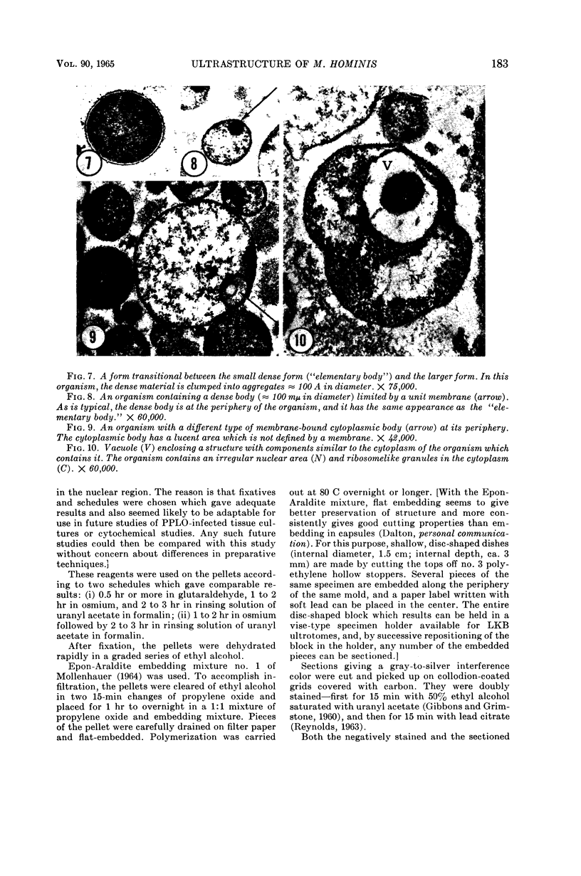
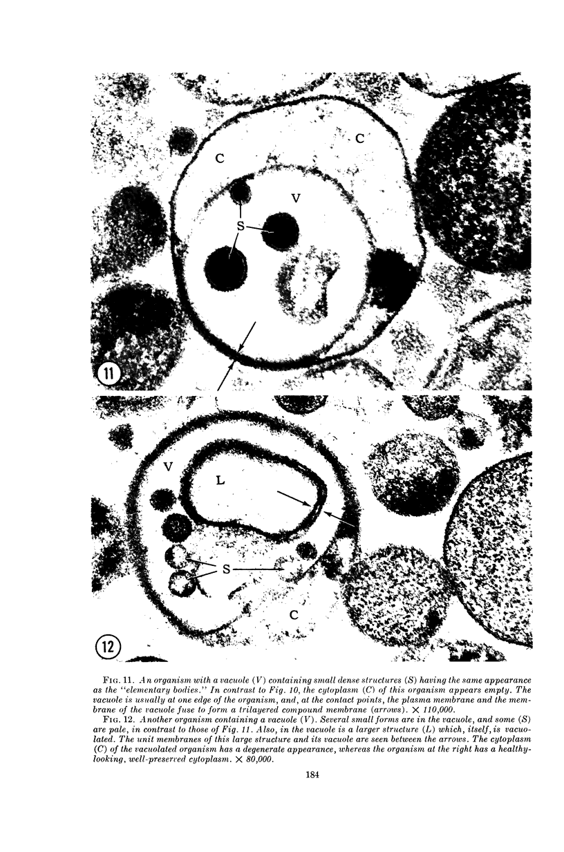
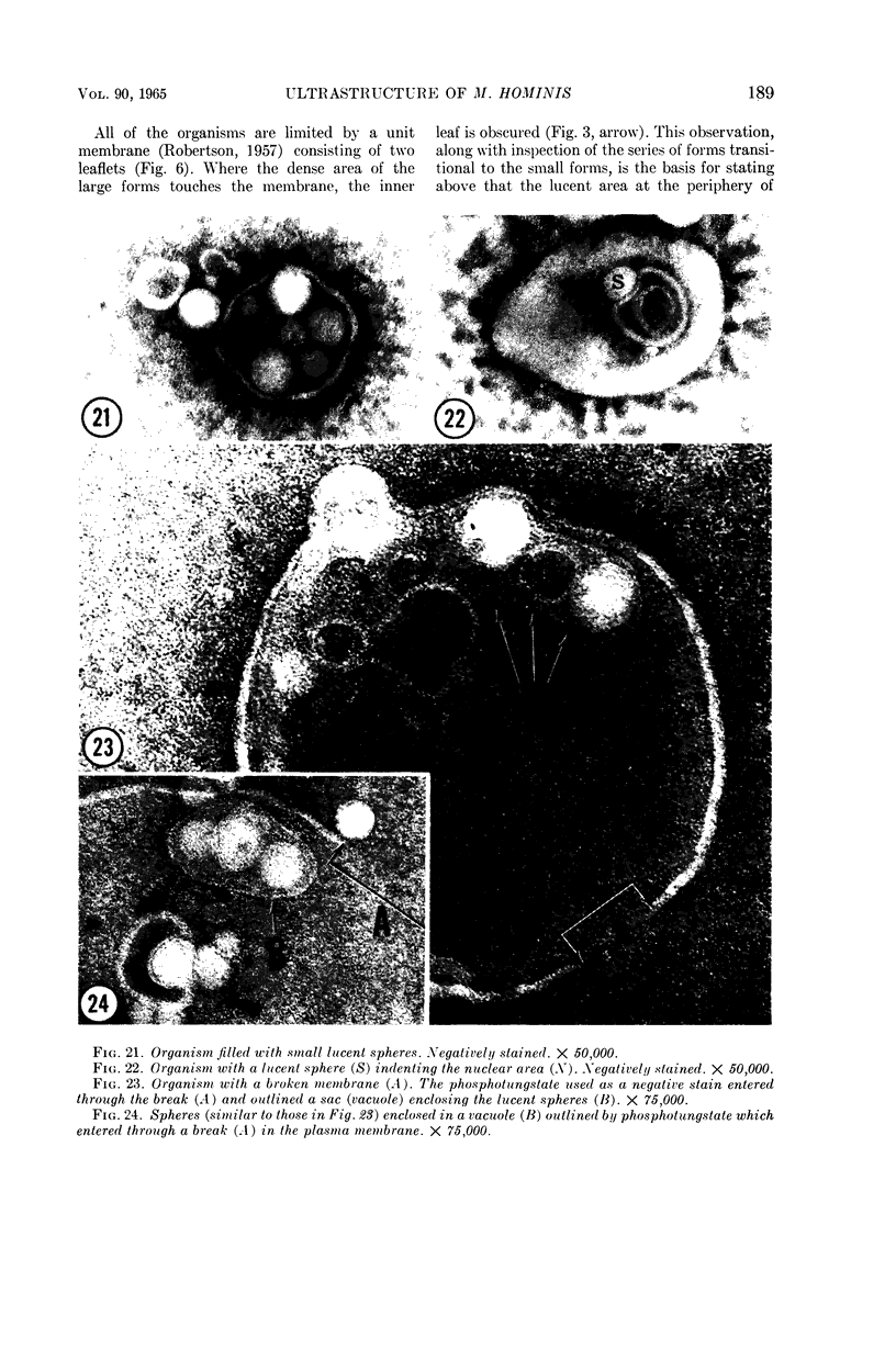
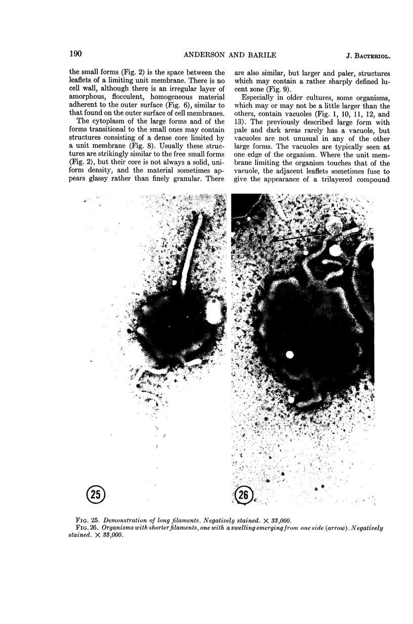
Anderson, Douglas R. (National Cancer Institute, Bethesda, Md.), and Michael F. Barile. Ultrastructure of Mycoplasma hominis. J. Bacteriol. 90:180–192. 1965.—Both thin-sectioning and negative staining were used in an electron microscopic study of the morphology of pleuropneumonia-like organism (PPLO) strain HEp-2 (Mycoplasma hominis, type I) grown in an artificial liquid medium. The morphology is quite variable and seems to depend, in part, on the age of the culture. The smallest form observed (“elementary body”) is 80 to 100 mμ in diameter. The internal components of the larger PPLO cells (0.5 to 1 μ) are variable—some have ribosomelike granules and nuclear areas of netlike strands, and others have only irregular dense areas in a pale groundplasm. Some of the forms have dense cytoplasmic bodies which look much like elementary bodies. Others have vacuoles which may contain structures which look like smaller organisms. Especially in older cultures, very large (10 μ) vacuolated organisms are seen, probably corresponding to the “large bodies” described by light microscopists. Filamented forms are also seen. These observations suggest several possible modes of reproduction, each perhaps operating under different cultural conditions or at different ages of the culture.
Full text
PDF
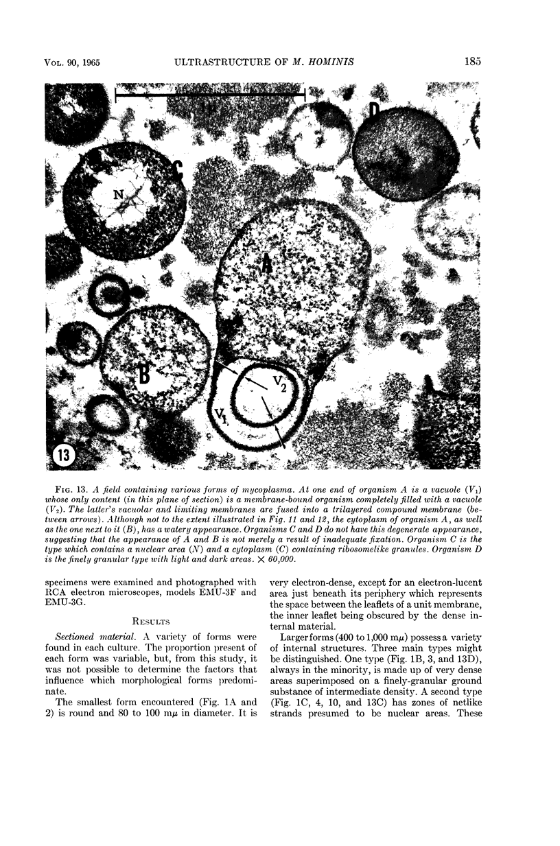
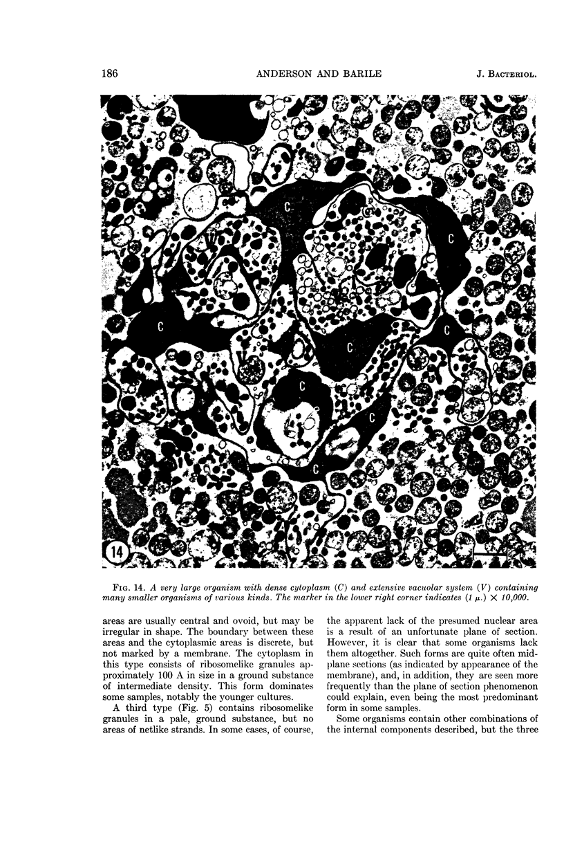
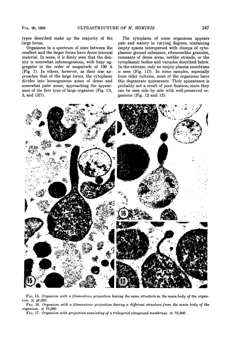
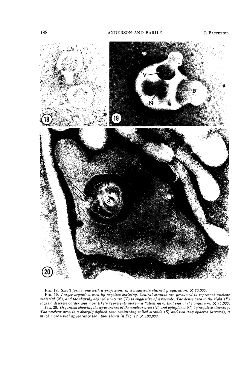
Images in this article
Selected References
These references are in PubMed. This may not be the complete list of references from this article.
- BARILE M. F., MALIZIA W. F., RIGGS D. B. Incidence and detection of pleuropneumonia-like organisms in cell cultures by fluorescent antibody and cultural procedures. J Bacteriol. 1962 Jul;84:130–136. doi: 10.1128/jb.84.1.130-136.1962. [DOI] [PMC free article] [PubMed] [Google Scholar]
- BRENNER S., HORNE R. W. A negative staining method for high resolution electron microscopy of viruses. Biochim Biophys Acta. 1959 Jul;34:103–110. doi: 10.1016/0006-3002(59)90237-9. [DOI] [PubMed] [Google Scholar]
- DOMERMUTH C. H., NIELSEN M. H., FREUNDT E. A., BIRCH-ANDERSEN A. ULTRASTRUCTURE OF MYCOPLASMA SPECIES. J Bacteriol. 1964 Sep;88:727–744. doi: 10.1128/jb.88.3.727-744.1964. [DOI] [PMC free article] [PubMed] [Google Scholar]
- EDWARDS G. A., FOGH J. Fine structure of pleuropneumonia-like organisms in pure culture and in infected tissue culture cells. J Bacteriol. 1960 Feb;79:267–276. doi: 10.1128/jb.79.2.267-276.1960. [DOI] [PMC free article] [PubMed] [Google Scholar]
- FREUNDT E. A. Morphology and classification of the PPLO. Ann N Y Acad Sci. 1960 Jan 15;79:312–325. doi: 10.1111/j.1749-6632.1960.tb42693.x. [DOI] [PubMed] [Google Scholar]
- GIBBONS I. R., GRIMSTONE A. V. On flagellar structure in certain flagellates. J Biophys Biochem Cytol. 1960 Jul;7:697–716. doi: 10.1083/jcb.7.4.697. [DOI] [PMC free article] [PubMed] [Google Scholar]
- MALIZIA W. F., BARILE M. F., RIGGS D. B. Immunofluorescence of pleuropneumonia-like organisms isolated from tissue cell cultures. Nature. 1961 Jul 8;191:190–191. doi: 10.1038/191190a0. [DOI] [PubMed] [Google Scholar]
- MOLLENHAUER H. H. PLASTIC EMBEDDING MIXTURES FOR USE IN ELECTRON MICROSCOPY. Stain Technol. 1964 Mar;39:111–114. [PubMed] [Google Scholar]
- PORTER G. H., 3rd, DALTON A. J., MOLONEY J. B., MITCHELL E. Z. ASSOCIATION OF ELECTRON-DENSE PARTICLES WITH HUMAN ACUTE LEUKEMIA. J Natl Cancer Inst. 1964 Sep;33:547–556. [PubMed] [Google Scholar]
- REYNOLDS E. S. The use of lead citrate at high pH as an electron-opaque stain in electron microscopy. J Cell Biol. 1963 Apr;17:208–212. doi: 10.1083/jcb.17.1.208. [DOI] [PMC free article] [PubMed] [Google Scholar]
- ROBERTSON J. D. New observations on the ultrastructure of the membranes of frog peripheral nerve fibers. J Biophys Biochem Cytol. 1957 Nov 25;3(6):1043–1048. doi: 10.1083/jcb.3.6.1043. [DOI] [PMC free article] [PubMed] [Google Scholar]
- ROBERTSON J. D. Structural alterations in nerve fibers produced by hypotonic and hypertonic solutions. J Biophys Biochem Cytol. 1958 Jul 25;4(4):349–364. doi: 10.1083/jcb.4.4.349. [DOI] [PMC free article] [PubMed] [Google Scholar]
- RYTER A., KELLENBERGER E., BIRCHANDERSEN A., MAALOE O. Etude au microscope électronique de plasmas contenant de l'acide désoxyribonucliéique. I. Les nucléoides des bactéries en croissance active. Z Naturforsch B. 1958 Sep;13B(9):597–605. [PubMed] [Google Scholar]
- SABATINI D. D., BENSCH K., BARRNETT R. J. Cytochemistry and electron microscopy. The preservation of cellular ultrastructure and enzymatic activity by aldehyde fixation. J Cell Biol. 1963 Apr;17:19–58. doi: 10.1083/jcb.17.1.19. [DOI] [PMC free article] [PubMed] [Google Scholar]
- SCHIMKE R. T., BARILE M. F. ARGININE METABOLISM IN PLEUROPNEUMONIA-LIKE ORGANISMS ISOLATED FROM MAMMALIAN CELL CULTURE. J Bacteriol. 1963 Aug;86:195–206. doi: 10.1128/jb.86.2.195-206.1963. [DOI] [PMC free article] [PubMed] [Google Scholar]
- SCHIMKE R. T., BARILE M. F. Arginine breakdown in mammalian cell culture contaminated with pleuropneumonia-like organisms (PPLO). Exp Cell Res. 1963 May;30:593–596. doi: 10.1016/0014-4827(63)90337-9. [DOI] [PubMed] [Google Scholar]
- SCHMIDT P. J., BARILE M. F., MCGINNISS M. H. MYCOPLASMA (PLEUROPNEUMONIA-LIKE ORGANISMS) AND BLOOD GROUP I; ASSOCIATIONS WITH NEOPLASTIC DISEASE. Nature. 1965 Jan 23;205:371–372. doi: 10.1038/205371a0. [DOI] [PubMed] [Google Scholar]
- van ITERSON, RUYS A. C. On the nature of P.P.L.O. II. Electron microscopy. Antonie Van Leeuwenhoek. 1960;26:9–22. doi: 10.1007/BF02538988. [DOI] [PubMed] [Google Scholar]
- van ITERSON, RUYS A. C. The fine structure of the Mycoplasmataceae (microorganisms of the pleuropneumonia group PPLO). 1. Mycoplasma hominis, M. fermentans and M. salivarium. J Ultrastruct Res. 1960 Feb;3:282–301. doi: 10.1016/s0022-5320(60)80015-9. [DOI] [PubMed] [Google Scholar]


