

Abstract
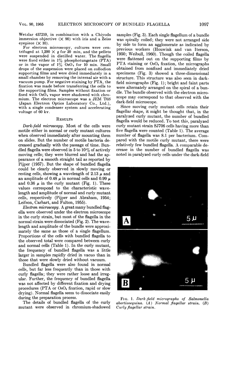
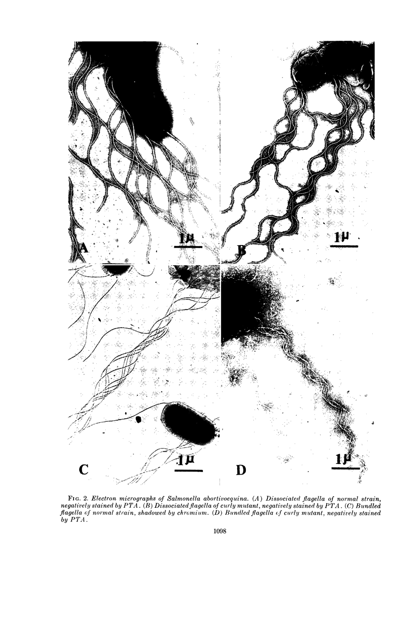
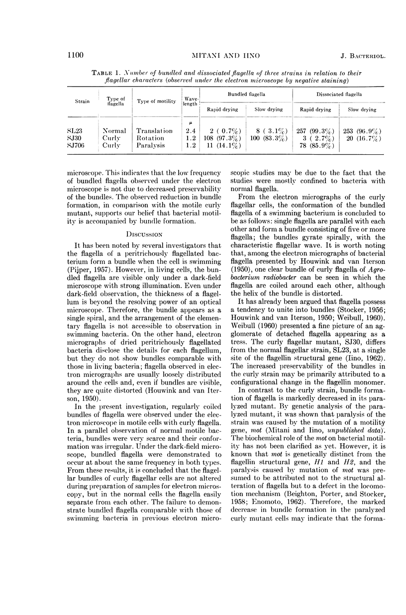
Mitani, Michiko (National Institute of Genetics, Mishima, Japan), and Tetsuo Iino. Electron microscopy of bundled flagella of the curly mutant of Salmonella abortivoequina. J. Bacteriol. 90:1096–1101. 1965.—The arrangement of flagella was observed by dark-field and electron microscopy in three strains of Salmonella abortivoequina, namely, normal flagellar, curly flagellar, and paralyzed curly flagellar strains. With dark-field microscopy, bundled flagella could be seen in 5 to 10% of actively moving normal or curly mutant cells. Under the electron microscope, a great many bundled flagella were observed in the curly mutant strain, but in the normal strain most of the flagella were dissociated or the bundles were rather loose and irregular. Normal flagella seem to separate easily during the process of preparation, but not the curly ones. Single flagella were found to run parallel with each other and to form a bundle consisting of five or more flagella; the bundle was spirally gyrating, with the characteristic flagellar wave. It is thought that the bundle observed with the electron microscope corresponds to that observed under the dark-field microscope. Further, the marked decrease of bundle formation in the paralyzed curly mutant cells suggests that bundle formation is not caused by curly flagellar structure per se, but corresponds to the mode of locomotion of peritrichously flagellated bacteria.
Full text
PDF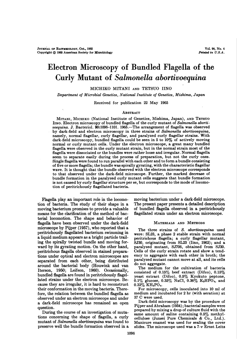
Images in this article
Selected References
These references are in PubMed. This may not be the complete list of references from this article.
- BEIGHTON E., PORTER A. M., STOCKER B. A. X-ray and related studies of the flagella of non-motile bacteria. Biochim Biophys Acta. 1958 Jul;29(1):8–13. doi: 10.1016/0006-3002(58)90139-2. [DOI] [PubMed] [Google Scholar]
- HOUWINK A. L., van ITERSON W. Electron microscopical observations on bacterial cytology; a study on flagellation. Biochim Biophys Acta. 1950 Mar;5(1):10–44. doi: 10.1016/0006-3002(50)90144-2. [DOI] [PubMed] [Google Scholar]
- LEIFSON E., CARHART S. R., FULTON M. Morphological characteristics of flagella of Proteus and related bacteria. J Bacteriol. 1955 Jan;69(1):73–82. doi: 10.1128/jb.69.1.73-82.1955. [DOI] [PMC free article] [PubMed] [Google Scholar]
- PIJPER A., ABRAHAM G. Wavelengths of bacterial flagella. J Gen Microbiol. 1954 Jun;10(3):452–456. doi: 10.1099/00221287-10-3-452. [DOI] [PubMed] [Google Scholar]
- PIJPER A. Bacterial flagella and motility. Ergeb Mikrobiol Immunitatsforsch Exp Ther. 1957;30:37–95. doi: 10.1007/978-3-662-25832-3_2. [DOI] [PubMed] [Google Scholar]