RPE65 is the isomerohydrolase in the visual cycle that isomerizes vitamin A to 11-cis isoform, which can be used as a chromophore of visual pigments. However, the phenotype of Rpe65−/− mice is different from that in vitamin A deficiency. This study systematically compares the ocular phenotypes of Rpe65−/− and vitamin-deficient mice.
Abstract
Purpose.
RPE65 gene knockout (Rpe65−/−) mice showed abolished isomerohydrolase activity in the visual cycle and were considered a model for vitamin A deficiency in the retina. The purpose of this study was to compare the retinal phenotypes between vitamin A–deficient (VAD) mice and Rpe65−/− mice under normal diet.
Methods.
The VAD mice were fed with a vitamin A–deprived diet after birth. The age-matched control mice and Rpe65−/− mice were maintained under normal diet. The structure of photoreceptor outer segment was compared using electron microscopy. Photoreceptor-specific gene expression was determined using real-time RT-PCR. The isomerohydrolase and lecithin-retinol acyltransferase (LRAT) activities were measured using an in vitro enzymatic activity assay. Endogenous retinoid profiles were analyzed by HPLC in mouse eyecup homogenates.
Results.
Compared to wild-type mice under normal diet, scanning and transmission electron microscopy showed that the outer segments of photoreceptors were disorganized in VAD mice and were not disorganized in Rpe65−/− mice, although they were shortened in the latter. VAD mice showed more prominent downregulation of middle wavelength cone opsin, whereas Rpe65−/− mice displayed more suppressed expression of short wavelength cone opsin. In vitro enzymatic activity assay and Western blot analysis showed that vitamin A deprivation downregulated LRAT expression and activity in the eyecup, but Rpe65−/− mice showed unchanged LRAT expression and activity. The depressed LRAT activity in VAD mice was partially rescued by the intraperitoneal injection of retinoic acid.
Conclusions.
VAD and Rpe65−/− mice are different in cone photoreceptor degeneration, photoreceptor-specific gene regulation, isomerohydrolase activity, endogenous retinoid profile, and LRAT activity.
Vitamin A is essential for the maintenance of epithelial cell growth, immune competence, embryonic growth and development, and visual function.1–3 There are several derivatives of vitamin A in living organisms, including retinol (alcohol form), retinal (aldehyde form), retinoic acid (acid form), and retinyl esters (ester forms).4 11-cis retinal is a polyene chromophore of visual pigments, and photoisomerization of 11-cis retinal triggers the phototransduction cascade and initiates vision. All-trans retinol is the major transportation form of vitamin A and serves as the precursor of chromophore of photosensitive pigments in both rods and cones5; it is essential for the epithelial cell RNA and glycoprotein synthesis on the ocular surface.6 Retinoic acid is an important embryonic growth factor,7 regulating the growth, differentiation, and morphology of RPE cells.8 Vitamin A deficiency caused by malnutrition, presenting with nyctalopia in humans,5 is the leading cause of night blindness by malnutrition in the developing world.9
RPE65, a membrane-associated protein predominantly expressed in the RPE, is essential for normal vision.10,11 Mutations in RPE65 are associated with inherited diseases involving severe vision loss, such as Leber's congenital amaurosis (LCA), RP, and early-onset severe rod–cone dystrophies.12–15 Homozygous RPE65 gene knockout (Rpe65−/−) mice lack isomerohydrolase activity and 11-cis retinoid products, revealing an interrupted visual cycle.16 Previous studies have shown that RPE65 is the isomerohydrolase in the visual cycle and is essential for the 11-cis retinal regeneration.17,18 Previously, we have reported that in Rpe65−/− mice, massive cone degeneration occurs at early ages, which is associated with significant downregulation of cone-specific genes, while rods remain intact at early ages.19
Several groups have used vitamin A–deficient (VAD) mice generated by a vitamin A deprivation diet in immunology, growth and development, and vision studies.20–23 Rpe65−/− mice were considered a model for vitamin A deficiency in the retina because they lack 11-cis retinal, the chromophore.18 However, the similarities and differences of vision between VAD mice and Rpe65−/− mice have never been compared. In the present study, we set out to compare VAD mice with Rpe65−/− mice in phenotypic similarities and differences, such as photoreceptor degeneration, photoreceptor-specific gene expression, endogenous retinoid profile, and isomerohydrolase and lecithin-retinol acyltransferase (LRAT) activity.
Methods
Experimental Animals
Animals were raised in a 12-hour light/dark cycle with an ambient light intensity of 85 ± 18 lux. Wild-type (WT) 129Sv mice were purchased from Jackson Laboratory (Bar Harbor, ME). Rpe65−/− mice were genotyped as described previously.18 The care, use, and treatment of the animals was in strict agreement with the ARVO Statement for the Use of Animals in Ophthalmic and Vision Research.
Electron Microscopy
The retinas for electron microscopy were processed as described.24 Ultrathin sections were cut using a diamond knife and stained with uranium and lead salts for electron microscopy. Tissue contrast was amplified by floating the specimens on 5% aqueous uranyl acetate for 10 minutes and Sato's lead stain for 3 minutes Sections were investigated using a scanning electron microscope (SEM; Model S-2250N, Hitachi, Tokyo, Japan).
Cone Cell Density Analysis
The retinas of the experimental animals were prepared as described previously.25 Briefly, the retina–lens complex was fixed in 4% formaldehyde solution in PBS (pH 7.4). After several washes in PBS, the retina–lens complex was incubated with FITC-conjugated peanut agglutinin (PNA; Sigma-Aldrich, Saint Louis, MO) overnight. After several washes in PBS, the retina was detached from the lens, flat-mounted, and covered by a coverslip after the application of several drops of antifade solution (Prolong; Molecular Probes, Eugene, OR). The slides were analyzed under a fluorescence microscope (Axioplan II; Carl Zeiss Meditec, Inc., Thornwood, NY) equipped with a digital camera. Images were captured (Spot-RT Camera with Spot software, version 3.0; Diagnostic Instruments, Sterling Heights, MI) and processed (Photoshop; Adobe Systems, Mountain View, CA). Ten random micrographs from each retina were taken in each animal, at 400× magnification. The cones in these micrographs were counted, and the number of cones per field was averaged and analyzed using the Student's t-test.
In Vitro Isomerohydrolase Activity Assay
After overnight dark adaptation, mice were killed and their eyes were enucleated. The anterior portion of the globe was removed, and the remaining eyecup (including the retina and RPE) from each mouse was homogenized individually for the assay. All-trans [11, 12-3H]-retinol in ethanol (1 mCi/mL, 52 Ci/mmol; Perkin Elmer, Waltham, MA) was dried under argon and resuspended in the same volume of dimethyl formamide (DMF). For each reaction, 2 μL of the nondiluted all-trans [11, 12-3H]-retinol in DMF and 250 μg of total protein from each homogenate were added to 200 μL of a reaction buffer (10 mM BTP [pH 8.0] 100 mM NaCl) containing 0.5% BSA and 25 μM cellular retinaldehyde-binding protein (CRALBP).26 After incubation in the dark at 37°C for 2 hours, the retinoids generated were extracted by the addition of 300 μL cold methanol and 300 μL hexane. The upper organic phase was collected and analyzed by normal phase HPLC coupled to a flow scintillation analyzer (Radiomatic 610TR; Perkin Elmer). Each retinoid was identified based on comparison to retention times of known retinoid standards. The activity was calculated from the area of the 11-cis [3H]-retinol peak using synthetic 11-cis [3H]-retinol as a standard for calibration.
Analysis of Endogenous Retinoids in the Mouse Eyecup
The eyecups (including the retina and RPE) from each mouse were combined and homogenized in 200 μL of PBS in a glass minigrinder, and retinoids were extracted under dim red light as described.19 After the addition of 300 μL cold methanol and 60 μL 1 M hydroxylamine in 0.2 M sodium phosphate buffer (pH 7.0), the resultant suspension was thoroughly vortexed for 30 seconds, and 300 μL of dichloromethane was added and vortexed for another 30 seconds. The solution was centrifuged at 10,000 g for 5 minutes The lower organic layer was collected with a Pasteur pipette. The residual suspension was extracted once more with an equal volume of dichloromethane. The combined organic layer was evaporated with oxygen-free argon. The residue was dissolved in 200 μL of HPLC mobile phase and applied to the HPLC column. The HPLC separation of retinoids and peak analyses were the same as described.27
Western Blot Analysis
The eyecups of each mouse were combined and homogenized. Protein concentration was measured by the Bradford assay.27 The equal amount (50 μg) of total protein from each sample was resolved by SDS-PAGE and electrotransferred onto a nitrocellulose membrane, as described previously.27 The membranes were blocked with 5% nonfat milk and separately blotted with a monoclonal anti-LRAT antibody and a polyclonal anti-RPE65 peptide antibody.28 After thorough washes, a peroxidase-conjugated goat anti-mice IgG (1:500) antibody was added, respectively, and incubated with the membranes. The signal was developed with the enhanced chemiluminescence system (Pierce, Rockford, IL).
LRAT Activity Assay
LRAT activity was measured as previously described.29 After overnight dark adaptation, mice were killed and their eyes were enucleated. The anterior chamber and retina were removed, and the remaining eyecup was homogenized and used for the LRAT activity assay. All-trans[11,12-3H]-retinol (NEN Life Science Products, Boston, MA) was diluted with cold all-trans-retinol to generate a specific radioactivity of 4 × 10⁷ dpm/nmol. For each assay, 300 μL of eyecup homogenate (0.14 mg of protein in 10 mM BTP, pH 8.0, 0.5% BSA) was added to 20 pmol of all-trans [3H]-retinol (dried under a stream of argon and dissolved in 5 μL of 10% BSA). The reaction mixture was incubated at 37°C, and 50-μL aliquots were removed at 5, 10, 15, 30, and 60 minutes. Each aliquot was immediately quenched with ice cold methanol (500 μL/sample) and H2O (100 μL), and hexane (500 μL) was added for extraction of retinoids, following a documented method.18 The extracted retinoids were analyzed using HPLC with a normal phase Lichrosphere SI-60 (Alltech, Deerfield, IL) 5-μm column and isocratic solvent of 11.2% ethyl acetate, 2.0% dioxane, and 1.4% octanol in hexane. Elution peaks were identified by spiking with authentic standards. Radioactive HPLC fractions were calculated as a percentage of the total radioactivity using Radiomatic 610TR software (Perkin Elmer, Waltham, MA).
Quantitative Real-Time RT-PCR
Six WT mice with vitamin A deficiency, Rpe65−/− mice, and age-matched WT control mice with normal diet were used for real-time RT-PCR. Total RNA was isolated from the retinas of each mouse individually. Reverse transcriptase (RT) reaction was performed as described previously.29 The RT products were diluted to 1:10; 2 μL of each diluted RT product was used for PCR, which included 3 pM of each primer in a final volume of 25 μL. The PCR included a denaturation and hot start at 95°C for 10 minutes, was followed by 45 cycles, with melting at 95°C for 15 seconds and elongation at 60°C for 60 seconds Sequences of PCR primers are shown in Table 1. Fluorescence changes were monitored after each cycle (SYBR Green; Applied Biosystems, Inc., Foster City, CA). Melting curve analysis was performed (0.5°C/sec increase from 55°C to 95°C, with continuous fluorescence readings) at the end of 45 cycles, to ensure that specific PCR products were obtained. Amplicon size and reaction specificity were confirmed by single-band product in agarose gel electrophoresis (data not shown). All reactions were performed in triplicate, and the results were analyzed by MyiQ software (Biorad, Hercules, CA). The average threshold cycle (CT) of fluorescence units was used for analysis. The mRNA level was normalized by the 18S rRNA level. Quantification was calculated using the CT of the target signal in comparison with the 18S rRNA signal in the same RNA sample. The mRNA levels were averaged in each group and expressed as percentages of that in age-matched WT control group.
Table 1.
Sequences of Primers for Quantitative RT-PCR
| Targets | Sequences | Amplicon Length (bp) |
|---|---|---|
| GNAT1 | F: GAGGATGCTGAGAAGGATGC | 209 |
| R: TGAATGTTGAGCGTGGTCAT | ||
| GNAT2 | F: GCATCAGTGCTGAGGACAAA | 192 |
| R: CTAGGCACTCTTCGGGTGAG | ||
| Rhodopsin | F: CAAGAATCCACTGGGAGATGA | 136 |
| R: GTGTGTGGGGACAGGAGACT | ||
| SWL opsin | F: TGTACATGGTCAACAATCGGA | 153 |
| R: ACACCATCTCCAGAATGCAAG | ||
| MWL opsin | F: CTCTGCTACCTCCAAGTGTGG | 154 |
| R: AAGTATAGGGTCCCCAGCAGA | ||
| 18S rRNA | F: TTTGTTGGTTTTCGGAACTGA | 199 |
| R: CGTTTATGGTCGGAACTACGA |
bp, base pair; F, forward; GNAT1, rod transducin α-subunits; GNAT2, cone transducin α-subunits; MWL, middle wavelength; R, reverse; SWL, short wavelength.
Results
Photoreceptor Outer Segments in VAD and Rpe65−/− Mice
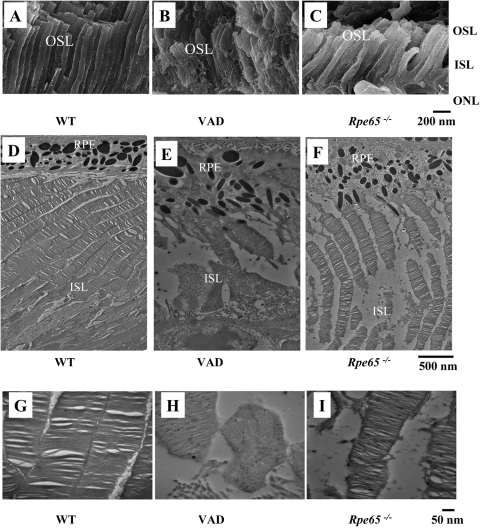
Retina SEM images were captured from WT mice under normal diet for 8 months, WT mice under vitamin A–deprived diet for 8 months, and Rpe65−/− mice under normal diet at 8 months of age. The outer segments of photoreceptors displayed irregular morphology and disorganized arrangement in VAD mice, whereas Rpe65−/− mice preserved the regular outer segment structure and arrangement, although the outer segments showed a reduced length and density compared to WT control mice under normal diet (Figs. 1A-C). The fine structure of photoreceptors was characterized further by transmission electron microscopy (TEM) images in lower (Figs. 1D-F) and higher magnifications (Figs. 1G-L). Disorganized outer segments, containing discs and whorls of various sizes that lacked the correct orientation, were observed in VAD but not Rpe65−/− mice.
Figure 1.
Histologic comparison of VAD and Rpe65−/− mice. (A-C) SEM images of photoreceptors in WT mice under normal diet (control, A) or a vitamin A–deficient diet (B), and Rpe65−/− mice (C) at 8 months of age. (D-F) TEM images of the same group of control mice (D), VAD mice (E), and Rpe65−/− mice (F). (G-I) High magnification TEM images from panels (D-F). ISL, inner segment layer; ONL, outer nuclear layer; OSL, outer segment layer; RPE, retinal pigment epithelium. Scale bar: (A-C), 200 nm; (D-F), 500 nm; (G-I), 50 nm.
Cone Degeneration in VAD and Rpe65−/− Mice
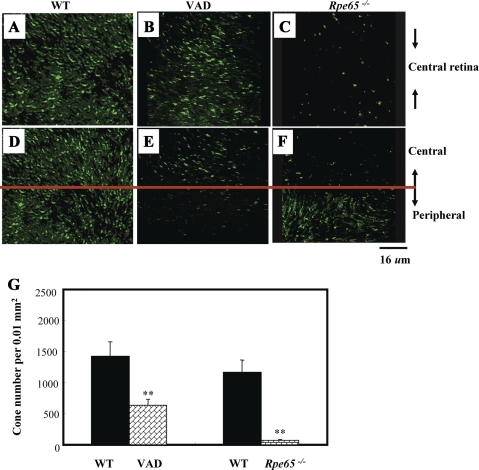
To assess cone density, cones were stained with fluorescent PNA, which labels both mid-wavelength (MWL) and short-wavelength (SWL) cones in the retina.30 In the flat-mounted retina from Rpe65−/− mice, significantly reduced cone densities were observed, especially in the central retina (Figs. 2A, C, D, F), consistent with our previous studies.31 In VAD mice, cone density in the central retina was higher than that in Rpe65−/− mice but lower in the peripheral retina (Figs. 2B, E). The labeled cones were counted in 10 random fields in each retina and compared. As shown in Figure 2C, the average number of cones was significantly reduced by 95.3% in Rpe65−/− mice and 52% in VAD mice, respectively, compared with age-matched WT mice (Fig. 2G).
Figure 2.
Cone degeneration in VAD and Rpe65−/− mice. Cones were labeled with FITC-PNA and visualized in retinal flat mounts from WT mice under normal diet (control, A and D) or under a vitamin A–deprived diet (B and E) at 3 months of age, and from age-matched Rpe65−/− mice under normal diet (C and F). (A-C) Representative images of the central retina; (D-F) representative images of the peripheral retina. (G) Cones were counted in the flat-mounted retina in 10 random fields (0.01 mm2) per retina, averaged in three mice from each group, and presented as cone number per field. **P < 0.01 compared with control. Scale bar, 16 μm.
Expression of Rod- and Cone-Specific Genes in the Retina of VAD and Rpe65−/− Mice
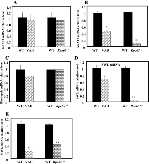
Previously, we found that cone opsins and cone transducin are downregulated at early ages of Rpe65−/− mice because of the lack of 11-cis retinal.31 To assess the effects of vitamin A deficiency on cone-specific gene expression in vivo, we quantified and compared mRNA levels of a subset of photoreceptor-specific genes, including SWL and MWL cone opsins, rhodopsin, and rod and cone transducin α-subunits (GNAT1 and GNAT2, respectively) in VAD and Rpe65−/− mice using quantitative real-time RT-PCR analysis and normalized by 18S rRNA levels. VAD mice had significantly decreased mRNA levels of SWL and MWL cone opsins and GNAT2 compared with that in age-matched normal mice, while the rhodopsin and rod transducin mRNAs remained unchanged (Figs. 3A-E). Similar changes were observed in Rpe65−/− mice, consistent with the previous finding that cones degenerate earlier than rods in Rpe65−/− mice. However, the mRNA levels of the MWL cone opsin had a more significant decline than that of the SWL cone opsin in VAD mice, whereas in Rpe65−/− mice, the SWL cone opsin mRNA levels showed a more prominent decrease than that of the MWL cone opsin.
Figure 3.
Expression of rod- and cone-specific genes in VAD and Rpe65−/− mice. Rod- and cone-specific mRNA levels in the retina were compared by real-time RT-PCR using an equal amount of total RNA from the retinas of the control WT mice with normal diet, VAD, and Rpe65−/− mice at 3 months of age. Average mRNA levels of the rod transducin α-subunit (GNAT1, A), cone transducin α-subunit (GNAT2, B), rhodopsin (C), short wavelength cone opsin (D), and middle wavelength cone opsin (E) were normalized by 18S rRNA levels, and mRNA levels in VAD and Rpe65−/− mice were expressed as a ratio of WT control (mean ± SD, n = 6). *P < 0.05, **P < 0.01 compared with WT controls.
Isomerohydrolase Activity in VAD and Rpe65−/− Mice
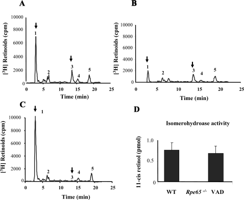
Isomerohydrolase activity in the mouse eyecup was measured using all-trans [3H]-retinol as a substrate. When the equal amount of eyecup homogenates was incubated with all-trans [3H]-retinol, both VAD and age-matched normal control mice eyecup homogenates generated 11-cis [3H]-retinol (Figs. 4A, B), while Rpe65−/− mice did not show any detectable isomerohydrolase activity (Fig. 4C). The isomerohydrolase activity was measured by quantifying the generated 11-cis [3H]-retinol with HPLC and averaged in six mice from each group. In VAD mice, isomerohydrolase activity in the eyecup homogenates was comparable to that in control mice under normal diet, whereas Rpe65−/− mice showed abolished isomerohydrolase activity (Fig. 4D), which is consistent with a previous report.27 These results indicated that VAD mice preserve isomerohydrolase activity.
Figure 4.
Isomerohydrolase activity differences in the eyecups of VAD and Rpe65−/− mice. At 6 months of age, the eyecups were homogenized, and the same amount (250 μg) of eyecup protein was incubated with all-trans [3H]-retinol for 1.5 hours. The generated retinoids were analyzed by HPLC. (A) WT mice, (B) VAD mice, (C) Rpe65−/− mice. Peak 1, retinyl esters; Peak 2, all-trans retinal; Peak 3, 11-cis retinol; Peak 4, 13-cis retinol; Peak 5, all-trans retinol. Peaks corresponding to retinyl esters and 11-cis retinol in the retina samples are labeled with arrows. (D) 11-cis retinol generated in the in vitro isomerohydrolase activity assay was quantified by measuring the area of the 11-cis retinol peak in the HPLC profile and averaged within the same group (mean ± SD, n = 6).
Endogenous Retinoid Profile in VAD and Rpe65−/− Eyecups
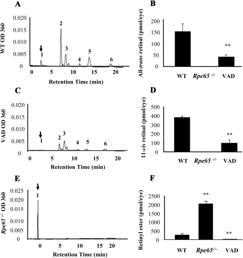
Rpe65−/− mice are known to lack any detectable 11-cis retinoids in the eyecups.19 To test whether vitamin A deficiency changes the normal profile of endogenous retinoids in the eye cups, endogenous retinoids were extracted from the eyecups of VAD mice and analyzed with HPLC. Each isoform of retinoids was identified based on the retention time of respective retinoid standard and quantified by measuring areas of peaks in HPLC profile. Consistent with a previous report,19 syn-11-cis retinal oxime and anti-11-cis retinal oxime were detected in eyecups from both VAD and control WT mice (Figs. 5A, C), whereas Rpe65−/− mice showed only increased levels of retinyl ester and no other forms of retinoids (Fig. 5E). Quantification of each isoform of retinoids showed that endogenous all-trans retinal and 11-cis retinal levels were significant decreased in VAD and absent in Rpe65−/− mice, compared with WT mice under normal diet (Figs. 5B, D). Retinyl ester was dramatically decreased in VAD mice, compared to normal controls, but was overaccumulated in Rpe65−/− mice (Fig. 5F).
Figure 5.
Endogenous retinoid profile alternations in VAD and Rpe65−/− eyecups. At 6 months of age, the eyecups were homogenized, and endogenous retinoids were extracted and analyzed by HPLC. Each panel is a representative of HPLC profiles from (A) WT, (C) VAD, and (E) Rpe65−/− mice. Peaks were identified by comparison to retinoid standards. Peak 1, retinyl esters; Peak 2, syn-11-cis retinal oxime; Peak 3, syn-all-trans retinal oxime; Peak 4, anti-13-cis retinal oxime; Peak 5, anti-11-cis retinal oxime; Peak 6, anti-all-trans retinal oxime. Peaks corresponding to retinyl esters in the retina samples are labeled with arrows. Amounts of all-trans retinal (B), 11-cis retinal (D), and retinyl esters (F) were quantified by measuring the peak areas and averaged within the group (mean ± SD, n = 3). *P < 0.05, **P < 0.01, compared with WT control.
Different LRAT Expression and Activities in VAD and Rpe65−/− Eyecups
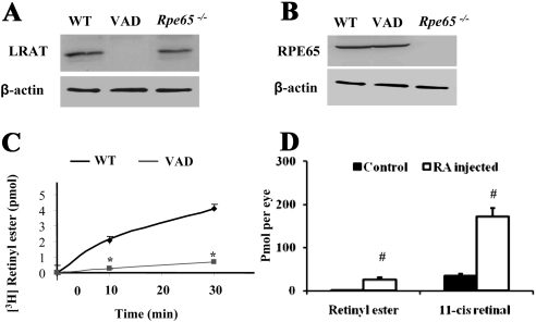
The protein levels of LRAT and RPE65 in the retina of VAD mice were compared with those in WT control and Rpe65−/− mice using Western blot analysis. Vitamin A deprivation resulted in a significant reduction of LRAT expression but no change in RPE65 expression compared with WT control mice, whereas RPE65 knockout did not affect LRAT expression in the retina (Figs. 6A, 6B). To determine the effect of vitamin A deprivation on LRAT enzymatic activity, we also measured LRAT activity by in vitro assay using mouse eyecup homogenates (Fig. 6C). When the same amount of eyecup proteins were used, VAD mice had significantly lower LRAT enzymatic activities compared to normal mice at both of the incubation durations (10 minutes and 30 minutes), whereas Rpe65−/− mice had LRAT activities similar to that in WT mice, consistent with our previous report.27
Figure 6.
LRAT expression and activity differences in VAD and Rpe65−/− eyecups. (A, B) Protein levels of LRAT and RPE65 in the eyecups were measured by Western blot analysis using an anti-LRAT antibody (A) and an anti-RPE65 antibody (B) in normal and VAD WT mice and Rpe65−/− mice at 6 months of age and normalized by β-actin levels. (C) Endogenous LRAT activities in WT control and VAD mice were measured. Equal amounts (250 μg) of eyecup protein were incubated with all-trans [3H]-retinol. The generated retinyl ester was analyzed by HPLC and quantified by measuring the area of the retinyl esters peaks in the HPLC profile after 10 and 30 minutes of incubation. All data were averaged within the group (mean ± SD, n = 3). (D) VAD mice were injected intraperitoneally with 200 μg retinoic acid (RA) or vehicle (control). Endogenous retinoid profiles were measured in the 24 hours postinjection. Retinyl esters and 11-cis retinal were quantified by measuring the peak areas and were averaged within the group (mean ± SD, n = 4). *P < 0.05 compared with WT control; #P < 0.05, compared with VAD mice with vehicle injection.
To further investigate the mechanism by which vitamin A deprivation leads to decreased LRAT expression, we intraperitoneally injected 200 μg retinoic acid (RA) and vehicle control into VAD mice. In vitro enzymatic activity assay showed that RA injection significantly increased LRAT activity by 48.7-fold (Fig. 6D), possibly through rescued LRAT expression. Consistently, level of 11-cis retinal also significantly increased by the injection, suggesting that the decreased LRAT activity induced by vitamin A deficiency can be caused by the lack of RA.
Discussion
Vitamin A plays essential roles in maintaining ocular functions.5 In the retina, vitamin A acts as a chromophore of visual pigments that serves as a trigger in the initiation of the visual phototransduction cascade.32 However, the exact pathogenesis of visual cycle interruption caused by vitamin A deficiency is not well understood. In the present study, we compared, for the first time, the phenotypes of VAD and Rpe65−/− mice in cone photoreceptor degeneration, photoreceptor-specific gene expression, isomerohydrolase activity, endogenous retinoid profile, and LRAT activity. This study revealed similarities and differences between VAD and Rpe65−/− mice.
Rpe65−/− mice are considered a model for vitamin A deficiency in the retina.18 However, it was reported that the Rpe65−/− photoreceptor outer segment (OS) were shorter in length than those of WT mice, but only slightly disorganized compared with those of WT mice.18 Consistently, the present study showed that Rpe65−/− mice have decreased densities of packing discs in photoreceptor OS and shorter and slightly disorganized OS. Interestingly, VAD mice had severe malformation and disorganization of photoreceptor OS. This observation suggests that OS formation is more sensitive to vitamin A deficiency than the lack of RPE65 expression. It is known that RA may play a critical role in photoreceptor cell morphology regulation.7 It remains to be determined whether the deprivation of retinol or/and RA in VAD mice is the cause of OS disorganization, which eventually leads to photoreceptor cell degeneration.
Our previous studies have shown that cone degeneration starts at early ages (2 weeks) in Rpe65−/− mice.31 Massive cone loss was observed, and a large area of central retina becomes cone-free at 4 weeks of age.31 In agreement, the expression of cone opsins and cone transducin was downregulated significantly in Rpe65−/− mice compared with WT mice. In contrast, the expression of rhodopsin and rod transducin was unchanged in young Rpe65−/− mice.19 The present study revealed that VAD mice have progressive cone degeneration, with massive cone loss in the peripheral retina, along with a lesser degree of cone loss in the central retina. Moreover, our data showed that vitamin A deficiency has a similar effect on photoreceptor-specific gene expression. However, in VAD mice, the expression of the MWL cone opsin was downregulated more than the SWL cone opsin, whereas in the Rpe65−/− mice, the SWL cone opsin downregulation was more prominent than that of the MWL cone opsin. The changes of cone opsin expression suggest that MWL and SWL cones may be affected differently by vitamin A deficiency and Rpe65−/− knockout.
Several studies have reported that Rpe65−/− mice lack isomerohydrolase activity and 11-cis retinoid products but overaccumulate retinyl ester in the eye.17,18 Moreover, our previous data showed that Rpe65−/− mice maintained the same LRAT activity as WT mice.27 The present study determined the effects of vitamin A deficiency on isomerohydrolase activity, profile of endogenous retinoids, and LRAT activity. The results show that VAD mice have unchanged RPE65 expression and therefore intact isomerohydrolase activity. Because vitamin A deprivation reduced all-trans retinyl ester, the substrate of RPE65, in the eye, VAD mice had lower levels of endogenous 11-cis retinoids in the retina and RPE compared to WT mice. Our study also revealed that both LRAT expression and LRAT activity were significantly decreased in VAD mice compared with WT mice, suggesting a downregulation in LRAT expression in VAD mice. A previous study has shown that supplementation with RA can significantly reverse retinal degeneration from VAD rats within a few days.33 In the present study, we injected RA intraperitoneally into VAD mice and found that the injection of RA partially restored LRAT activity. This suggests that the decreased LRAT activity induced by vitamin A deficiency is caused by the lack of RA in the eye. In agreement, several previous studies have shown that RA treatment activates the transcription of the LRAT gene in human prostate cancer cells,34 and that RA synergizes with vitamin A to enhance LRAT expression in the neonatal lung.35
Homozygous LRAT knockout (Lrat−/−) mice have a severe loss of cone visual functions at an early age (8 weeks).36 The rapid cone degeneration is caused by cone opsin mistrafficking to the outer segments.37 In addition, rod outer segments were significantly shorter in Lrat−/− mice at 6 to 8 weeks of age,36 which shows different morphologic features than that in VAD mice (Fig. 1B). Lrat−/− mice have only trace levels of all-trans retinyl esters and no 11-cis retinal in the eye, indicating that the visual cycle was interrupted at the esterification step.37 Our data showed that the esterification step interruption is only one of the combination effects of vitamin A deficiency on the visual cycle.
In summary, our results showed the differences of VAD and Rpe65−/− mice in cone photoreceptor degeneration, photoreceptor-specific gene regulation, isomerohydrolase activity, endogenous retinoid profile, and LRAT activity. These results suggest that the visual cycle inhibition by vitamin A deficiency is a progressive process, possibly affecting more processes in addition to the visual cycle.
Footnotes
Supported by National Institutes of Health Grants EY018659, EY012231, EY019309, and P20RR024215, and a research award from the American Diabetes Association.
Disclosures: Y. Hu, None; Y. Chen, None; G. Moiseyev, None; Y. Takahashi, None; R. Mott, None; J. Ma, None.
References
- 1. Chopra DP, Klinger MM, Sullivan JK. Effects of vitamin A on growth and differentiation of human tracheobronchial epithelial cell cultures in serum-free medium. J Cell Sci. 1989;93(Pt 1):133–142 [DOI] [PubMed] [Google Scholar]
- 2. Antipatis C, Grant G, Ashworth CJ. Moderate maternal vitamin A deficiency affects perinatal organ growth and development in rats. Br J Nutr. 2000;84:125–132 [PubMed] [Google Scholar]
- 3. Ruhl R. [Retinoids, vitamin A and pro-vitamin A carotenoids. Regulation of the immune system and allergies]. Pharm Unserer Zeit. 2009;38:126–131 [DOI] [PubMed] [Google Scholar]
- 4. Wolf G. The discovery of the visual function of vitamin A. J Nutr. 2001;131:1647–1650 [DOI] [PubMed] [Google Scholar]
- 5. Smith J, Steinemann TL. Vitamin A deficiency and the eye. Int Ophthalmol Clin. 2000;40:83–91 [DOI] [PubMed] [Google Scholar]
- 6. Sommer A. Effects of vitamin A deficiency on the ocular surface. Ophthalmology. 1983;90:592–600 [DOI] [PubMed] [Google Scholar]
- 7. Kramer B, Penny C. Regulation of embryonic chick insulin cells: effect of retinoic acid and insulin-like growth factor 1. Cells Tissues Organs. 2001;169:42–48 [DOI] [PubMed] [Google Scholar]
- 8. Campochiaro PA, Hackett SF, Conway BP. Retinoic acid promotes density-dependent growth arrest in human retinal pigment epithelial cells. Invest Ophthalmol Vis Sci. 1991;32:65–72 [PubMed] [Google Scholar]
- 9. Reddy V. Control of vitamin A deficiency and blindness. Acta Paediatr Scand Suppl. 1991;374:30–37 [DOI] [PubMed] [Google Scholar]
- 10. Mata NL, Moghrabi WN, Lee JS, et al. Rpe65 is a retinyl ester binding protein that presents insoluble substrate to the isomerase in retinal pigment epithelial cells. J Biol Chem. 2004;279:635–643 [DOI] [PubMed] [Google Scholar]
- 11. Gollapalli DR, Maiti P, Rando RR. RPE65 operates in the vertebrate visual cycle by stereospecifically binding all-trans-retinyl esters. Biochemistry. 2003;42:11824–11830 [DOI] [PubMed] [Google Scholar]
- 12. Gu SM, Thompson DA, Srikumari CR, et al. Mutations in RPE65 cause autosomal recessive childhood-onset severe retinal dystrophy. Nat Genet. 1997;17:194–197 [DOI] [PubMed] [Google Scholar]
- 13. Marlhens F, Bareil C, Griffoin JM, et al. Mutations in RPE65 cause Leber's congenital amaurosis. Nat Genet. 1997;17:139–141 [DOI] [PubMed] [Google Scholar]
- 14. Morimura H, Fishman GA, Grover SA, Fulton AB, Berson EL, Dryja TP. Mutations in the RPE65 gene in patients with autosomal recessive retinitis pigmentosa or Leber congenital amaurosis. Proc Natl Acad Sci U S A. 1998;95:3088–3093 [DOI] [PMC free article] [PubMed] [Google Scholar]
- 15. Thompson DA, Gyurus P, Fleischer LL, et al. Genetics and phenotypes of RPE65 mutations in inherited retinal degeneration. Invest Ophthalmol Vis Sci. 2000;41:4293–4299 [PubMed] [Google Scholar]
- 16. Ekesten B, Gouras P, Salchow DJ. Ultraviolet and middle wavelength sensitive cone responses in the electroretinogram (ERG) of normal and Rpe65 −/− mice. Vision Res. 2001;41:2425–2433 [DOI] [PubMed] [Google Scholar]
- 17. Moiseyev G, Chen Y, Takahashi Y, Wu BX, Ma JX. RPE65 is the isomerohydrolase in the retinoid visual cycle. Proc Natl Acad Sci U S A. 2005;102:12413–12418 [DOI] [PMC free article] [PubMed] [Google Scholar]
- 18. Redmond TM, Yu S, Lee E, et al. Rpe65 is necessary for production of 11-cis-vitamin A in the retinal visual cycle. Nat Genet. 1998;20:344–351 [DOI] [PubMed] [Google Scholar]
- 19. Chen Y, Moiseyev G, Takahashi Y, Ma JX. RPE65 gene delivery restores isomerohydrolase activity and prevents early cone loss in Rpe65−/− mice. Invest Ophthalmol Vis Sci. 2006;47:1177–1184 [DOI] [PubMed] [Google Scholar]
- 20. Smith SM, Levy NS, Hayes CE. Impaired immunity in vitamin A-deficient mice. J Nutr. 1987;117:857–865 [DOI] [PubMed] [Google Scholar]
- 21. Etchamendy N, Enderlin V, Marighetto A, Pallet V, Higueret P, Jaffard R. Vitamin A deficiency and relational memory deficit in adult mice: relationships with changes in brain retinoid signalling. Behav Brain Res. 2003;145:37–49 [DOI] [PubMed] [Google Scholar]
- 22. Kuwata T, Wang IM, Tamura T, et al. Vitamin A deficiency in mice causes a systemic expansion of myeloid cells. Blood. 2000;95:3349–3356 [PubMed] [Google Scholar]
- 23. Moore T, Holmes PD. The production of experimental vitamin A deficiency in rats and mice. Lab Anim. 1971;5:239–250 [DOI] [PubMed] [Google Scholar]
- 24. Kedzierski W, Lloyd M, Birch DG, Bok D, Travis GH. Generation and analysis of transgenic mice expressing P216L-substituted rds/peripherin in rod photoreceptors. Invest Ophthalmol Vis Sci. 1997;38:498–509 [PubMed] [Google Scholar]
- 25. Znoiko SL, Crouch RK, Moiseyev G, Ma JX. Identification of the RPE65 protein in mammalian cone photoreceptors. Invest Ophthalmol Vis Sci. 2002;43:1604–1609 [PubMed] [Google Scholar]
- 26. Crabb JW, Chen Y, Goldflam S, West K, Kapron J. Methods for producing recombinant human cellular retinaldehyde-binding protein. Methods Mol Biol. 1998;89:91–104 [DOI] [PubMed] [Google Scholar]
- 27. Moiseyev G, Crouch RK, Goletz P, Oatis J, Jr, Redmond TM, Ma JX. Retinyl esters are the substrate for isomerohydrolase. Biochemistry. 2003;42:2229–2238 [DOI] [PubMed] [Google Scholar]
- 28. Ma J, Zhang J, Othersen KL, et al. Expression, purification, and MALDI analysis of RPE65. Invest Ophthalmol Vis Sci. 2001;42:1429–1435 [PubMed] [Google Scholar]
- 29. Ruiz A, Winston A, Lim YH, Gilbert BA, Rando RR, Bok D. Molecular and biochemical characterization of lecithin retinol acyltransferase. J Biol Chem. 1999;274:3834–3841 [DOI] [PubMed] [Google Scholar]
- 30. Blanks JC, Johnson LV. Specific binding of peanut lectin to a class of retinal photoreceptor cells. A species comparison. Invest Ophthalmol Vis Sci. 1984;25:546–557 [PubMed] [Google Scholar]
- 31. Znoiko SL, Rohrer B, Lu K, Lohr HR, Crouch RK, Ma JX. Downregulation of cone-specific gene expression and degeneration of cone photoreceptors in the Rpe65−/− mouse at early ages. Invest Ophthalmol Vis Sci. 2005;46:1473–1479 [DOI] [PubMed] [Google Scholar]
- 32. Deterre P, Pfister C, Bigay J, Chabre M. The retinal phototransduction process: enzymatic cascade and regulation. Biochimie (Paris). 1987;69:365–370 [DOI] [PubMed] [Google Scholar]
- 33. Kraft SP, Parker JA, Matuk Y, Rao AV. The rat electroretinogram in combined zinc and vitamin A deficiency. Invest Ophthalmol Vis Sci. 1987;28:975–984 [PubMed] [Google Scholar]
- 34. Cai K, Gudas LJ. Retinoic acid receptors and GATA transcription factors activate the transcription of the human lecithin:retinol acyltransferase gene. Int J Biochem Cell Biol. 2009;41:546–553 [DOI] [PMC free article] [PubMed] [Google Scholar]
- 35. Wu L, Ross AC. Acidic retinoids synergize with vitamin A to enhance retinol uptake and STRA6, LRAT, and CYP26B1 expression in neonatal lung. J Lipid Res. 2010;51:378–387 [DOI] [PMC free article] [PubMed] [Google Scholar]
- 36. Batten ML, Imanishi Y, Maeda T, et al. Lecithin-retinol acyltransferase is essential for accumulation of all-trans-retinyl esters in the eye and in the liver. J Biol Chem. 2004;279:10422–10432 [DOI] [PMC free article] [PubMed] [Google Scholar]
- 37. Fan J, Rohrer B, Frederick JM, Baehr W, Crouch RK. Rpe65−/− and Lrat−/− mice: comparable models of leber congenital amaurosis. Invest Ophthalmol Vis Sci. 2008;49:2384–2389 [DOI] [PMC free article] [PubMed] [Google Scholar]


