Abstract
Background
The present study was designed to test the hypothesis that the increased duration of analgesia caused by adding dexmedetomidine to local anesthetic results from blockade of the hyperpolarization-activated cation (Ih)current.
Methods
In this randomized, blinded, controlled study, the analgesic effects of peripheral nerve blocks using 0.5% ropivacaine alone or 0.5% ropivacaine plus dexmedetomidine (34 μM or 6 μg/kg) were assessed with or without the pretreatment of α1- and α2-adrenoceptor antagonists (prazosin and idazoxan, respectively) and antagonists and agonists of the Ih current (ZD 7288 and forskolin, respectively). Sciatic nerve blocks were performed, and analgesia was measured by paw withdrawal latency to a thermal stimulus every 30 min for 300 min post-block.
Results
The analgesic effect of dexmedetomidine added to ropivacaine was not reversed by either prazosin or idazoxan. There were no additive or attenuated effects from the pretreatment with ZD 7288 (Ih current) when compared with dexmedetomidine added to ropivacaine. When forskolin was administered as a pretreatment to ropivacaine plus dexmedetomidine, there were statistically significant reductions in duration of analgesia at time points 90–180 min (p < 0.0001 for each individual comparison). The duration of blockade for the forskolin (768 μM) followed by ropivacaine plus dexmedetomidine group mirrored the pattern of the ropivacaine alone group, thereby implying a reversal effect.
Conclusion
Dexmedetomidine added to ropivacaine caused approximately a 175% increase in the duration of analgesia, which was reversed by pretreatment with an Ih current enhancer. The analgesic effect of dexmedetomidine was not reversed by an ∝2-adrenoceptor antagonist.
Introduction
Single-shot peripheral nerve blocks are routinely performed as an alternative to general anesthesia and in order to decrease postoperative pain and opioid requirement. The most commonly used long-acting local anesthetic, ropivacaine, provides sufficient analgesia for 9–14 h.1,2 Due to the timing of the placement of most nerve blocks for surgery, many patients complain of pain for the first time during the nighttime hours. Especially for outpatient surgery, resources for treating acute pain after the waning of a nerve block can be limited and can cause frustration for patients and clinicians. As such, many surgeons, anesthesiologists, and nurses recommend that patients take opioids prior to the waning of the nerve block to preempt the pain. Side effects of opioids can be an independent source of morbidity, causing nausea, vomiting, sedation, constipation, and sleep disturbance.3,4 Therefore, there has been considerable interest in new medications or combinations of existing medications that will allow for longer durations of analgesia to provide better postoperative pain relief, especially for the first full 24 h after surgery.
The efficacy of peripheral perineural dexmedetomidine added to bupivacaine and ropivacaine for sciatic nerve blocks in rat has been established.5–7 The increase in duration of analgesia is dose-dependent,7 and the effect is peripheral (i.e., not due to centrally-mediated or systemic analgesia).5 Human studies in greater palatine and axillary brachial plexus nerve blocks have subsequently demonstrated that increased duration of sensory blockade can be achieved by adding dexmedetomidine to bupivacaine and levobupivacaine, respectively.8,9
Previous studies demonstrated a peripheral site of dexmedetomidine action,5 but the mechanism underlying the prolongation of sensory blockade of the peripheral nerve has heretofore been unknown. Dexmedetomidine is an agonist of the ∝2-adrenoceptor. An in vitro study of frog sciatic nerve conduction with high-concentration dexmedetomidine (published after the completion of the present study) found that the reduction of the compound action potential was concentration-dependent and not ∝2-mediated.10 Through in vivo and in vitro studies, the effects of clonidine, another ∝2-adrenoceptor agonist, on the peripheral nerve were found to be likely mediated through blockade of the hyperpolarization-activated cation current (Ih current), not due to agonism of the ∝2-adrenoceptor.11–14 An in vitro study of the effects of dexmedetomidine in the paraventricular nucleus neurons found that dexmedetomidine acts in part through inhibition of the Ih current.15 The present study was designed to test the hypothesis that the increased duration of analgesia from dexmedetomidine added to local anesthetic in an in vivo model of a peripheral nerve block in rat is due to blockade of the Ih current and not due to ∝1- or ∝2-adrenoceptor agonism.
Materials and Methods
The study followed the American Physiological Society and National Institutes of Health guidelines and was approved by the University of Michigan Committee for the Use and Care of Animals (Ann Arbor, Michigan). Male, Sprague Dawley rats (n = 74) purchased from Charles River Laboratories (Wilmington, MA) ranging from 250–300 g were housed in a 12–12 h light-dark cycle facility. Rats were allowed access to food and water ad libitum. Subjects were conditioned to the Hargreaves chamber and neurobehavioral monitoring for 60 min each morning for 1 week prior to surgery. Five preoperative baseline paw withdrawal latency measurements were taken 24 h prior to experimental testing, and five postoperative measurements were recorded 24 h after surgery.
Drug solutions
Pharmaceutical grade dexmedetomidine (Precedex®, Hospira Inc., Lake Forest, IL) at a concentration of 100 μg/ml, and pharmaceutical grade 0.75% ropivacaine (Naropin®, APP Pharmaceuticals, LLC, Schaumburg, IL) were used to create drug solutions. Based on a previous study, 34 μM (approximately 6 μg/kg) dexmedetomidine added to 0.5% ropivacaine significantly enhanced the duration of sensory sciatic nerve blockade, without appreciable systemic effect (no differences between treatment groups in the analgesia of the unblocked contralateral paw).7 Therefore, the concentration of dexmedetomidine was fixed at 34 μM in all groups, and the concentration of ropivacaine was fixed at 0.5%. Both the ropivacaine alone and ropivacaine plus dexmedetomidine groups were brought to volume with 0.9% preservative-free normal saline.
Due to the poor solubility of prazosin and forskolin in saline, dimethyl sulfoxide (DMSO) was used as the solvent for all pretreatment injections. Prazosin hydrochloride (Sigma-Aldrich, St. Louis, MO) was first dissolved in dimethyl sulfoxide to a concentration of 10 mg/ml. This was then diluted 100-fold in acidic preservative-free saline (pH 4) for a final concentration of 100 μg/ml in 1% DMSO (238.2 μM or 70.4 μg/kg). Idazoxan hydrochloride (Sigma-Aldrich) was dissolved in DMSO to a concentration of 1.5 mg/ml; a subsequent 1:100 dilution in saline yielded a final concentration of 15 μg/ml in 1% DMSO (62.3 μM or 10.56 μg/kg). ZD 7288 (Tocris Bioscience, Ellisville, MO) was diluted directly into saline with 1% DMSO to a concentration of 1.625 mg/ml (5.55 mM or 1.14 mg/kg) due to its uncomplicated solubility and in order to minimize waste. Forskolin (Sigma-Aldrich) was diluted in DMSO to a concentration of 15.75 mg/ml, from which a 1:100 dilution was made into saline for a final concentration of 157.5 μg/ml in 1% DMSO (384 μM or 110.9 μg/kg). Two additional dosages of forskolin were also tested, 150% and 200% the original dose, 576 μM (166.4 μg/kg) and 768 μM (221.8 μg/kg) respectively. These were diluted directly into saline with 1.5% and 2.0% DMSO, respectively. All solutions were prepared immediately before each experiment and brought to a pH of 5.69 ± 0.05.
Dosage Justifications
The dosages of prazosin and idazoxan were determined by calculating the amount of drug required to antagonize the specified 34 μM (approximately 6 μg/kg; average rat weight 284 g) dose of dexmedetomidine. The dose of prazosin selected was previously demonstrated to antagonize the analgesic effects of epinephrine added to local anesthetic (lidocaine).13 The dose of idazoxan selected exceeded that which antagonized the analgesic effects of epidural dexmedetomidine.16 Doses of prazosin and idazoxan were fixed at 20 μg and 3 μg per 0.2 ml injection, respectively. Therefore, each 0.2 ml injection of prazosin solution contained a concentration of 238.2 μM (70.4 μg/kg) prazosin, and each 0.2 ml injection of idazoxan solution contained 62.3 μM (10.56 μg/kg) idazoxan. The previously discussed dosages of ZD 7288 and forskolin were based on a previous in vivo sciatic nerve block study of clonidine and lidocaine.13
Surgery
Subjects were assigned to a treatment group using simple random sampling without replacement, and the experimenter (FSA) conducting the nerve block and neurobehavioral monitoring was blinded to the treatment conditions. General anesthesia was induced using 3.0% isoflurane in an acrylic chamber until righting reflex was lost. The left leg and hip were shaved, and anesthesia was maintained with isoflurane. Respiratory rate, body temperature and inspired isoflurane concentration were recorded. An incision was made on the left hip posterior to the femur, and the muscle fascia was separated by blunt dissection to expose the sciatic nerve, as previously described.5–7 Two separate injections were made 10 min apart. The first injection was the agonist/antagonist or placebo (preservative-free normal saline), and the second injection was the local anesthetic combination (table 1).
Table 1.
Group Assignments
| First perineural injection | Second perineural injection (10 min after first injection) | |
|---|---|---|
| Group 1 (n = 8) Standard of care | Saline | Ropiv |
| Group 2 (n = 7) Study (DEX) group | Saline | Ropiv + DEX |
| Group 3 (n = 8) ∝1-antagonist | Prazosin (238.2 μM) | Ropiv + DEX |
| Group 4 (n = 8) ∝2-antagonist | Idazoxan (62.3 μM) | Ropiv + DEX |
| Group 5 (n = 8) Ih blocker | ZD 7288 (5.55 mM) | Ropiv |
| Group 6 (n = 7) Ih blocker + DEX | ZD 7288 (5.55 mM) | Ropiv + DEX |
| Group 7 (n = 8) Ih enhancer | Forskolin (768 μM) | Ropiv |
| Group 8 (n = 8, 4, 8) Ih enhancer + DEX | Forskolin (384 μM) | Ropiv + DEX |
| Forskolin (576 μM) | ||
| Forskolin (768 μM) |
In a randomized, blinded fashion, rats received two sciatic nerve blocks separated by 10 min. The effects on the duration of analgesia following nerve block were assessed. Each rat received a pretreatment (first sciatic nerve block) of one of the following: 1) an ∝1- or ∝2-adrenoceptor antagonist, 2) an Ih agonist, 3) an Ih antagonist, or 4) preservative-free saline. Blocks were achieved using either 0.5% ropivacaine alone or 0.5% ropivacaine plus a fixed dose of 34 μM dexmedetomidine. For Group 8, three different concentrations of forskolin were studied, and the highest concentration (768 μM) was used for all between-group comparisons (fig. 3). DEX = dexmedetomidine; Ropiv = ropivacaine; Saline = preservative free normal saline.
Experiments Using ∝1- and ∝2-Adrenoceptor Antagonists
After nerve exposure, the first injection of 0.2 ml saline, 238.2 μM (70.4 μg/kg) prazosin, or 62.3 μM (10.56 μg/kg) idazoxan, was administered into the perineural space beneath the fascial plane covering the nerve and perineural tissue with a 30 g needle and tuberculin syringe under direct visualization as previously described.5–7 Ten minutes after the first injection, the second injection of 0.2 mL containing either 0.5% ropivacaine, or 34 μM dexmedetomidine in 0.5% ropivacaine, was injected perineurally. A suture was placed in the muscle fascia directly above the injection, and three sutures were used to close the skin incision. Isoflurane was then discontinued and the subject was returned to its cage for recovery. Upon regaining consciousness, as indicated by the resumption of righting reflex, the subject was placed in the paw withdrawal chamber for subsequent neurobehavioral testing. Previous work demonstrated that ∝1- and ∝2-adrenoceptor antagonists added to local anesthetic do not have any analgesic benefit or notable sensory effects,13 therefore prazosin and idazoxan were not studied with ropivacaine alone.
Ih Current Agonist and Antagonist Studies
The analgesic effect of peripheral perineural dexmedetomidine was previously shown to be due to enhancement of the hyperpolarization-activated cation current (Ih current), which prevents the nerve from returning from a hyperpolarized state to resting membrane potential for subsequent firing.11,13 In the present experiments, the effects of ZD 7288 (Ih current blocker) and forskolin (Ih current enhancer) were studied individually (table 1). Previous studies have pre-administered ZD 7288 60 min prior to the local anesthetic injection due to the long onset time.13 These studies, however, were conducted with a short-acting local anesthetic (lidocaine). Preliminary data from our group (not shown), indicated that the analgesic effects of ZD 7288 added to ropivacaine were prolonged when administered 10 min prior to the local anesthetic injection when compared with the 60-min pretreatment. As such, a 10-min pretreatment with 5.55 mM (1.14 mg/kg) ZD 7288 was selected to allow for direct comparison with the dexmedetomidine coadministration and to ensure that the investigator performing the surgery and neurobehavioral monitoring remained blinded.
The selected dose of 384 μM of forskolin was derived from previous doses shown to antagonize the peripheral perineural analgesic effects of clonidine added to lidocaine.13 Preliminary data using forskolin (110.9 μg/kg) with dexmedetomidine plus ropivacaine found a partial reversal. As such, 1.5 and 2 fold increases in the dose, 576 μM (166.4 μg/kg) and 786 μM (221.8 μg/kg) forskolin, respectively, were also studied (table 1). Forskolin has effects on multiple organ systems, including cardiac, vascular, and respiratory. Although the finding that forskolin attenuates dexmedetomidine-induced analgesia is consistent with that which has been shown with clonidine,13 we cannot rule out the possibility that this reversal was due to other systemic effects.
Paw Withdrawal Latency Measurement
The level of analgesia achieved by the sciatic nerve block in the operative limb was measured using thermal antinociceptive testing.5–7 The subjects were placed on an elevated glass base in the same compartments of a six-compartment acrylic chamber in which their conditioning took place (Model 400, IITC Life Science Inc., Woodland Hills, CA). The glass plate was set to 30 degrees Celsius in order to minimize variations in temperature across the plate.17 Using a mounted mirror to visualize underneath the glass base, a blinded experimenter focused a beam of light (idle intensity 10%, active intensity 40%) on the plantar aspect of the hind paw of the operative leg. The time between light source activation and retraction of the heated paw away from the beam of light was recorded as the paw withdrawal latency and was measured to the nearest 0.01 s by the IITC Life Science Plantar Analgesia Meter (Series 8 Model 336T, IITC Life Science Inc.). A maximum activation time of 15.00 s was set to prevent tissue damage under conditions of complete nerve blockade. The procedure was repeated on the nonoperative paw to serve as a control. Three measurements taken on each paw at every time point, and the mean values at each time point were used in the analysis. Measurements were recorded every 30 min, beginning 30 min after the second perineural injection, and were carried out to 300 min postinjection.
Statistics
A previous study demonstrated a statistically significant increase in the duration of analgesia from the addition of dexmedetomidine (34 μM) to 0.5% ropivacaine when compared with ropivacaine alone. The difference in the time to return to normal sensation was 150 ± 45 min in the dexmedetomidine plus ropivacaine group versus 90 ± 21 min in the ropivacaine alone group. Based on these data, we estimated a sample size of six rats per group (two-sided, ∝ = 0.05, β = 0.01). An additional two rats per group were included due to the potential for technical failure during the peripheral nerve block. Therefore, there were eight rats included in each group.
Data were analyzed using SAS 9.2 (SAS Institute Inc., Cary, NC) and GraphPad Prism 5.0a (GraphPad Software Inc., La Jolla, CA). The distribution for each set of data was systematically examined to insure that it met the assumptions of the underlying statistical models. Time course data were analyzed using a two-way repeated measures analysis of variance with post hoc tests to compare between groups at individual time points. A Bonferroni correction for multiple comparisons was used (∝ = 0.00833 for each comparison). For the comparison of the time to return to normal sensation between the ropivacaine dexmedetomidine group and the three forskolin pretreatment followed by ropivacaine plus dexmedetomidine groups, a Kruskal-Wallis test was conducted followed by the Dunn’s Multiple Comparison test. Data are presented as mean ± SEM.
Results
∝1- and ∝2-Adrenoceptor Antagonists
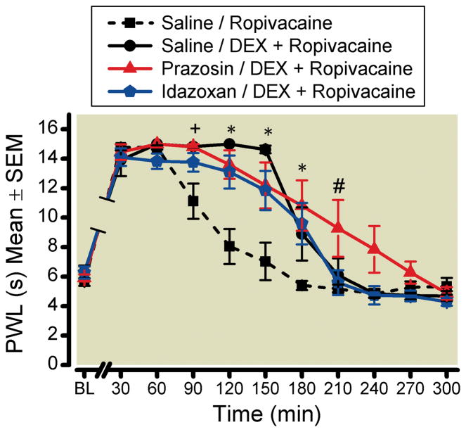
Neither pretreatment with prazosin (∝1-adrenoceptor antagonist) or idazoxan (∝2-adrenoceptor antagonist) attenuated the increased duration of sensory blockade caused by the addition of dexmedetomidine to ropivacaine (fig. 1). There were significant time (p < 0.001), drug (p = 0.0028), and time-by-drug (p < 0.0001) differences for the four groups. In post hoc analyses, there were multiple individual time points of statistical significance when all of the dexmedetomidine groups were compared with the ropivacaine alone group, regardless of the pretreatment (saline, prazosin or idazoxan; p < 0.005 for each significant comparison noted in fig. 1). Only at the 210-min time point was the prazosin pretreatment group significantly different from the idazoxan pretreatment group (p = 0.0019). Otherwise, there were no significant differences between the different individual time point assessments between the three dexmedetomidine groups. There were no significant time by drug interactions found in the control (unblocked) paws between the groups (p = 0.47, control paw data not shown).
Figure 1. Prazosin and Idazoxan Did Not Attenuate the Analgesic Effects of Perineural Dexmedetomidine Added to Ropivacaine.
Each rat received a pretreatment perineural injection followed by a second injection 10 min later. The duration of sensory blockade was measured by assessing paw withdrawal latency (PWL) to a heat stimulus every 30 min following the nerve block with a maximum response of 15 s. All of the groups containing dexmedetomidine plus ropivacaine had a longer duration of sensory blockade when compared with ropivacaine alone.
Groups are noted in the upper right by the “Pretreatment/Second Injection.”
∝ = 0.00833
* indicates a significant difference for all three DEX groups when compared with the Saline/Ropiv control group.
+ indicates Saline/DEX+Ropiv and Prazosin/DEX+Ropiv are each significantly different from the Saline/Ropiv control group.
# indicates Prazosin/DEX+Ropiv significantly differs from both Idazoxan/DEX+Ropiv and the Saline/Ropiv control group.
BL = baseline; DEX = dexmedetomidine; Ropiv = ropivacaine
Ih Current Antagonist (ZD 7288)
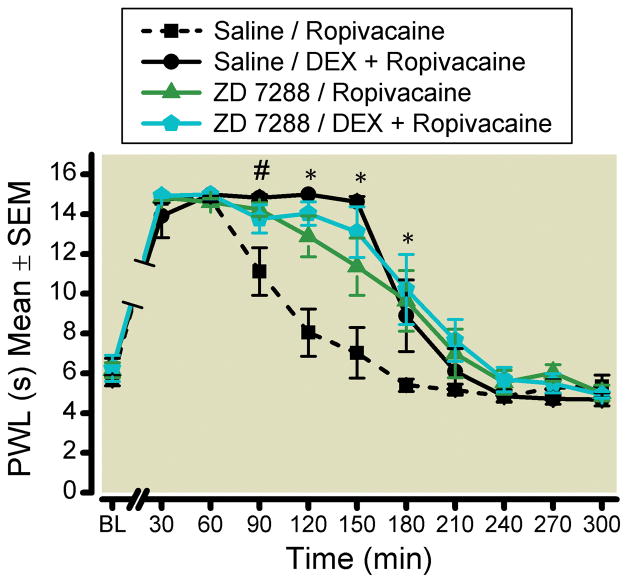
Pretreatment with ZD 7288 did not enhance or attenuate the duration of analgesia for dexmedetomidine added to ropivacaine (fig. 2). There were time (p < 0.0001), drug (p = 0.0006) and time-by-drug (p < 0.0001) effects. Post hoc analyses of time points 90 – 180 min between groups ZD 7288, dexmedetomidine, and the combination of ZD 7288 plus dexmedetomidine showed significant increases in the duration of analgesia when compared with ropivacaine alone (p < 0.004 for each significant comparison noted in fig. 2). There were no statistically significant differences between the ZD 7288 group and the dexmedetomidine group, and the combination of the two together with ropivacaine did not enhance or attenuate the sensory effects of dexmedetomidine (p = 0.17 – 0.39 between groups at time points 90 – 180). There were no significant time-by-drug interactions in the control paw between groups (p = 0.06; control paw data not shown).
Figure 2. The Ih Blocker ZD 7288 Did Not Increase the Duration of Dexmedetomidine-Mediated Analgesia.
The duration of the sciatic nerve block was measured by paw withdrawal latency (PWL) to a thermal stimulus every 30 min. Both ZD 7288 and dexmedetomidine increased the duration of sensory blockade when compared with the Saline/Ropivacaine control group. There was no additive effect when both ZD 7288 and dexmedetomidine were coadministered with ropivacaine.
Groups are noted in the upper right by the “Pretreatment/Second Injection.”
∝ = 0.00833
* indicates the Saline/Ropiv control group is significantly different from the other three groups. At 150 min, there is also a significant difference between Saline/DEX+Ropiv and ZD 7288/Ropiv.
# indicates Saline/DEX+Ropiv and ZD 7288/Ropiv are each significantly different from the Saline/Ropiv control group.
BL = baseline; DEX = dexmedetomidine; Ropiv = ropivacaine
Ih Current Agonist (Forskolin)
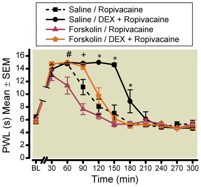
Pretreatment with the highest dose of forskolin (786 μM) attenuated the analgesic effects of dexmedetomidine added to ropivacaine (fig. 3). There were significant time (p < 0.0001), drug (p < 0.0001), and time-by-drug (p < 0.0001) interactions. Post hoc analyses demonstrated a significantly longer duration of analgesia in the dexmedetomidine plus ropivacaine group when compared with the other three groups at time points 90 – 180 min (p < 0.0001 for each significant time point noted in fig. 3). Individual time point analyses from 90 – 210 min between the ropivacaine alone group and the group receiving forskolin pretreatment followed by dexmedetomidine plus ropivacaine showed differences only at the 90-min time point (p = 0.0002), but there were otherwise no significant differences between the groups (p = 0.05, 0.35, 0.71, 0.89 for time points 120, 150, 180, 210 min, respectively). Pretreatment of forskolin with ropivacaine alone decreased the duration of analgesia when compared with ropivacaine alone with a saline (placebo) pretreatment. Significant differences were detected at time points 60 (p = 0.0001) and 90 (p < 0.0001). There was not a significant time-by-drug interaction found in the control (unblocked) paw between groups (p = 0.068, control paw data not shown).
Figure 3. Forskolin Attenuated the Analgesic Effects of Dexmedetomidine Added to Ropivacaine.
When the Ih agonist, forskolin, was given as a pretreatment to the dexmedetomidine plus ropivacaine combination, the duration of analgesia mirrored that of ropivacaine alone and was significantly different than the Saline/DEX+Ropiv group. Forskolin pretreatment also attenuated the analgesic effects of ropivacaine alone, as noted by the differences between the Forskolin/Ropiv and Saline/Ropiv groups.
Groups are noted in the upper right by the “Pretreatment/Second Injection.”
∝ = 0.00833
# indicates that the Forskolin/Ropiv group is significantly different from the other three groups.
+ indicates that the Saline/Ropiv control group is significantly different from the other three groups. Forskolin/Ropiv is also significantly different from the remaining two groups.
* indicates that the Saline/DEX+Ropiv group is significantly different from the other three groups. At 120 min, there is also a significant difference between the two Forskolin groups. BL = baseline; DEX = dexmedetomidine; PWL = paw withdrawal latency; Ropiv = ropivacaine
Three doses of forskolin (384, 576 and 768 μM) doses followed by ropivacaine plus dexmedetomidine were studied. The highest dose of forskolin (768 μM) significantly attenuated the dexmedetomidine-induced duration of sensory blockade when compared with the ropivacaine plus dexmedetomidine group (median 180 min [25th, 75th interquartile range 180, 210] vs. 150 [127.5, 150]). While the study was not powered to detect a difference between the forskolin doses, there was a downward, but not statistically significant, trend in the duration of analgesia with each increasing dose of forskolin pretreatment (384, 576, and 768 μM forskolin; 180 [150, 180], 150 [127.5, 172.5], 150 [127.5, 150] min, respectively).
Neurobehavioral Assessments 24 Hours After Nerve Block
There were no differences between any of the groups for operative or control paw withdrawal latency when reassessed 24 h after the placement of the nerve block. There were no gross motor abnormalities or wound infections noted the day after the nerve block.
Discussion
Perineural Dexmedetomidine Acts Peripherally by Blocking the Ih Current
In this blinded, randomized, controlled study, the effects of dexmedetomidine were shown to be likely due to the blockade of the hyperpolarization-activated cation current (figs. 2 and 3). This is the first in vivo study to investigate the mechanism of action for peripheral perineural dexmedetomidine. In a recent study, we demonstrated that the analgesic effects of dexmedetomidine were significantly greater when administered perineurally versus systemically (subcutaneously) in a ropivacaine sciatic nerve block in rat model;5 however, the underlying mechanism was still not known. The present study design employed was comparable to Kroin et al.13 As with clonidine, a blocker of the Ih current (ZD 7288) did not have any effect on the duration of analgesia associated with the addition of dexmedetomidine to ropivacaine, while an enhancer of the Ih current (forskolin) attenuated dexmedetomidine’s analgesic effects. Interestingly, the dose of forskolin required to fully attenuate the effects of dexmedetomidine added to ropivacaine (fig. 3) was higher than that which reversed clonidine added to lidocaine. The present results encourage future clinical studies designed to determine whether more potent inhibition of the Ih current by dexmedetomidine will cause a longer duration of analgesia when compared with clonidine.
Perineural Dexmedetomidine Not Reversed by ∝1- or ∝2-Adrenoceptor Antagonists
The peripheral analgesic effects of dexmedetomidine were not reversed by either ∝1- or ∝2-adrenoceptor antagonists (prazosin and idazoxan, respectively; fig. 1). Dexmedetomidine is approved for intravenous administration for sedation and analgesia in intubated and mechanically ventilated patients in the intensive care unit and for non-intubated patients for surgical and other procedures.18 The described mechanism of action for intravenous dexmedetomidine is ∝2-mediated. The antinociceptive properties of neuraxial dexmedetomidine were previously demonstrated to be ∝2-mediated and were reversed by a proportionally much lower dose of idazoxan than that which was used in the present study.16,19,20 Consistent with our findings, an in vitro study of frog sciatic nerves demonstrated a dose-dependent decrease of compound action potentials with dexmedetomidine, which was not reversed by ∝2-adrenoceptor antagonists.10 When compared using the same compound action potential model, the effects of clonidine were only 20% of that which was seen with dexmedetomidine.
Mechanism of Action for Peripheral Perineural Dexmedetomidine Similar to Clonidine
The present study indicates that the mechanism of action for peripheral perineural dexmedetomidine is similar to clonidine.12–14,21,22 Clonidine has been used as an additive to local anesthetics for peripheral nerve blocks for many years, dating back to the first human descriptions in the early 1990s.23–25 Despite demonstrating clear efficacy with short- and intermediate-acting local anesthetics, the effects of clonidine added to long-acting local anesthetics for peripheral nerve blocks has been questioned by some experts and the data are conflicting.26,27 Based on the available data, it was concluded in a meta-analysis that clonidine increases the duration of long-acting local anesthetics by about 2 h.27 The limited analgesic benefit, potential side effects (hypotension, bradycardia, and sedation), and current cost ($46.93 for a 1,000 μg vial at the University of Michigan) have tempered the widespread use of clonidine in clinical practice. Some institutions, such as ours, have elected to have pharmacists prepare ten sterile 100 μg clonidine syringes from a 1,000 μg vial to control cost; however, this again has issues of labor costs and resources. Dexmedetomidine is currently more expensive than clonidine ($68.86 for a 200 μg vial at the University of Michigan), therefore efficacy greater than clonidine should be established to justify the additional cost.
Laboratory studies of perineural dexmedetomidine conducted have all shown impressive efficacy when combined with long-acting local anesthetics (bupivacaine and ropivacaine),5–7 whereas the preclinical clonidine data have been confined to combinations with short-acting local anesthetics (lidocaine).12,13 There are limited human data to date using dexmedetomidine in peripheral nerve blocks, but both studies have clearly demonstrated efficacy when added to long-acting local anesthetics.8,9 In a randomized, double-blind, controlled trial, Obayah et al.9 found a statistically significant increase in the time to first analgesic request from the addition of 1 μg/kg of dexmedetomidine added to bupivacaine versus bupivacaine alone in greater palatine nerve blocks for cleft palate repair in children (mean 22 h [range 20.6–23.7] vs. 14.2 h [13–15], respectively). There were no noted differences in sedation, heart rate or blood pressure between the groups. More recently, in a randomized, double blind, control trial, Esmaoglu et al.8 studied 150 μg dexmedetomidine added to levobupivacaine compared with levobupivacaine alone for axillary brachial plexus blocks. The investigators used a nerve stimulation technique for the axillary block and large volumes (40 ml). Dexmedetomidine prolonged the duration of analgesia and motor blockade, while also decreasing the time to onset of sensory and motor blockade. Systolic blood pressures and heart rate were lower in the dexmedetomidine group for the first 2 h; however, there were no adverse events. While the dexmedetomidine group was clearly superior to the control group, the analgesic effects may have been undervalued in this study by the use of high volumes (lower concentration of dexmedetomidine) and failure to use ultrasound. Advances in ultrasound technology allows for placement of the injectate closer to the peripheral nerve, thereby concentrating the drug(s) at the site of action and decreasing the required volume.28 The side effects of dexmedetomidine clinically are likely to be dependent on the total dose and systemic absorption rate, rather than concentration, while the analgesic effects appear to be concentration-dependent.7
Dexmedetomidine is not approved for neuraxial or perineural administration. To our knowledge, there have been eight human studies on the subject to date, including two peripheral perineural,8,9 one intrathecal,29 and five epidural studies.30–34 All of the studies have demonstrated efficacy. Side effects have been noted, but the data are not consistent. Some of the studies have noted dexmedetomidine-associated decreases in heart rate and blood pressure8,34 and increased sedation.32,33 The other four studies found no differences in side effects associated with dexmedetomidine. The sedation associated with intravenous dexmedetomidine has been termed “cooperative sedation,” which may mean that any sedation from perineural administration may be beneficial, especially when compared with the type of sedation associated with benzodiazepines and opioids.35
Limitations
The analgesic effects of dexmedetomidine were attenuated by pretreatment with forskolin; however, forskolin has many effects in addition to blockade of the Ih current and other membrane transport and channel proteins. Forskolin stimulates adenylyl cyclase and thereby increases intracellular cyclic adenosine monophosphate.36 Forskolin can inhibit platelet aggregation and mast cell degranulation, and it can increase cardiac contractility, insulin secretion, thyroid function, and lypolysis. In addition, forskolin can cause vasodilation, which could affect the duration of the nerve block independent of its effects on the Ih current. As with clonidine,13 pretreatment with a known blocker of the Ih current (ZD 7288) provided a comparable duration of analgesia to dexmedetomidine without additive or synergistic effects when combined (fig. 2). Whereas there are multiple in vivo and in vitro studies demonstrating Ih current blocking effects of clonidine and dexmedetomidine,11–15 it is possible that other actions of forskolin reversed the analgesic effects of dexmedetomidine.
The primary outcome measure of the study was the duration of sensory blockade as measured by the time to paw withdrawal to a thermal stimulus every 30 min after a sciatic nerve block. The duration of motor blockade was not assessed, as it would have required handling of the rats and handling would have confounded the sensory testing. The duration of motor blockade in regional anesthesia is important and should be included in clinical trials, as a selective sensory block without motor blockade offers clear advantages in facilitating comfortable postoperative rehabilitation. There are preclinical data to support that clonidine more selectively blocks C fibers (pain fibers) than A∝ fibers (motor neurons).22 Whether this is also true for dexmedetomidine is unknown, and is open to future investigation.
What we already know about this topic
Although dexemedetomidine prolongs and/or intensifies peripheral nerve blockade from local anesthetics, the mechanisms by which it does so are unclear
What this article tells us that is new
Using sciatic nerve block in rats, dexmedetomidine more than doubled the duration of ropivacaine block, and this effect was reversed by an Ih channel enhancer, but not by an α2-adrenoceptor antagonist
Acknowledgments
We thank Kevin K. Tremper, M.D., Ph.D. (Professor, Department of Anesthesiology, University of Michigan) for support and guidance. For statistical support, we thank Kathleen B. Welch, M.A., M.P.H. (Senior Statistician, Center for Statistical Consultation and Research, University of Michigan). For technical support, we thank members of the research laboratory at the University of Michigan, Department of Anesthesiology: Mary Norat, B.S. (Laboratory Manager), Christopher Watson, Ph.D. (Research Investigator), Sarah L. Watson, B.S. (Research Associate).
Financial Support: Dr. Brummett is supported by grant UL1RR024986 from the National Center for Research Resources (National Institutes of Health, Bethesda, Maryland). Elizabeth Hong was supported by the Foundation for Anesthesia Education and Research Fellowship (Rochester, Minnesota). Ralph Lydic, Ph.D. is supported by National Institutes of Health grants HL-40881 and HL-65272 (Bethesda, Maryland). Additional support from the Department of Anesthesiology, University of Michigan, Ann Arbor, Michigan. The content is solely the responsibility of the authors and does not necessarily represent the official views of National Center for Research Resources or the National Institutes of Health.
Footnotes
Study performed at: Department of Anesthesiology, University of Michigan, Ann Arbor, Michigan
Disclosures: The University of Michigan has filed for a patent application covering the subject matter of this publication. Brummett CM, Inventor; The Regents of the University of Michigan, Assignee; Anesthetic Methods and Compositions; US patent application US 12/791,506; June 1, 2010.
References
- 1.Casati A, Fanelli G, Albertin A, Deni F, Anelati D, Antonino FA, Beccaria P. Interscalene brachial plexus anesthesia with either 0.5% ropivacaine or 0.5% bupivacaine. Minerva Anestesiol. 2000;66:39–44. [PubMed] [Google Scholar]
- 2.Casati A, Fanelli G, Aldegheri G, Berti M, Colnaghi E, Cedrati V, Torri G. Interscalene brachial plexus anaesthesia with 0.5%, 0.75% or 1% ropivacaine: A double-blind comparison with 2% mepivacaine. Br J Anaesth. 1999;83:872–5. doi: 10.1093/bja/83.6.872. [DOI] [PubMed] [Google Scholar]
- 3.Apfelbaum JL, Gan TJ, Zhao S, Hanna DB, Chen C. Reliability and validity of the perioperative opioid-related symptom distress scale. Anesth Analg. 2004;99:699–709. doi: 10.1213/01.ANE.0000133143.60584.38. [DOI] [PubMed] [Google Scholar]
- 4.Lydic R, Baghdoyan HA. In: Neurochemical mechanisms mediating opioid-induced REM sleep disruption, Sleep and Pain. Lavigne G, Sessle BJ, Choiniere M, Soja PJ, editors. Seattle: International Association for the Study of Pain (IASP) Press; 2007. pp. 99–122. [Google Scholar]
- 5.Brummett CM, Amodeo FS, Janda AM, Padda AK, Lydic R. Perineural dexmedetomidine provides an increased duration of analgesia to a thermal stimulus when compared with a systemic control in a rat sciatic nerve block. Reg Anesth Pain Med. 2010;35:427–31. doi: 10.1097/AAP.0b013e3181ef4cf0. [DOI] [PubMed] [Google Scholar]
- 6.Brummett CM, Norat MA, Palmisano JM, Lydic R. Perineural administration of dexmedetomidine in combination with bupivacaine enhances sensory and motor blockade in sciatic nerve block without inducing neurotoxicity in rat. Anesthesiology. 2008;109:502–11. doi: 10.1097/ALN.0b013e318182c26b. [DOI] [PMC free article] [PubMed] [Google Scholar]
- 7.Brummett CM, Padda AK, Amodeo FS, Welch KB, Lydic R. Perineural dexmedetomidine added to ropivacaine causes a dose-dependent increase in the duration of thermal antinociception in sciatic nerve block in rat. Anesthesiology. 2009;111:1111–9. doi: 10.1097/ALN.0b013e3181bbcc26. [DOI] [PMC free article] [PubMed] [Google Scholar]
- 8.Esmaoglu A, Yegenoglu F, Akin A, Turk CY. Dexmedetomidine added to levobupivacaine prolongs axillary brachial plexus block. Anesth Analg. 2010;111:1548–51. doi: 10.1213/ANE.0b013e3181fa3095. [DOI] [PubMed] [Google Scholar]
- 9.Obayah GM, Refaie A, Aboushanab O, Ibraheem N, Abdelazees M. Addition of dexmedetomidine to bupivacaine for greater palatine nerve block prolongs postoperative analgesia after cleft palate repair. Eur J Anaesthesiol. 2010;27:280–4. doi: 10.1097/EJA.0b013e3283347c15. [DOI] [PubMed] [Google Scholar]
- 10.Kosugi T, Mizuta K, Fujita T, Nakashima M, Kumamoto E. High concentrations of dexmedetomidine inhibit compound action potentials in frog sciatic nerves without alpha(2) adrenoceptor activation. Br J Pharmacol. 2010;160:1662–76. doi: 10.1111/j.1476-5381.2010.00833.x. [DOI] [PMC free article] [PubMed] [Google Scholar]
- 11.Dalle C, Schneider M, Clergue F, Bretton C, Jirounek P. Inhibition of the I(h) current in isolated peripheral nerve: A novel mode of peripheral antinociception? Muscle Nerve. 2001;24:254–61. doi: 10.1002/1097-4598(200102)24:2<254::aid-mus110>3.0.co;2-#. [DOI] [PubMed] [Google Scholar]
- 12.Gaumann DM, Brunet PC, Jirounek P. Hyperpolarizing afterpotentials in C fibers and local anesthetic effects of clonidine and lidocaine. Pharmacology. 1994;48:21–9. doi: 10.1159/000139158. [DOI] [PubMed] [Google Scholar]
- 13.Kroin JS, Buvanendran A, Beck DR, Topic JE, Watts DE, Tuman KJ. Clonidine prolongation of lidocaine analgesia after sciatic nerve block in rats is mediated via the hyperpolarization-activated cation current, not by α-adrenoreceptors. Anesthesiology. 2004;101:488–94. doi: 10.1097/00000542-200408000-00031. [DOI] [PubMed] [Google Scholar]
- 14.Erne-Brand F, Jirounek P, Drewe J, Hampl K, Schneider MC. Mechanism of antinociceptive action of clonidine in nonmyelinated nerve fibres. Eur J Pharmacol. 1999;383:1–8. doi: 10.1016/s0014-2999(99)00620-2. [DOI] [PubMed] [Google Scholar]
- 15.Shirasaka T, Kannan H, Takasaki M. Activation of a G protein-coupled inwardly rectifying K+ current and suppression of Ih contribute to dexmedetomidine-induced inhibition of rat hypothalamic paraventricular nucleus neurons. Anesthesiology. 2007;107:605–15. doi: 10.1097/01.anes.0000281916.65365.4e. [DOI] [PubMed] [Google Scholar]
- 16.Fisher B, Zornow MH, Yaksh TL, Peterson BM. Antinociceptive properties of intrathecal dexmedetomidine in rats. Eur J Pharmacol. 1991;192:221–5. doi: 10.1016/0014-2999(91)90046-s. [DOI] [PubMed] [Google Scholar]
- 17.Dirig DM, Salami A, Rathbun ML, Ozaki GT, Yaksh TL. Characterization of variables defining hindpaw withdrawal latency evoked by radiant thermal stimuli. J Neurosci Methods. 1997;76:183–91. doi: 10.1016/s0165-0270(97)00097-6. [DOI] [PubMed] [Google Scholar]
- 18.Gerlach AT, Murphy CV, Dasta JF. An updated focused review of dexmedetomidine in adults. Ann Pharmacother. 2009;43:2064–74. doi: 10.1345/aph.1M310. [DOI] [PubMed] [Google Scholar]
- 19.Sabbe MB, Penning JP, Ozaki GT, Yaksh TL. Spinal and systemic action of the α2 receptor agonist dexmedetomidine in dogs. Antinociception and carbon dioxide response. Anesthesiology. 1994;80:1057–72. doi: 10.1097/00000542-199405000-00015. [DOI] [PubMed] [Google Scholar]
- 20.Nazarian A, Christianson CA, Hua XY, Yaksh TL. Dexmedetomidine and ST-91 analgesia in the formalin model is mediated by alpha2A-adrenoceptors: A mechanism of action distinct from morphine. Br J Pharmacol. 2008;155:1117–26. doi: 10.1038/bjp.2008.341. [DOI] [PMC free article] [PubMed] [Google Scholar]
- 21.Gaumann DM, Brunet PC, Jirounek P. Clonidine enhances the effects of lidocaine on C-fiber action potential. Anesth Analg. 1992;74:719–25. doi: 10.1213/00000539-199205000-00017. [DOI] [PubMed] [Google Scholar]
- 22.Butterworth JF, Strichartz GR. The α2-adrenergic agonists clonidine and guanfacine produce tonic and phasic block of conduction in rat sciatic nerve fibers. Anesth Analg. 1993;76:295–301. [PubMed] [Google Scholar]
- 23.Eledjam JJ, Deschodt J, Viel EJ, Lubrano JF, Charavel P, d’Athis F, du Cailar J. Brachial plexus block with bupivacaine: Effects of added alpha-adrenergic agonists: Comparison between clonidine and epinephrine. Can J Anaesth. 1991;38:870–5. doi: 10.1007/BF03036962. [DOI] [PubMed] [Google Scholar]
- 24.Gaumann D, Forster A, Griessen M, Habre W, Poinsot O, Della Santa D. Comparison between clonidine and epinephrine admixture to lidocaine in brachial plexus block. Anesth Analg. 1992;75:69–74. doi: 10.1213/00000539-199207000-00013. [DOI] [PubMed] [Google Scholar]
- 25.Singelyn FJ, Dangoisse M, Bartholomee S, Gouverneur JM. Adding clonidine to mepivacaine prolongs the duration of anesthesia and analgesia after axillary brachial plexus block. Reg Anesth. 1992;17:148–50. [PubMed] [Google Scholar]
- 26.McCartney CJ, Duggan E, Apatu E. Should we add clonidine to local anesthetic for peripheral nerve blockade? A qualitative systematic review of the literature. Reg Anesth Pain Med. 2007;32:330–8. doi: 10.1016/j.rapm.2007.02.010. [DOI] [PubMed] [Google Scholar]
- 27.Popping DM, Elia N, Marret E, Wenk M, Tramer MR. Clonidine as an adjuvant to local anesthetics for peripheral nerve and plexus blocks: A meta-analysis of randomized trials. Anesthesiology. 2009;111:406–15. doi: 10.1097/ALN.0b013e3181aae897. [DOI] [PubMed] [Google Scholar]
- 28.Riazi S, Carmichael N, Awad I, Holtby RM, McCartney CJ. Effect of local anaesthetic volume (20 vs 5 ml) on the efficacy and respiratory consequences of ultrasound-guided interscalene brachial plexus block. Br J Anaesth. 2008;101:549–56. doi: 10.1093/bja/aen229. [DOI] [PubMed] [Google Scholar]
- 29.Kanazi GE, Aouad MT, Jabbour-Khoury SI, Al Jazzar MD, Alameddine MM, Al-Yaman R, Bulbul M, Baraka AS. Effect of low-dose dexmedetomidine or clonidine on the characteristics of bupivacaine spinal block. Acta Anaesthesiol Scand. 2006;50:222–7. doi: 10.1111/j.1399-6576.2006.00919.x. [DOI] [PubMed] [Google Scholar]
- 30.El-Hennawy AM, Abd-Elwahab AM, Abd-Elmaksoud AM, El-Ozairy HS, Boulis SR. Addition of clonidine or dexmedetomidine to bupivacaine prolongs caudal analgesia in children. Br J Anaesth. 2009;103:268–74. doi: 10.1093/bja/aep159. [DOI] [PubMed] [Google Scholar]
- 31.Elhakim M, Abdelhamid D, Abdelfattach H, Magdy H, Elsayed A, Elshafei M. Effect of epidural dexmedetomidine on intraoperative awareness and postoperative pain after one-lung ventilation. Acta Anaesthesiol Scand. 2010;54:703–9. doi: 10.1111/j.1399-6576.2009.02199.x. [DOI] [PubMed] [Google Scholar]
- 32.Vieira AM, Schnaider TB, Brandao AC, Pereira FA, Costa ED, Fonseca CE. Epidural clonidine or dexmedetomidine for post-cholecystectomy analgesia and sedation. Rev Bras Anestesiol. 2004;54:473–8. doi: 10.1590/s0034-70942004000400003. [DOI] [PubMed] [Google Scholar]
- 33.Saadawy I, Boker A, Elshahawy MA, Almazrooa A, Melibary S, Abdellatif AA, Afifi W. Effect of dexmedetomidine on the characteristics of bupivacaine in a caudal block in pediatrics. Acta Anaesthesiol Scand. 2009;53:251–6. doi: 10.1111/j.1399-6576.2008.01818.x. [DOI] [PubMed] [Google Scholar]
- 34.Schnaider TB, Vieira AM, Brandao AC, Lobo MV. Intraoperative analgesic effect of epidural ketamine, clonidine or dexmedetomidine for upper abdominal surgery. Rev Bras Anestesiol. 2005;55:525–31. doi: 10.1590/s0034-70942005000500007. [DOI] [PubMed] [Google Scholar]
- 35.Bekker A, Sturaitis MK. Dexmedetomidine for neurological surgery. Neurosurgery. 2005;57:1–10. doi: 10.1227/01.neu.0000163476.42034.a1. [DOI] [PubMed] [Google Scholar]
- 36.Laurenza A, Sutkowski EM, Seamon KB. Forskolin: A specific stimulator of adenylyl cyclase or a diterpene with multiple sites of action? Trends Pharmacol Sci. 1989;10:442–7. doi: 10.1016/S0165-6147(89)80008-2. [DOI] [PubMed] [Google Scholar]


