Dear Editor,
Embryonic stem cell (ES cell) lines were first generated by culturing mouse inner cell mass (ICM) on feeder layers in 1981 1. However, in large domestic animals, attempts to establish ES cell lines from ICM of blastocysts or the later epiblast have not been successful. This has hindered the efficient production of genetically modified livestocks by using ES-based approaches. Recently, it was found that ectopic expression of various combinations of transcription factors is able to reprogram somatic cells to a pluripotent state 2, 3, 4, 5. These induced pluripotent stem (iPS) cells show similarities to embryo-derived ES cells and can be used to produce viable mice through tetraploid complementation 6, 7. So far, iPS cells of several mammalian species have been successfully generated 2, 3, 8, 9, 10, 11, 12. In this letter, we report the first establishment of bovine iPS cells using defined transcription factors and a modified culture medium.
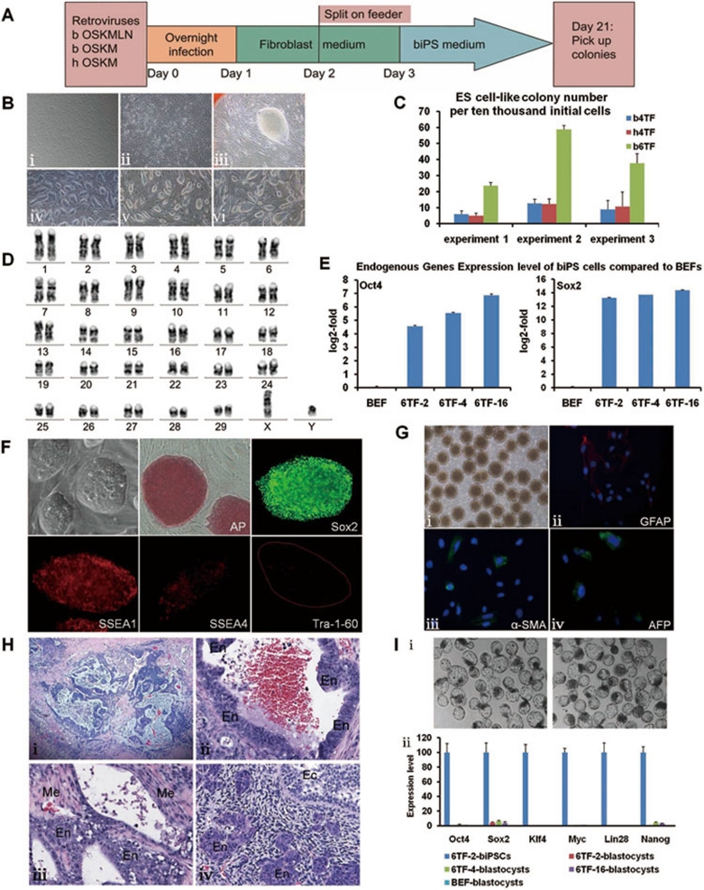
cDNAs coding for the bovine OCT4 (also named POU5F1), SOX2, KLF4, MYC, LIN28, and NANOG genes were cloned into pMXs retroviral vector. The pMXs plasmids containing human OCT4, SOX2, KLF4, and c-MYC genes were all purchased from Addgene. GP2-293 cells were used as the packaging cell line for retroviral production. Bovine fibroblasts used in this study were derived from an E55 Western Shandong Yellow cattle fetus. Three sets of factors, termed b4TF, b6TF, and h4TF, were used to transduce cells by overnight retroviral infection, respectively. Whereas the former two included only bovine factors (b4TF: bOCT4, bSOX2, bMYC, bKLF4; b6TF: bOCT4, bSOX2, bKLF4, bMYC, bLIN28, bNANOG), the latter employed only human factors (hOCT4, hSOX2, hc-MYC, hKLF4). On day 2, the infected cells were harvested by trypsinization and plated onto mitomycin C-treated MEF feeders at a density of 1 × 10⁴ cells per 100-mm dish. The next day after being seeded onto feeders, growing cells were cultured in iPS media (Figure 1A and Supplementary information, Table S1). Bovine iPS (hereinafter named biPS) cells with a mouse ES-like morphology were detected after ∼3 weeks (Figure 1B). On day 21-35, colonies were isolated mechanically using a 200 μl pipette and transferred to feeder-coated tissue culture dishes. The biPS cells were split with trypsin at a ratio of 1:10 every 4-5 days afterwards (Supplementary information, Data S1). A total of 26 b6TF-derived colonies have been expanded into biPS cell lines. These lines maintained good ES-like morphology for more than 16 passages. However, none of the colonies generated by b4TF or h4TF could be passaged more than six times. Importantly, we showed that the combination of six transcription factors (b6TF set) significantly increased the efficiency of iPS cell generation (by three-fold) compared to the other two combinations (Figure 1C).
Figure 1.
Generation of bovine iPS cells from embryonic fibroblast cells. (A) Scheme depicting the generation of bovine iPS cell lines. (B) Morphology of bovine iPS cells. (B-i) Bovine embryonic fibroblast cells (BEFs). (B-ii) Fibroblast cells 6 days after six-bovine-transcription factor transduction. (B-iii) Primary colony appearing on feeder cells 25 days after infection. (B-iv) b6TF-2 iPS cells at passage 12. (B-v) b4TF-2 iPS cells at passage 4. (B-vi) h4TF-3 iPS cells at passage 3. (C) The numbers of ES-like colonies derived from BEFs (1 × 10⁴ per 100-mm dish) transduced with retrovirus carrying four bovine transcription factors (OCT4, SOX2, KLF4, and MYC), four human factors (OCT4, SOX2, KLF4, and c-MYC), or six bovine factors (OCT4, SOX2, NANOG, LIN28, MYC, and KLF4) on day 28. (D) The karyotype of b6TF-2 iPS cells is 60, XY. (E) Endogenous expression levels of OCT4 and SOX2 in biPS cells compared to BEFs. (F) Alkaline phosphatase (AP) staining and ES-cell-specific marker immunocytochemistry (ICC). (G) In vitro differentiation of bovine iPS cells to form embryoid bodies (EBs). (G-i) After 5 days under differentiation conditions (iPS medium without bFGF). biPSCs can effectively differentiate in vitro into cells of the three germ layers, including neural cells (ectoderm, GFAP+, red), smooth muscle cells (mesoderm, α-SMA+, green), and endodermal cells (AFP+, green). (G-ii, G-iii and G-iv) Show the immunostaining results for EBs transferred onto tissue-culture dishes for 5 days. DAPI was used to stain the cell nuclei (blue). (H) A representative teratoma developed from one injection site. Tissues were analyzed by hematoxylin and eosin staining. (H-i) Shows cell types in one teratoma. (H-ii, H-iii and H-iv) Show that our biPS cells formed teratoma consisting of three basic germ layers (Ec: Ectoderm; Me: Mesoderm; En: Endoderm). (I) SCNT blastocysts. (I-i) Morphology of biPSCs-derived SCNT blastocysts (left) and BEFs-derived ones (right). (I-ii) All the exogenous transgenes were silenced in biPSCs-derived SCNT blastocysts. Error bars represent SD (n = 3).
We tested eight different types of biPS culture media (Supplementary information, Table S1) by assessing the numbers of ES-like colonies obtained from b6TF-transduced BEFs on day 28. Three out of the eight media could efficiently generate biPS cells (Supplementary information, Figure S1). Our results suggested that knockout serum replacement and basic fibroblast growth factor are optimal for biPS cell generation. In addition, we found that DMEM as basic medium could greatly improve the reprogramming efficiency. When DMEM is replaced with knockout DMEM, we observed decreased efficiency of ES-like colony formation (data not shown). In summary, the optimal culture medium for biPS cell generation is composed of DMEM supplemented with 20% knockout serum replacement, 2 mM ℒ-glutamine, 1% non-essential amino acids, 0.1 mM β-mercaptoethanol and 4 ng/ml bFGF.
Karyotype analysis showed that our biPS cells maintained a normal karyotype: 60, XY. The pluripotent characteristics of our biPS cells were clearly not associated with accumulation of chromosomal abnormalities (Figure 1D). As revealed by quantitative PCR, expression of endogenous OCT4 and SOX2 was reactivated in our biPS cells (Figure 1E). We did not see any significant increase in endogenous NANOG expression (Supplementary information, Figure S2). Moreover, we noticed that the exogenous transgenes continued to be co-expressed along with their bovine orthologs in reprogrammed biPS cells (data not shown). This is consistent with other studies that also described the incomplete transgene silencing in pig iPS cells 10. The biPS cells are positive for alkaline phosphatase, SSEA1, NANOG, and SOX2, but are weakly positive for SSEA4, and are negative for Tra-1-60 and TRA-1-81 (Figure 1F and Supplementary information, Data S1). These immunofluorescent staining results suggest that our biPS cells are more similar to mouse ES cells than human ES cells. The expression levels of several other genes associated with the pluripotent state were analyzed by real-time PCR, and were shown to be increased to various degree (Supplementary information, Figure S3).
We then examined the differentiation ability of the biPS cells in vitro and in vivo to confirm their potential as pluripotent stem cells (Supplementary information, Data S1). We found that the biPS cells cultured in biPS medium without LIF and bFGF on a non-adhesive petri dish (Figure 1G-i) were able to form typical embryoid bodies (EBs). After growing in suspension for 5 days, the EBs were replated in adherent conditions to induce further differentiation. Immunostaining on the differentiated structures showed that biPS cells could differentiate into endoderm (AFP), mesoderm (α-SMA), and ectoderm (GFAP) derivatives (Figure 1G-ii, 1G-iii and 1G-iv). Real-time PCR analysis of EBs confirmed the differentiation ability of the biPS cells in vitro (Supplementary information, Figure S4). To test pluripotency in vivo, we transplanted the biPS cells into the renal capsule of NOD/SCID mice. At 9 weeks after injection, we observed tumor formation. Histological examination showed that biPS cells formed teratoma consisting of three basic germ layers (Figure 1H).
Since biPS cells have long lifespan when cultured in vitro, it could provide a better nucleus-donor resource for the production of genetically modified cattle. We used somatic cell nuclear transfer (SCNT) technique to test this potential. We succeeded in using three biPS cell lines as donors to produce reconstructed embryos (Supplementary information, Data S1). There was no significant difference between biPSCs-derived SCNT blastocysts (named 6TF-2-blastocysts, 6TF-4-blastocysts, and 6TF-16-blastocysts, respectively) and BEFs-derived SCNT blastocysts (BEF-blastocysts) in morphology (Figure 1I-i), and cell numbers of 7.5-day embryos (170 ± 20.46 vs 159 ± 20.44, n > 5, P > 0.05). We tested the expression levels of exogenous genes in cloned embryos and found that all the transgenes were shut down (Figure 1I-ii). This suggested that our biPS cells could be used as nuclear-donor resource to produce cloned animal breeds.
In summary, we have successfully generated biPS cell lines from bovine embryonic fibroblast cells by the transduction of six bovine transcription factors. Knockout serum replacement and basic fibroblast growth factor are optimal for the induction. Our biPS cells exhibit a mouse ES-like morphology. They are alkaline phosphatase positive, and express pluripotent markers such as SSEA1, SOX2, and NANOG. Karyotyping analysis demonstrated that biPS cells showed a normal chromosome number. Furthermore, the biPS cells can differentiate to three basic germ layers in vitro and in vivo. For practical applications, our biPS cells can be used as SCNT donor cells to produce genetically modified breeds. Finally, the successful generation of biPS cells will facilitate the establishment of bovine ES cell lines in the future.
Acknowledgments
We are grateful to Dr Rogers Keith (Institute of Molecular and Cell Biology, Singapore) for assisting in the identification of tissue types in teratoma and Dr Fillon Valérie (UMR de Genetique Cellulaire, INRA Toulouse, France) for advice on karyotype alignment. This study was supported in part by grants from the Hi-Tech Research and Development Program of China (2010AA100504), the China National Basic Research Program (2009CB941003, 2010CB945404, 2011CBA01000, 2011CBA01102), and A-Star Singapore. We would like to thank our colleagues from the Stem Cell and Developmental Biology group in the Genome Institute of Singapore for their assistance and suggestions.
Footnotes
(Supplementary information is linked to the online version of the paper on the Cell Research website.)
Supplementary Material
The composition of the 8 different iPS media
Materials and Methods
Numbers of ES-like colonies obtained from b6TF transduced BEFs cultured in different media (1 × 10⁴ cells per 100 mm dish).
(i) Endogenous expression levels of OCT4, SOX2 and NANOG in BEFs.
Other pluripotent gene expression levels of biPS cells.
Real time-PCR analysis of relative transcript concentrations of lineage-specific genes in biPSC lines (6TF-2, 6TF-4 and 6TF-16), BEFs, and biPSCs differentiated into EB by using either FBS or retinoic acid (RA).
References
- Evans MJ, Kaufman MH. Establishment in culture of pluripotential cells from mouse embryos. Nature. 1981;292:154–156. doi: 10.1038/292154a0. [DOI] [PubMed] [Google Scholar]
- Takahashi K, Yamanaka S. Induction of pluripotent stem cells from mouse embryonic and adult fibroblast cultures by defined factors. Cell. 2006;126:663–676. doi: 10.1016/j.cell.2006.07.024. [DOI] [PubMed] [Google Scholar]
- Yu J, Vodyanik MA, Smuga-Otto K, et al. Induced pluripotent stem cell lines derived from human somatic cells. Science. 2007;318:1917–1920. doi: 10.1126/science.1151526. [DOI] [PubMed] [Google Scholar]
- Takahashi K, Tanabe K, Ohnuki M, et al. Induction of pluripotent stem cells from adult human fibroblasts by defined factors. Cell. 2007;131:861–872. doi: 10.1016/j.cell.2007.11.019. [DOI] [PubMed] [Google Scholar]
- Blelloch R, Venere M, Yen J, Ramalho-Santos M. Generation of induced pluripotent stem cells in the absence of drug selection. Cell Stem Cell. 2007;1:245–247. doi: 10.1016/j.stem.2007.08.008. [DOI] [PMC free article] [PubMed] [Google Scholar]
- Kang L, Wang J, Zhang Y, Kou Z, Gao S. iPS cells can support full-term development of tetraploid blastocyst-complemented embryos. Cell Stem Cell. 2009;5:135–138. doi: 10.1016/j.stem.2009.07.001. [DOI] [PubMed] [Google Scholar]
- Zhao XY, Li W, Lv Z, et al. iPS cells produce viable mice through tetraploid complementation. Nature. 2009;461:86–90. doi: 10.1038/nature08267. [DOI] [PubMed] [Google Scholar]
- Liu H, Zhu F, Yong J, et al. Generation of induced pluripotent stem cells from adult rhesus monkey fibroblasts. Cell Stem Cell. 2008;3:587–590. doi: 10.1016/j.stem.2008.10.014. [DOI] [PubMed] [Google Scholar]
- Liao J, Cui C, Chen S, et al. Generation of induced pluripotent stem cell lines from adult rat cells. Cell Stem Cell. 2009;4:11–15. doi: 10.1016/j.stem.2008.11.013. [DOI] [PubMed] [Google Scholar]
- Esteban MA, Xu J, Yang J, et al. Generation of induced pluripotent stem cell lines from Tibetan miniature pig. J Biol Chem. 2009;284:17634–17640. doi: 10.1074/jbc.M109.008938. [DOI] [PMC free article] [PubMed] [Google Scholar]
- Shimada H, Nakada A, Hashimoto Y, Shigeno K, Shionoya Y, Nakamura T. Generation of canine induced pluripotent stem cells by retroviral transduction and chemical inhibitors. Mol Reprod Dev. 2010;77:2. doi: 10.1002/mrd.21117. [DOI] [PubMed] [Google Scholar]
- Honda A, Hirose M, Hatori M, et al. Generation of induced pluripotent stem cells in rabbits: potential experimental models for human regenerative medicine. J Biol Chem. 2010;285:31362–31369. doi: 10.1074/jbc.M110.150540. [DOI] [PMC free article] [PubMed] [Google Scholar]
Associated Data
This section collects any data citations, data availability statements, or supplementary materials included in this article.
Supplementary Materials
The composition of the 8 different iPS media
Materials and Methods
Numbers of ES-like colonies obtained from b6TF transduced BEFs cultured in different media (1 × 10⁴ cells per 100 mm dish).
(i) Endogenous expression levels of OCT4, SOX2 and NANOG in BEFs.
Other pluripotent gene expression levels of biPS cells.
Real time-PCR analysis of relative transcript concentrations of lineage-specific genes in biPSC lines (6TF-2, 6TF-4 and 6TF-16), BEFs, and biPSCs differentiated into EB by using either FBS or retinoic acid (RA).


