

Abstract
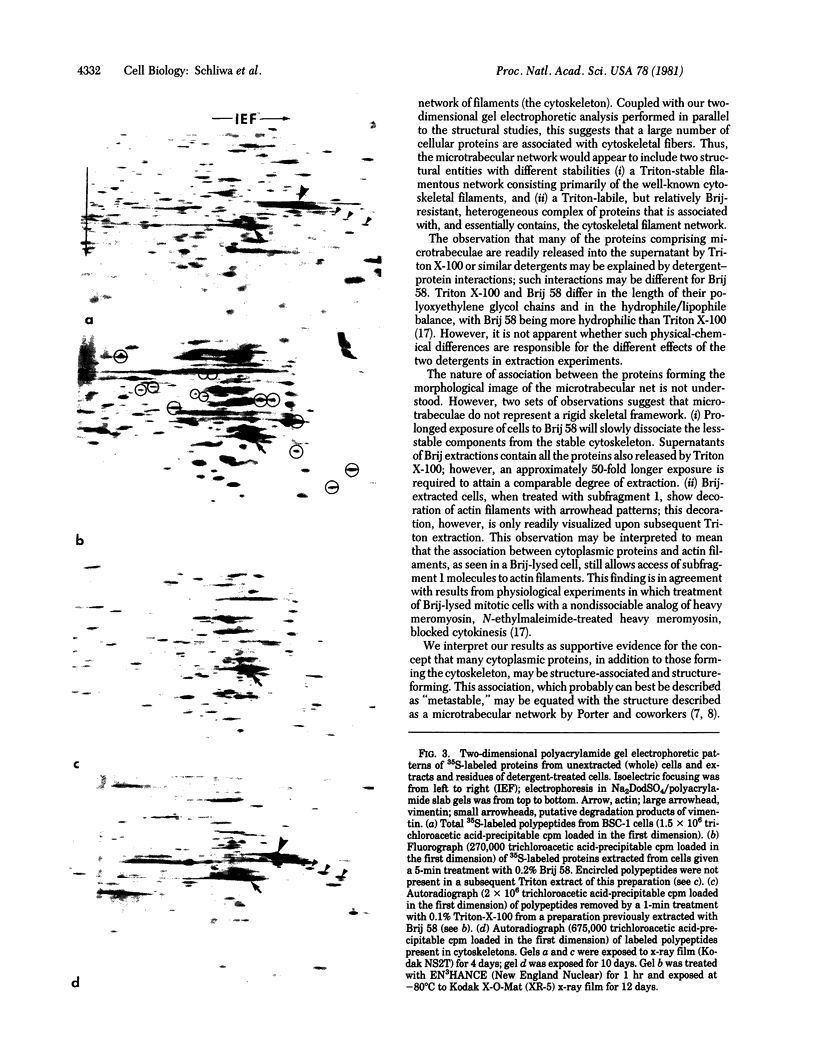
Treatment of epithelial BSC-1 cells with low concentrations of the detergent Brij 58 results in partial or complete removal of the plasmalemma and partial extraction of internal membrane-bound organelles without causing massive release of "cytosolic" proteins from the cytoplasmic ground substance. Stereoscopic high-voltage electron microscopy of such extracted and fixed cells demonstrates a system of slender (4-20 nm) strands in a three-dimensional "microtrabecular" arrangement similar to that observed in unextracted whole-mount preparations. Extraction of Brij-extracted cells with Triton X-100 dissolves many of the microtrabecular strands, leaving, as a more stable structure, a characteristic cytoskeletal network composed of various filaments and microtubules. Two-dimensional polyacrylamide gel electrophoresis of 35S-labeled polypeptides performed concurrently with the morphological studies demonstrates that Triton extraction of Brij-extracted cells releases a large number of polypeptides. This release parallels the loss of structural components observed by electron microscopy. Labeling of Brij-extracted cells with heavy meromyosin subfragment 1 decorates actin filaments with characteristic arrowhead complexes which are readily visualized only after subsequent Triton extraction. These observations support the concept that many cytoplasmic proteins are structure-bound and, in addition to the components comprising the cytoskeleton, are structure-forming. We conclude that a metastable association of various proteins of the cytoplasmic ground substance exists whose morphological integrity is maintained, at lest temporarily, after removal of the plasmalemma in solutions containing Brij 58.
Full text
PDF


Images in this article


Selected References
These references are in PubMed. This may not be the complete list of references from this article.
- Brown S., Levinson W., Spudich J. A. Cytoskeletal elements of chick embryo fibroblasts revealed by detergent extraction. J Supramol Struct. 1976;5(2):119–130. doi: 10.1002/jss.400050203. [DOI] [PubMed] [Google Scholar]
- Byers H. R., Porter K. R. Transformations in the structure of the cytoplasmic ground substance in erythrophores during pigment aggregation and dispersion. I. A study using whole-cell preparations in stereo high voltage electron microscopy. J Cell Biol. 1977 Nov;75(2 Pt 1):541–558. doi: 10.1083/jcb.75.2.541. [DOI] [PMC free article] [PubMed] [Google Scholar]
- Cande W. Z. A permeabilized cell model for studying cytokinesis using mammalian tissue culture cells. J Cell Biol. 1980 Nov;87(2 Pt 1):326–335. doi: 10.1083/jcb.87.2.326. [DOI] [PMC free article] [PubMed] [Google Scholar]
- Cande W. Z., Snyder J., Smith D., Summers K., McIntosh J. R. A functional mitotic spindle prepared from mammalian cells in culture. Proc Natl Acad Sci U S A. 1974 Apr;71(4):1559–1563. doi: 10.1073/pnas.71.4.1559. [DOI] [PMC free article] [PubMed] [Google Scholar]
- Cande W. Z., Wolniak S. M. Chromosome movement in lysed mitotic cells is inhibited by vanadate. J Cell Biol. 1978 Nov;79(2 Pt 1):573–580. doi: 10.1083/jcb.79.2.573. [DOI] [PMC free article] [PubMed] [Google Scholar]
- Franke W. W., Schmid E., Vandekerckhove J., Weber K. Permanently proliferating rat vascular smooth muscle cell with maintained expression of smooth muscle characteristics, including actin of the vascular smooth muscle type. J Cell Biol. 1980 Dec;87(3 Pt 1):594–600. doi: 10.1083/jcb.87.3.594. [DOI] [PMC free article] [PubMed] [Google Scholar]
- Helenius A., Simons K. Solubilization of membranes by detergents. Biochim Biophys Acta. 1975 Mar 25;415(1):29–79. doi: 10.1016/0304-4157(75)90016-7. [DOI] [PubMed] [Google Scholar]
- Henderson D., Weber K. Three-dimensional organization of microfilaments and microtubules in the cytoskeleton. Immunoperoxidase labelling and stereo-electron microscopy of detergent-extracted cells. Exp Cell Res. 1979 Dec;124(2):301–316. doi: 10.1016/0014-4827(79)90206-4. [DOI] [PubMed] [Google Scholar]
- Heuser J. E., Kirschner M. W. Filament organization revealed in platinum replicas of freeze-dried cytoskeletons. J Cell Biol. 1980 Jul;86(1):212–234. doi: 10.1083/jcb.86.1.212. [DOI] [PMC free article] [PubMed] [Google Scholar]
- Lenk R., Ransom L., Kaufmann Y., Penman S. A cytoskeletal structure with associated polyribosomes obtained from HeLa cells. Cell. 1977 Jan;10(1):67–78. doi: 10.1016/0092-8674(77)90141-6. [DOI] [PubMed] [Google Scholar]
- Meeusen R. L., Cande W. Z. N-ethylmaleimide-modified heavy meromyosin. A probe for actomyosin interactions. J Cell Biol. 1979 Jul;82(1):57–65. doi: 10.1083/jcb.82.1.57. [DOI] [PMC free article] [PubMed] [Google Scholar]
- O'Farrell P. H. High resolution two-dimensional electrophoresis of proteins. J Biol Chem. 1975 May 25;250(10):4007–4021. [PMC free article] [PubMed] [Google Scholar]
- Osborn M., Weber K. The detertent-resistant cytoskeleton of tissue culture cells includes the nucleus and the microfilament bundles. Exp Cell Res. 1977 May;106(2):339–349. doi: 10.1016/0014-4827(77)90179-3. [DOI] [PubMed] [Google Scholar]
- Schliwa M., Euteneuer U., Bulinski J. C., Izant J. G. Calcium lability of cytoplasmic microtubules and its modulation by microtubule-associated proteins. Proc Natl Acad Sci U S A. 1981 Feb;78(2):1037–1041. doi: 10.1073/pnas.78.2.1037. [DOI] [PMC free article] [PubMed] [Google Scholar]
- Small J. V., Celis J. E. Direct visualization of the 10-nm (100-A)-filament network in whole and enucleated cultured cells. J Cell Sci. 1978 Jun;31:393–409. doi: 10.1242/jcs.31.1.393. [DOI] [PubMed] [Google Scholar]
- Trotter J. A., Foerder B. A., Keller J. M. Intracellular fibres in cultured cells: analysis by scanning and transmission electron microscopy and by SDS-polyacrylamide gel electrophoresis. J Cell Sci. 1978 Jun;31:369–392. doi: 10.1242/jcs.31.1.369. [DOI] [PubMed] [Google Scholar]
- Webster R. E., Henderson D., Osborn M., Weber K. Three-dimensional electron microscopical visualization of the cytoskeleton of animal cells: immunoferritin identification of actin- and tubulin-containing structures. Proc Natl Acad Sci U S A. 1978 Nov;75(11):5511–5515. doi: 10.1073/pnas.75.11.5511. [DOI] [PMC free article] [PubMed] [Google Scholar]
- Wolosewick J. J., Porter K. R. Microtrabecular lattice of the cytoplasmic ground substance. Artifact or reality. J Cell Biol. 1979 Jul;82(1):114–139. doi: 10.1083/jcb.82.1.114. [DOI] [PMC free article] [PubMed] [Google Scholar]
- Wolosewick J. J., Porter K. R. Stereo high-voltage electron microscopy of whole cells of the human diploid line, WI-38. Am J Anat. 1976 Nov;147(3):303–323. doi: 10.1002/aja.1001470305. [DOI] [PubMed] [Google Scholar]