Abstract
Rift Valley fever virus (RVFV) is a mosquito-borne zoonotic bunyavirus of the genus Phlebovirus and a serious human and veterinary pathogen. RVFV contains a three-segmented RNA genome, which is comprised of the large (L), medium (M), and small (S) segments. The proteins that are essential for genome replication are encoded by the L and S segments, whereas the structural glycoproteins are encoded by the M segment. We have produced BHK replicon cell lines (BHK-Rep) that maintain replicating L and S genome segments. Transfection of BHK-Rep cells with a plasmid encoding the structural glycoproteins results in the efficient production of RVFV replicon particles (RRPs). To facilitate monitoring of infection, the NSs gene was replaced with an enhanced green fluorescent protein gene. RRPs are infectious for both mammalian and insect cells but are incapable of autonomous spreading, rendering their application outside biosafety containment completely safe. We demonstrate that a single intramuscular vaccination with RRPs protects mice from a lethal dose of RVFV and show that RRPs can be used for rapid virus neutralization tests that do not require biocontainment facilities. The methods reported here will greatly facilitate vaccine and drug development as well as fundamental studies on RVFV biology. Moreover, it may be possible to develop similar systems for other members of the bunyavirus family as well.
INTRODUCTION
The family Bunyaviridae is divided into five genera, of which four (Orthobunyavirus, Nairovirus, Phlebovirus, and Hantavirus) include numerous virus species capable of causing severe disease in both animals and humans. Well-known examples are hantavirus (HTNV; genus Hantavirus), Crimean-Congo hemorrhagic fever virus (CCHFV; genus Nairovirus), and Rift Valley fever virus (RVFV; genus Phlebovirus). Although RVFV, HTNV, and CCHFV cause severe disease with high fatality rates, no vaccines are available for the prevention of these diseases in humans, and no antiviral agents are registered for postexposure treatment. The development of such control tools is complicated by the fact that these viruses must be handled under high-level biosafety containment conditions.
In the veterinary field, RVFV is the most feared bunyavirus. The mortality rate in adult ruminants can be up to 20%, whereas fatality rates in unborn and young animals can be even more dramatic, approaching 100% (16, 17). The human fatality rate is historically estimated to be below 1%, although considerably higher mortality rates have been reported (1, 2, 19). RVFV is currently largely confined to the African continent and the Arabian Peninsula, but mosquitoes capable of transmitting RVFV are not restricted to these areas (33, 43, 44). This explains the growing concern for RVFV incursions into previously unaffected areas, including Europe, Australasia, and the Americas (9, 10, 12, 22, 37).
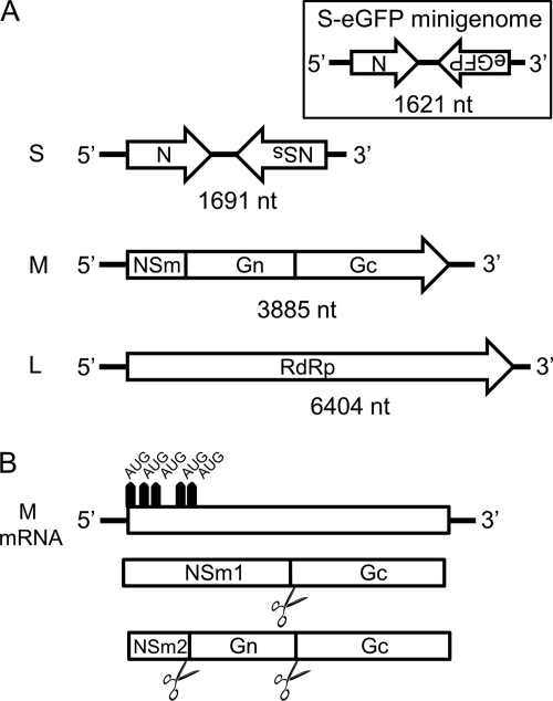
Like other bunyavirus family members, RVFV contains a three-segmented RNA genome, comprising a large (L), medium (M), and small (S) segment (21). The L segment encodes the viral RNA polymerase. The M segment contains five in-frame start codons which give rise to the structural glycoproteins Gn and Gc and two major nonstructural proteins, referred to as NSm1 and NSm2 (Fig. 1). The S segment encodes the nonstructural NSs protein and the nucleocapsid (N) protein. The NSs protein suppresses host innate immune responses and was shown to be the primary virulence factor of the virus (5, 13, 25, 34).
Fig. 1.
RVFV genome organization and expression strategy of the M genome segment. (A) Schematic representation of the RVFV small (S), medium (M), and large (L) genome segments in antigenomic orientation. (Inset) S-based minigenome containing the eGFP gene used in this work. (B) Coding strategy of the M genome segment. The mRNA encoded by the M segment is translated into a polyprotein that is processed into the NSm1, NSm2, Gn, and Gc proteins.
The recent establishment of a reverse-genetics system for RVFV has provided important new insights into its biology (7, 12, 23, 26). A few years after the first successful rescue of RVFV from cloned cDNA, the packaging of a reporter minigenome into virus-like particles (VLPs) was reported (24). The VLPs were produced by transient expression of the NSm, Gn, Gc, N, and L proteins in the presence of the reporter minigenome. In this system, the N and L proteins produced from protein expression plasmids facilitate replication of the minigenome. The minigenome is subsequently packaged by the structural glycoproteins into so-called infectious VLPs (iVLPs), which are able to transport the RNA to receiving cells. Whereas primary transcription occurs in these cells, replication of the minigenome and high-level reporter gene expression depend on de novo production of N and L proteins from transfected plasmids (24).
A recent study reported the copackaging of the M and S genome segments into VLPs (42). Packaging of the L genome segment into VLPs was not accomplished in these studies, and it was proposed that the M genome segment, either alone or in a coordinated action with the S segment, is essential for packaging of the L segment. Here, we describe efficient methods for producing large amounts of virus particles that contain both S and L genome segments and thereby demonstrate that the M segment is not essential for this process. By virtue of the L and S genome segments, the particles are capable of autonomous genome replication and high-level gene expression. However, since the M genome segment is absent, the particles are incapable of autonomous spread. The so-called RVFV replicon particles (RRPs) were produced to titers exceeding 107 infectious particles/ml.
We propose that RVFV RRPs optimally combine the efficacy of live vaccines with the safety of inactivated vaccines and support this notion by demonstrating that a single intramuscular vaccination with 106 RRPs completely protects mice from a lethal dose of RVFV. We also show that RRPs can be used for rapid virus neutralization tests that do not require biosafety containment facilities.
The methods described here will greatly facilitate both fundamental and applied research on RVFV and can potentially be established for other members of the bunyavirus family as well.
MATERIALS AND METHODS
Cells and growth conditions.
BSR-T7/5 cells were kindly provided by K. Conzelmann (Max von Pettenkofer-Institut, Munich, Germany). BSR-T7/5 cells, BHK cells, and derivatives were grown in Glasgow minimum essential medium (GMEM; Invitrogen, Carlsbad, CA) supplemented with 4% tryptose phosphate broth (Invitrogen), 1% minimum essential medium nonessential amino acids (MEM NEAA, Invitrogen), and 5 to 10% fetal bovine serum (FBS; Bodinco, Alkmaar, The Netherlands). For maintenance of stable cell lines, Geneticin (G-418; Promega, Madison, WI) was used at a concentration of 1 mg/ml. For the production of RRPs for the vaccination-challenge trial, cells were grown in Optimem (Invitrogen) supplemented with 2% FBS. Cells were grown at 37°C and 5% CO2. Drosophila (S2) cells were grown in Schneider's medium (Invitrogen) at 28°C. Aedes albopictus C6/36 cells were grown in Dulbecco's modified Eagle medium (Invitrogen) supplemented with 10% FBS at 28°C and 5% CO2.
Plasmids and viruses.
Plasmid pCIneo-GnGc contains the open reading frame of the M segment of RVFV strain 35/74, starting at the fourth methionine codon. The Gn/Gc-coding sequence was codon optimized for optimal expression in mammalian cells and synthesized by the GenScript Corporation (Piscataway, NJ). Plasmids pCIneo-M and pCAGGS-M contain cDNA of the authentic RNA sequence of the M segment starting at the first methionine codon. Expression of genes from pCIneo is controlled by a cytomegalovirus (CMV) immediate-early enhancer/promoter, whereas the expression of genes from the pCAGGS plasmid is controlled by a CMV immediate enhancer/ß-actin (CAG) promoter (36).
RVFV strain 35/74 was isolated from a liver of a sheep that died during an RVFV outbreak in the Free State province of South Africa in 1974 (3). The virus was passaged four times in mouse brain and three times in BHK cells. Amplification of the genome was performed by a one-step RT-PCR using primers previously described by Bird et al. (8). PCR products were purified from agarose gels and used for GS FLX sequencing at Inqaba Biotec (Pretoria, South Africa) essentially as described previously (40). The consensus sequences corresponding to each genome segment were synthesized and cloned in pUC57, a standard cloning vector of the GenScript Corporation (Piscataway, NJ). pUC57-L, pUC57-M, and pUC57-S encode the RVFV L, M, and S genome segments, respectively, in antigenomic orientation. The complete sequence of the L, M, and S genome sequences can be found in GenBank under the accession numbers JF784386, JF784387, and JF784388, respectively. Of note, the cDNA of the M segment present in plasmid pUC57-M contains a silent A-to-G mutation at position 182. The transcription plasmids each contain a complete copy of the viral RNA segments and are flanked by a minimal T7 promoter and a hepatitis delta virus ribozyme sequence. In pUC57-S-eGFP, the NSs gene is replaced by the gene encoding enhanced green fluorescent protein (eGFP) (Fig. 1, inset).
Rescue of recombinant RVFV strain 35/74.
BSR-T7/5 cells were seeded in six-well plates and were cotransfected with 1 μg of plasmids pUC57-L, pUC57-M, and pUC57-S using jetPEI transfection reagent according to the instructions of the manufacturer (Polyplus-transfection SA, Illkirch, France). After 6 days of incubation, the medium was collected and used for virus titration on BHK cells.
Alternatively, BHK cells were infected with a recombinant fowlpox virus (FPV) that produces T7 polymerase (15, 18). This virus, named fpEFLT7pol (referred to here as FP-T7), was previously kindly provided by the Institute for Animal Health (IAH, Compton, United Kingdom). After incubation with FP-T7 for 1 h and recovery for another hour, the cells were treated as described for BSR-T7/5 cells. Virus titers were determined as 50% tissue culture infective doses (TCID50) using BHK cells and were calculated using the Spearman-Kärber method (27, 41).
Production of RRPs.
For the production of RRPs using the three-plasmid system, BHK or BHK-GnGc cells were seeded in six-well plates and incubated with FP-T7 for 1.5 to 2 h at 37°C. Medium was refreshed, and cells were allowed to recover for 1 h. Cells were subsequently transfected with 600 ng each of pUC57-L, pUC57-S-eGFP, and pCAGGS-M. The medium was refreshed the next day. Supernatants were harvested after 72 h, precleared by low-speed centrifugation at room temperature, and stored at 4°C until further use.
For the production of RRPs using the one-plasmid system, HEK293T cells were seeded in six-well plates and infected with RRPs at a multiplicity of infection (MOI) of 3. Three days later, the cells were passaged in the wells of a 6-well plate and transfected with 1 μg of pCAGGS-M. RRPs were collected from the culture medium at 72 h posttransfection. Alternatively, BHK-Rep cells were transfected with pCAGGS-M, and RRPs were collected at different time points.
The titers of RRPs were determined by TCID50 assays using BHK cells and were calculated using the Spearman-Kärber method (27, 41). Infectivity was detected by monitoring eGFP expression using a Zeiss fluorescence microscope.
For the production of RRPs for vaccination of mice, BHK-Rep cells were grown in Optimem supplemented with 2% FBS. The cells were transfected with pCAGGS-M, and after 24 h the medium was collected. The RRPs in the collected medium were concentrated using Amicon filters (Millipore, Billerica, MA) and subsequently diluted in PBS to a titer of 107.3 TCID50/ml.
Northern blotting.
RNA probes were prepared by T7-based in vitro transcription in the presence of digoxigenin-11-UTP according to the instructions provided with the DIG Northern starter kit (Roche, Woerden, The Netherlands). Templates for in vitro transcription were produced by PCR using cDNA of the S, M, and L segments as the template and dedicated primers containing the T7 promoter sequence. The S probe comprised nucleotides 1 to 378 of the S segment, the M probe comprised nucleotides 3545 to 3885, and the L probe comprised nucleotides 1 to 320 (all in antigenomic-sense orientation).
Viral RNA of recombinant RVFV strain 35/74 (r35/74) was extracted with a High Pure viral RNA kit (Roche) according to the instructions of the manufacturer. RVFV RNA from the BHK-Rep and BHK-Rep2 cells was extracted using the RNeasy kit according to the instructions of the manufacturer (Qiagen, Hilden, Germany). RNA was separated by electrophoresis using the glyoxal-dimethyl sulfoxide system provided with the Northern-Max-Gly kit (Ambion, Austin, TX). Size-fractionated RNA was transferred by blotting onto positively charged nylon membranes (Roche) using the transfer buffer of the Northern-Max-Gly kit. RNA was fixed on the membrane by baking at 80°C for 30 min. Hybridization of digoxigenin (DIG)-labeled RNA probes was performed using UltraHyb hybridization buffer and wash solutions according to the recommendations of the manufacturer (Ambion).
Detection of hybridized probes was performed using the DIG Northern starter kit (Roche) according to the manufacturer's protocol. Briefly, blots were incubated in blocking solution for 30 min and subsequently incubated with alkaline phosphatase-conjugated anti-DIG antibody for 30 min, followed by washing twice in washing buffer. After equilibration in detection buffer, blots were incubated with the chemiluminescent substrate supplied by the DIG kit (CDP-Star) and exposed to X-ray films (Amersham Hyperfilm).
Polyacrylamide gel electrophoresis (PAGE) and Western blotting.
Proteins were separated in bis-Tris gradient gels (Invitrogen) and analyzed by Western blotting as described previously (28). For Western blot analysis of Gn and Gc, rabbit peptide antisera were used (20). Monoclonal antibody (MAb) F1D11 (kindly provided by Alejandro Brun, CISA-INIA, Madrid, Spain) was used for the detection of the N protein.
IPMAs.
Immunoperoxidase monolayer assays (IPMAs) were performed as described previously (20). For the detection of the Gn and Gc proteins, a polyclonal sheep antiserum was used (28).
Flow cytometry.
Flow cytometry was performed using a CyAn ADP flow cytometer (Beckman Coulter) equipped with a 488 nm laser. For the analysis of the data, Summit v4.3 software was used.
Transmission electron microscopy (TEM).
RRPs were applied to carbon-coated copper Formvar grids (Stork Veco BV, Eerbeek, The Netherlands). The grids were stained with 1% phosphotungstate (PTA) at pH 7 (Merck, Darmstadt, Germany). Images were recorded at a calibrated magnification of ×60,000 using a FEI Tecnai 12 electron microscope.
VNTs using RRPs.
Classical and RRP virus neutralization tests (VNTs) were performed with sera from lambs that had previously been experimentally infected with the 35/74 virus. To confirm the presence of RVFV-specific antibodies, the sera were analyzed with a recombinant N (recN) RVFV enzyme-linked immunosorbent assay (ELISA) (BDSL, Irvine, Ayrshire, Scotland, United Kingdom) prior to analysis by VNT. The classical VNT was performed as described previously (20). For the RRP VNT, serum dilutions were prepared in 96-well plates in 50 μl GMEM supplemented with 5% FBS, 4% tryptose phosphate broth (TPB), 1% MEM NEAA, and 1% penicillin-streptomycin. Culture medium containing ∼200 RRPs in a 50-μl volume was added to the serum dilutions and incubated for 1.5 h at room temperature. Next, 50 μl of growth medium containing 40 000 BHK cells was added to each well. Plates were incubated at 37°C and 5% CO2. After 36 to 48 h, the neutralization titer was calculated by the Spearman-Kärber method (27, 41).
Vaccination and challenge of mice.
Female BALB/c mice (Charles River Laboratories, Maastricht, The Netherlands) were housed in groups of five animals and kept under biosafety level 3 containment. Groups of 10 mice were vaccinated via the intramuscular or subcutaneous route either once, on day 21, or twice, on days 0 and 21, with 106 TCID50 of RRPs in 50 μl PBS. One group of nine mice was left untreated. The body weights of the mice were monitored weekly. On day 42, all mice were challenged via the intraperitoneal route with 102.7 TCID50 of RVFV strain 35/74 in 0.5 ml culture medium. Challenged mice were monitored daily for visual signs of illness and mortality.
This experiment was approved by the Ethics Committee for Animal Experiments of the Central Veterinary Institute of Wageningen University and Research Centre.
RESULTS
Establishment of a three-plasmid system for the production of RRPs.
RVFV strain 35/74 was readily rescued from cDNA by transfection of BSR-T7/5 cells with plasmids pUC57-S, pUC57-M, and pUC57-L encoding the viral RNA segments in antigenomic orientation. Our next aim was to produce only the L and S genome segments and to package these segments into RRPs by providing the structural glycoproteins Gn and Gc in trans from a protein expression plasmid. To facilitate monitoring of genome replication, a reporter minigenome was produced in which the nonessential NSs gene of the S genome segment is replaced by the enhanced green fluorescent protein (eGFP) gene (Fig. 1, inset). The resulting plasmid was named pUC57-S-eGFP. Cotransfection of pUC57-L and pUC57-S-eGFP into BSR-T7/5 cells resulted in only very few eGFP-positive cells, and cotransfection with a plasmid providing the NSm, Gn, and Gc proteins (i.e., pCAGGS-M) did not result in the production of RRPs.
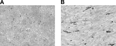
To improve the system, we evaluated the use of a recombinant fowlpox virus as a source of T7 polymerase (15, 18). The pUC57-S plasmid was transfected on its own into BSR-T7/5 cells and into BHK cells that had been infected with FP-T7 prior to transfection. Whereas the N protein was not detected in BSR-T7/5 cells transfected with pUC57-S, FP-T7-infected BHK cells that were transfected with this plasmid stained intensely with a monoclonal antibody (MAb) specific for the N protein (Fig. 2).
Fig. 2.
Expression of the N protein from the antigenomic-sense S segment. BSR-T7/5 cells (A) or FP-T7-infected BHK-21 cells (B) were transfected with plasmid pUC57-S, containing the RVFV S genome segment in antigenomic-sense orientation. Expression of the RVFV N protein was detected using an N-protein-specific MAb and horseradish peroxidase-conjugated anti-mouse IgG antibodies.
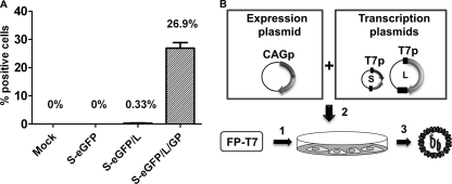
BHK cells were infected with the fowlpox virus (FP-T7) and subsequently transfected with pUC57-S-eGFP only, cotransfected with pUC57-S-eGFP and pUC57-L, or cotransfected with pUC57-S-eGFP, pUC57-L, and pCAGGS-M. Transfection with only pUC57-S-eGFP did not result in eGFP expression (Fig. 3A). After 72 h, eGFP expression was observed in a small percentage (0.33%) of cells that were cotransfected with pUC57-S-eGFP and pUC57-L. However, when pCAGGS-M was added to the transfection mixture, about one quarter of the cells expressed eGFP (Fig. 3A). This finding suggested that infectious particles were formed upon introduction of the pCAGGS-M plasmid, resulting in an increase in the number of eGFP-expressing cells. Collected supernatant was added to BHK cell monolayers, and after 36 h, infection was monitored by fluorescence microscopy. Only cells incubated with the supernatant obtained from cells transfected with all three plasmids exhibited eGFP expression. This confirmed that we were successful in producing RVFV replicon particles (RRPs). Three independently performed experiments yielded an average RRP titer of 104.8 TCID50/ml. The three-plasmid system for the production of RRPs is schematically depicted in Fig. 3B.
Fig. 3.
Production of RRPs by the three-plasmid system. (A) BHK cells were infected with FP-T7 and subsequently remained untreated (mock) or were transfected with plasmid pUC57-S-eGFP (S-eGFP) only, in combination with plasmid pUC57-L encoding the RVFV L genome segment (S-eGFP/L), or with the aforementioned plasmids and pCAGGS-M (GP), encoding the structural glycoproteins (S-eGFP/L/GP). The percentage of eGFP-positive cells was determined by flow cytometry (values are means and standard deviations for three replicates). (B) Schematic representation of the three-plasmid system. BHK cells were first infected with FP-T7 (step 1) and subsequently transfected with transcription plasmids pUC57-L (L) and pUC57-S-eGFP (S) and expression plasmid pCAGGS-M (step 2). After 24 h, the culture medium containing the RRPs was collected (step 3). Transcription from the expression plasmid is controlled by a CAG promoter (CAGp), and transcription from the transcription plasmids is controlled by a T7 promoter (T7p). Untranslated regions are depicted as black boxes.
Establishment of a one-plasmid system for the production of RRPs.
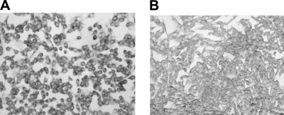
The RRP titers obtained with the three-plasmid system never exceeded 105 TCID50/ml. To develop a system for the continuous production of high-titer RRPs, we created stable BHK cell lines that constitutively produce the Gn and Gc proteins. BHK cells were transfected with pCIneo-GnGc, and clones with integrated plasmids were grown in the presence of G-418. Whereas an anti-Gn/Gc serum revealed clear glycoprotein expression 1 to 2 days after transfection, after cloning of the cells, only very few selected clones revealed Gn/Gc expression by IPMA, and in all cases, expression seemed very low. One clone that revealed the most intense staining in IPMAs (Fig. 4) was selected and named BHK-GnGc.
Fig. 4.
(A) RVFV glycoprotein expression in BHK-GnGc cells. BHK cells were transfected with plasmid pCIneo-GnGc, encoding the RVFV structural glycoproteins Gn and Gc. The BHK-GnGc cells were cloned by limiting dilution. Expression of Gn and Gc was detected by staining of BHK-GnGc cells with polyclonal antibodies specific for the Gn and Gc proteins. (B) Similarly treated BHK parent cells.
It was previously demonstrated that expression of the Gn and Gc proteins in both mammalian (31) and insect cells (20) results in the production of VLPs. To determine if VLPs were produced by BHK-GnGc cells, supernatants were ultracentrifuged (100,000 × g, 2 h), and the proteins in the collected pellets were analyzed by Western blotting. Neither the Gn nor the Gc protein was detected in the pellet fractions (data not shown), suggesting either that no VLPs were produced or, more likely, that glycoprotein production was too low to allow detection.
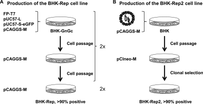
Although the BHK-GnGc cells apparently produced only very limited amounts of Gn and Gc, we hypothesized that these cells might tolerate Gn/Gc expression from plasmids to a greater extent than normal BHK cells and therefore be more suitable for RRP production. To substantiate this hypothesis, BHK-GnGc cells were infected with FP-T7 and subsequently cotransfected with the three plasmids. In an attempt to increase the number of eGFP-expressing cells, the cells were next transfected with pCAGGS-M only. This whole procedure was then repeated, followed by two cell passages and introduction of the pCAGGS-M plasmid (Fig. 5A). At this point, flow cytometry demonstrated that >90% of the BHK-GnGc cells were positive for eGFP expression. The resulting cell line, which maintains the L genome segment and the S-eGFP minigenome, was designated BHK-Rep. Transfection of BHK-Rep cells with pCAGGS-M after cell passages 83, 103, and 105 yielded an average RRP titer of 106.7 TCID50/ml. It is important to note that we also used a similar protocol starting with wild-type BHK cells instead of BHK-GnGc cells. In these experiments, reporter gene expression did not continue to increase, and we were therefore unable to produce a cell line with >90% reporter gene expression using this procedure.
Fig. 5.
Cartoon representing the construction of the BHK-Rep (A) and BHK-Rep2 (B) cell lines. Construction of the BHK-Rep cells started with a BHK cell line constitutively expressing small amounts of the Gn and Gc proteins (BHK-GnGc), whereas wild-type BHK cells were used for production of the BHK-Rep2 cell line. Stable glycoprotein expression was achieved in these cells by introducing the pCIneo-M plasmid. The S-eGFP and L genome segments were introduced in the BHK-Rep cell line by FP-T7-driven transcription from plasmids, whereas these genome segments were introduced into the BHK-Rep2 cells by infection with RRPs. Transfection with pCAGGS-M was used to produce RRPs, which assist in spread of the genome segments among cells.
The NSm protein was previously reported to suppress virus-induced apoptosis (45), so we reasoned that a cell line constitutively expressing not only Gn and Gc but also the NSm proteins could be more efficient in the constitutive production of RRPs by suppressing apoptosis. To this end, we aimed to produce a BHK-Rep cell line with a stably integrated plasmid encoding the NSm, Gn, and Gc proteins (i.e., pCIneo-M). For convenience, the L and S-eGFP genome segments were introduced into BHK cells not by transfection of plasmids but instead by infection of the cells with RRPs combined with transfection with pCAGGS-M. Cells were passaged and subsequently transfected with pCIneo-M and grown in the presence of G-418. In this way, BHK-Rep cells were produced without introducing FP-T7 and the transcription plasmids. The cells were used for cloning by endpoint dilution, and a selected clone was named BHK-Rep2. The steps followed to produce the BHK-Rep2 cell line are depicted in Fig. 5B. Although this cell line also did not constitutively produce RRPs, transfection of this cell line with the pCAGGS-M plasmid yielded an average RRP titer of 107.2 TCID50/ml (n = 6).
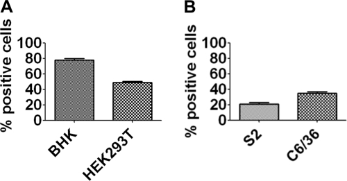
To determine if other mammalian and insect cells can be infected with RRPs, human embryonic kidney 293 cells (HEK293T), Drosophila S2 cells, and Aedes albopictus C6/36 cells were infected with RRPs at an MOI of 1 (calculated using the titer determined on BHK cells). This experiment demonstrated that both mammalian and insect cells can be readily infected with RRPs (Fig. 6). Expression of eGFP in mammalian cells and insect cells was optimal at 42 and 72 h postinfection, respectively.
Fig. 6.
RRP infection of mammalian cells (A) and insect cells (B). BHK cells, human embryonic kidney 293T cells (HEK293T), Drosophila S2 cells, and Aedes albopictus C6/36 cells were infected with RRPs at an MOI of 1. The number of positive cells was determined by flow cytometry at 42 (BHK and HEK293T) or 72 (S2 and C6/36) h postinfection. Histograms show averaged results of three independent measurements, with standard deviations.
It is important to note that the one-plasmid system is not restricted to BHK-Rep cells. Transfection of RRP-infected HEK293T cells with the pCAGGS-M plasmid yielded an RRP titer of 106.5 TCID50/ml. This demonstrates that alternative cell types can be used for the production of RRPs by combining an RRP infection with a transfection of the pCAGGS-M plasmid (Fig. 7).
Fig. 7.
Schematic representation of the one-plasmid systems. Transfection of BHK-Rep cells with pCAGGS-M results in the production of RRPs. Alternatively, RRPs can be produced by infection of potentially any type of mammalian cell, such as HEK293T cells, by infection with RRPs followed by transfection with pCAGGS-M.
Characterization of BHK-Rep cells.
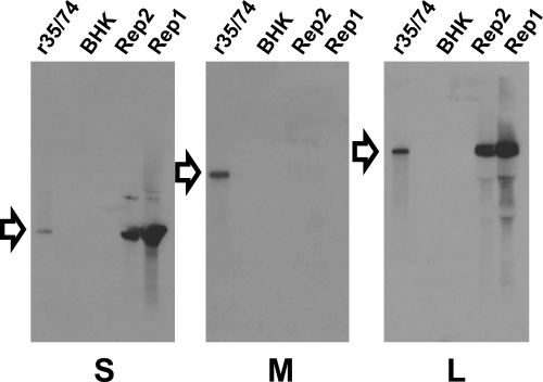
Persistence of RVFV RNA in cells was previously reported by Billecocq et al. (6). Those authors detected defective interfering (DI) RNAs with large internal deletions and suggested that these DI RNAs could be responsible for the persistent infection. Considering this, we determined whether abnormal viral RNA segments were present in the BHK-Rep and BHK-Rep2 cells. Northern blotting was performed with total RNA extracted from BHK-Rep and BHK-Rep2 cells at cell passage 13 and 45, respectively. S-, M-, and L-specific probes revealed only the S and L segments, confirming that no M segment is present in the BHK-Rep cells (Fig. 8). The S-eGFP and L segments were detected at the same positions as the S and L segments of the recombinant virus, suggesting that no significant levels of DI RNAs were present.
Fig. 8.
Northern blot analysis of the viral RNA segments present in BHK-Rep cells. Total RNA of BHK-Rep and BHK-Rep2 cells was extracted, separated by electrophoresis using the glyoxal-dimethyl sulfoxide system, and transferred to positively charged nylon membranes. Hybridization was performed with DIG-labeled S, M, and L probes, and hybridization was detected by phosphatase-conjugated anti-DIG antibodies. RNA extracted from recombinant RVFV 35/74 was used as a positive control and a reference for size. Wild-type BHK cells were used as a negative control. The specificity of the probe used for hybridization of each blot is indicated below each panel. The positions of the S, M, and L segments are indicated by arrows.
To determine if mutations were introduced upon replication of the L and S-eGFP genome segments in BHK-Rep cells, cDNA was produced and sequenced, with the exception of the primer binding regions on the extreme 3′ and 5′ ends, using standard techniques. Only a single mutation (G1181→A) was detected in the L genome segment present in the BHK-Rep cells, resulting in an amino acid change from glycine to glutamic acid at position 388 of the L protein. This mutation was also detected in the BHK-Rep2 cells, together with a silent mutation in the L gene (A1602→C) and two mutations in the 5′ untranslated region (T6325→C and T6367→C). In the S-eGFP segments, only a single silent mutation was detected in the BHK-Rep2 cells (C65→T). The number of the mutations corresponds to their position in the antigenomic-sense RNA. We did not study the effect of these mutations further.
The viral RNA segments were introduced in BHK-Rep cells by plasmids that were transcribed by the FP-T7 virus. Although it is well established that FPV does not replicate in mammalian cells (15), the BHK-Rep and BHK-Rep2 cells were tested for the presence of FP-T7 by PCR. No FPV DNA was detected in these assays (data not shown).
Characterization of RRPs.
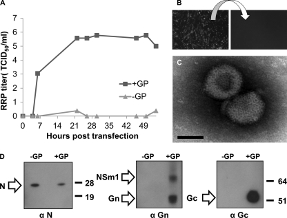
To establish the kinetics of RRP production, BHK-rep cells were transfected with pCAGGS-M, and the culture medium was collected at different time points posttransfection. This experiment demonstrated that the titer was already close to 106 TCID50/ml after 22 h (Fig. 9A).
Fig. 9.
Characterization of RRPs. (A) BHK-Rep cells were either left untreated (−GP) or transfected with pCAGGS-M (+GP), and RRP titers in the collected supernatant were determined at different time points posttransfection. (B) To demonstrate that RRPs are nonspreading, BHK cells were infected with RRPs, and after 2 days, eGFP expression was observed in infected cells (left). Fresh BHK cells were incubated with the collected supernatant and monitored for eGFP expression after 3 days (right). (C) Electron micrograph of RRPs. Concentrated RRPs were stained with 1% PTA and analyzed by TEM. Bar, 50 nm. (D) To visualize RRP proteins, culture medium of BHK-Rep cells (−GP) or of BHK-Rep cells transfected with pCAGGS-M (+GP) was ultracentrifuged at 100,000 × g for 2 h. The proteins present in the pellets were separated in 4 to 12% bis-Tris gels and subsequently transferred to nitrocellulose blots. Specific proteins were detected by an anti-Gn (α Gn) or anti-Gc (α Gc) peptide antiserum or a MAb specific for the N protein (α N). The positions of the NSm, Gn, Gc, and N proteins are indicated by arrows. Molecular weight standards are indicated on the right, in thousands.
To demonstrate that RRPs are incapable of autonomous spread, BHK cells were infected with RRPs at an MOI of 1. After 2 days, eGFP expression was observed by fluorescence microscopy (Fig. 9B, left). BHK cells were incubated with the collected precleared supernatant, and after 3 days, cells were monitored for eGFP expression. No eGFP expression was observed, demonstrating that no progeny infectious particles were produced by the RRP-infected BHK cells (Fig. 9B, right).
To visualize RRPs by transmission electron microscopy (TEM), BHK-Rep2 cells were transfected with pCAGGS-M. After 30 h, the RRPs present in precleared culture supernatant were concentrated using Amicon filters and subsequently used to coat carbon-coated Formvar grids. Grids were stained with 1% PTA and analyzed by TEM. Representative particles of 80 ± 2 nm are depicted in Fig. 9C. The average particle size varied from 80 to 100 nm.
To visualize RRP proteins, RRPs were pelleted by ultracentrifugation. The proteins were separated in polyacrylamide gels, transferred to nitrocellulose membranes, and detected using peptide antisera specific for the Gn and Gc proteins or a MAb specific for the N protein. Analysis of the supernatant obtained from nontransfected BHK-Rep cells revealed only the N protein (Fig. 9D). This result suggests that the RVFV N protein is released from cells, resembling results previously obtained in studies on CCHFV (4). Analysis of supernatant from BHK-Rep cells transfected with pCAGGS-M revealed the NSm1 protein, the Gn and Gc proteins, and the N protein (Fig. 9D).
Establishment of a novel VNT.
To determine if RRPs can be used in VNTs, sera obtained from experimentally infected lambs were used in the classical VNT as described previously (20) and used in a novel VNT that uses RRPs instead of live virus. The sera were also tested by ELISA. As is done with the complete virus in the classical VNT, serum dilutions were preincubated with RRPs, and the mixtures were subsequently incubated with BHK cells. Whereas in the classical VNT, the lack of neutralization is detected by cytopathic effect, in the RRP VNT, eGFP expression demonstrates lack of neutralization. Titers are determined in both assays using the Spearman-Kärber method (27, 41).
The experiment revealed that the so-called RRP VNT has an optimal readout between 36 and 48 h and has a sensitivity equal to, if not higher than, that of the classical VNT (Table 1).
Table 1.
Comparison of classical VNT and RRP VNT
| Serum sample | Neutralization titer in VNTa |
ELISA resultb | |
|---|---|---|---|
| Classical | RRP | ||
| 4308 | 3.56 | 3.94 | Pos |
| 4309 | 4.09 | 4.16 | Pos |
| 4310 | 0 | 0 | Neg |
| 4311 | 4.01 | 4.24 | Pos |
| 4312 | 3.71 | 4.76 | Pos |
| 4314 | 3.56 | 4.46 | Pos |
| 4315 | 3.71 | 4.39 | Pos |
| 4318 | 4.16 | 4.39 | Pos |
| 4321 | 0 | 0 | Neg |
| 4324 | 4.24 | 4.31 | Pos |
| 4328 | 4.01 | 4.69 | Pos |
Reported as log10 50% endpoint titers.
Sera were analyzed by the recN ELISA (BDSL).
Vaccination and challenge of mice.
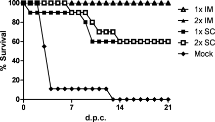
To study the vaccine efficacy of RRPs, groups of 10 mice were immunized with 50 μl of an inoculum containing 106 TCID50 of RRPs, via either the subcutaneous or intramuscular route, either once or twice with a 3-week interval. One group of 9 nonvaccinated mice was added as a control group. The mice were challenged on day 42 with a known lethal dose of RVFV strain 35/74. All nonvaccinated mice displayed overt clinical signs and weight loss, and eight of a total of nine nonvaccinated mice succumbed to the infection within 4 days after challenge. One mouse survived for 12 days but eventually died. The survival rate in the groups of mice vaccinated either once or twice via the subcutaneous route was 60%. In contrast, 100% of the mice vaccinated via the intramuscular route, either once or twice, survived the challenge (Fig. 10). These mice did not show any clinical signs or weight loss throughout the experiment. This demonstrates that a single intramuscular vaccination with 106 RRPs can protect mice from a lethal dose of RVFV.
Fig. 10.
Vaccine efficacy of RRPs. Mice were either left unvaccinated (n = 9; mock) or vaccinated (n = 10) either once (1×) or twice (2×) via the intramuscular route (IM) or subcutaneous route (SC) with 106 TCID50 of RRPs. Mice were challenged with a known lethal dose of RVFV strain 35/74 via the intraperitoneal route. The mortality rates were determined until 21 days postchallenge (d.p.c.).
DISCUSSION
Here, we report the creation of a nonspreading bunyavirus. Critical to the production of the RVFV replicon particles (RRPs) was the use of FPV as a source of T7 polymerase. Despite much effort, production of RRPs using BSR-T7/5 cells, which are routinely used for bunyavirus reverse genetics, never succeeded.
Rescue of a bunyavirus requires only three T7 transcripts representing the viral RNA molecules in antigenomic-sense orientation. This suggests that the antigenomes act not only as replication intermediates but also as mRNAs (30). Although in our experiments, expression of the N protein from antigenomic-sense S RNA was not detectable in BSR-T7/5 cells, expression was readily detected in FP-T7-infected cells. T7-based transcription in BSR-T7/5 cells results in uncapped RNAs, whereas the FP-T7 virus provides its own capping enzyme (15). We propose that this explains the improved gene expression from antigenomic viral RNAs and thereby the superiority of FP-T7-infected cells over BSR-T7/5 cells to drive minigenome expression.
It is interesting that the first rescue of a bunyavirus (i.e., Bunyamwera virus [BUNV]) from cDNA was performed using another poxvirus, namely, vaccinia virus (VV), as a source of T7 polymerase (i.e., vTF7-3) (14). The protocol for BUNV rescue was later adapted to use BSR-T7/5 cells, which circumvented the need to remove the VV from the culture medium after bunyavirus rescue (30). In our studies, we preferred FPV over VV for providing T7 polymerase, since the former not only is much less cytopathic but also does not replicate in mammalian cells (15). There is thus no FPV to be removed from the culture medium after rescue of a bunyavirus.
Although production of RRPs using the initially established three-plasmid system is highly reproducible, yields of RRPs using this system never exceeded 105 TCID50/ml. We therefore aimed to develop a system that allows the constitutive production of higher titers of RRPs. Stable cell lines were created that constitutively produce the structural glycoproteins Gn and Gc, but the levels of expression were too low for efficient trans-complementation. However, it was remarkable to find that repeated passage of these cells when containing replicating L and S-eGFP genome segments did not result in cytopathic effect or loss of reporter gene expression. Reporter gene expression in the resulting BHK-Rep cells was maintained for at least 100 cell passages. Importantly, when the RVFV genome segments were introduced into wild-type BHK cells, expression of the reporter was quickly lost upon passage of the cells. Analysis of the culture medium of BHK-Rep cells was occasionally found to contain low titers of RRPs, varying from undetectable levels to 100 TCID50/ml. From this, we first hypothesized that the small amounts of Gn and Gc endogenously produced by the BHK-Rep cells ensures that cells that lose the genome segments are reinfected with RRPs, thereby reintroducing these segments. This hypothesis, however, was rejected, since passage of the BHK-Rep cells in the presence of a neutralizing serum did not result in a decrease in the number of eGFP-positive cells (data not shown). It therefore remains unclear at this point how BHK-Rep cells efficiently maintain the RVFV genome segments.
By establishing the BHK-Rep cells, a one-plasmid system for the production of RRPs was provided that can reproducibly yield titers of up to 107 TCID50/ml even after 100 cell passages. We also demonstrate that the one-plasmid system is not restricted to using BHK-Rep cells. When combining an infection with RRPs with a transfection of the plasmid encoding the structural glycoproteins, any mammalian or insect cell can potentially be used for the production of RRPs and the use of FP-T7 and the transcription plasmids is no longer required. To underscore this notion, we show that HEK293T cells can be used for the efficient production of RRPs.
The VNT is the gold-standard diagnostic assay for the serologic confirmation of RVFV infection and the prescribed test for animal trade (36a). The classical VNT requires handling of live RVFV and must therefore be performed in appropriate biosafety containment facilities. Another drawback of the classical VNT is that the assay requires 5 to 7 days for completion. We show that eGFP-expressing RRPs can be used for VNTs that do not require biocontainment facilities and that this so-called RRP-VNT requires only 24 to 48 h for completion. Apart from the RRPs developed in the current work, attenuated viruses containing the eGFP gene can potentially be similarly applied.
Finally, we demonstrate that RRPs can be used as a highly effective vaccine. We and others have previously reported efficient VLP production systems and VLPs comprising only the Gn and Gc proteins are highly effective vaccine candidates (20, 29, 31, 32, 35, 38). Mandell et al. demonstrated that including the N protein in VLPs improves vaccine efficacy (31), which could be due to stabilization of the particle and/or to partial protective efficacy mediated by the N protein alone (11). It was subsequently reported that so-called infectious VLPs capable of primary transcription of the N gene are remarkably effective, providing complete protection in mice after a single vaccination (38). The RRPs produced in the current work could be even more efficacious, since the particles are capable of autonomous genome replication and high-level gene expression. Replication of the RRP genome in vivo could induce interferon and other innate immune responses and in vivo production of the L and N proteins could also induce adaptive cellular immune responses. Although additional studies are required to characterize in detail the innate and adaptive immune responses elicited by RRPs, we already demonstrate here that a single intramuscular vaccination with RRPs protects mice against a lethal challenge dose. Experiments are planned to study the immune response elicited by RRP vaccination in detail and to study the vaccine efficacy of RRPs in sheep.
Apart from applied research, our methods will also facilitate studies on RVFV genome replication and packaging. In this respect, it is interesting that the current work already demonstrates that the M genome segment is not required for packaging of the L segment, contrasting a recent suggestion (42). Our system is also particularly useful for functional studies on the structural glycoproteins Gn and Gc. Mutations can be easily introduced in the pCAGGS-M plasmid, and their effects on host cell attachment, entry, and fusion can be studied independently. Since the system does not depend on live virus, even the nature of lethal mutations can be studied. An alternative system that can be used for such studies is the previously described system for producing iVLPs (24, 38, 39). Reporter gene expression in iVLP-infected cells, however, requires cotransfection with helper plasmids, which likely renders this system more prone to experimental variation than the one-plasmid system described here.
In summary, the methods reported here allow both fundamental and applied research on RVFV to be conducted outside biocontainment facilities and will therefore greatly facilitate the development of novel therapeutics and vaccines for the control of RVFV. A newly identified member of the phlebovirus genus causing severe fever with thrombocytopenia syndrome (SFTS) in humans recently emerged in China (46), and members of other genera of the bunyavirus family, such as CCHFV and HTNV, also continue to be of serious public health concern. The methods described here could facilitate the future control of these highly pathogenic bunyaviruses as well.
ACKNOWLEDGMENTS
We thank K. Conzelmann (Max von Pettenkofer-Institut, München, Germany) for providing the BSR-T7/5 cells and Geoff Oldham (Institute for Animal Health, Compton, United Kingdom) for providing FP-T7. We thank Isabel Wright (ARC-OVI) and Jet Kant (CVI-WUR) for technical assistance and Alejandro Brun (CISA-INIA, Madrid, Spain) for providing the N-specific MAb. We thank Adriaan Antonis and the animal technicians for performing the animal trials, and we thank Peter Rottier, Berend-Jan Bosch, and Matthijn de Boer for useful discussions and critically reviewing the manuscript. We thank Matthijn de Boer for performing electron microscopic analysis and George Posthuma of the Cell Microscopy Centre (Department of Cell Biology, University Medical Center Utrecht) for technical assistance. We thank Alice Vogel for performing the Northern blots and sequencing analysis.
This work was supported by the Dutch Ministry of Economic Affairs, Agriculture and Innovation, project codes BO-10-006-084 and BO-08-010-023.
Footnotes
Published ahead of print on 28 September 2011.
REFERENCES
- 1. Adam A. A., Karsany M. S., Adam I. 2010. Manifestations of severe Rift Valley fever in Sudan. Int. J. Infect. Dis. 14:e179–180 [DOI] [PubMed] [Google Scholar]
- 2. Al-Hazmi M., et al. 2003. Epidemic Rift Valley fever in Saudi Arabia: a clinical study of severe illness in humans. Clin. Infect. Dis. 36:245–252 [DOI] [PubMed] [Google Scholar]
- 3. Barnard B. J. 1979. Rift Valley fever vaccine—antibody and immune response in cattle to a live and an inactivated vaccine. J. S. Afr. Vet. Assoc. 50:155–157 [PubMed] [Google Scholar]
- 4. Bergeron E., Vincent M. J., Nichol S. T. 2007. Crimean-Congo hemorrhagic fever virus glycoprotein processing by the endoprotease SKI-1/S1P is critical for virus infectivity. J. Virol. 81:13271–13276 [DOI] [PMC free article] [PubMed] [Google Scholar]
- 5. Billecocq A., et al. 2004. NSs protein of Rift Valley fever virus blocks interferon production by inhibiting host gene transcription. J. Virol. 78:9798–9806 [DOI] [PMC free article] [PubMed] [Google Scholar]
- 6. Billecocq A., Vialat P., Bouloy M. 1996. Persistent infection of mammalian cells by Rift Valley fever virus. J. Gen. Virol. 77:3053–3062 [DOI] [PubMed] [Google Scholar]
- 7. Bird B. H., Albarino C. G., Nichol S. T. 2007. Rift Valley fever virus lacking NSm proteins retains high virulence in vivo and may provide a model of human delayed onset neurologic disease. Virology 362:10–15 [DOI] [PubMed] [Google Scholar]
- 8. Bird B. H., Khristova M. L., Rollin P. E., Ksiazek T. G., Nichol S. T. 2007. Complete genome analysis of 33 ecologically and biologically diverse Rift Valley fever virus strains reveals widespread virus movement and low genetic diversity due to recent common ancestry. J. Virol. 81:2805–2816 [DOI] [PMC free article] [PubMed] [Google Scholar]
- 9. Bird B. H., Ksiazek T. G., Nichol S. T., MacLachlan N. J. 2009. Rift Valley fever virus. J. Am. Vet. Med. Assoc. 234:883–893 [DOI] [PubMed] [Google Scholar]
- 10. Boshra H., Lorenzo G., Busquets N., Brun A. 2011. Rift valley Fever: recent insights into pathogenesis and prevention. J. Virol. 85:6098–6105 [DOI] [PMC free article] [PubMed] [Google Scholar]
- 11. Boshra H., Lorenzo G., Rodriguez F., Brun A. 2011. A DNA vaccine encoding ubiquitinated Rift Valley fever virus nucleoprotein provides consistent immunity and protects IFNAR−/− mice upon lethal virus challenge. Vaccine 29:4469–4475 [DOI] [PubMed] [Google Scholar]
- 12. Bouloy M., Flick R. 2009. Reverse genetics technology for Rift Valley fever virus: current and future applications for the development of therapeutics and vaccines. Antiviral Res. 84:101–118 [DOI] [PMC free article] [PubMed] [Google Scholar]
- 13. Bouloy M., et al. 2001. Genetic evidence for an interferon-antagonistic function of rift valley fever virus nonstructural protein NSs. J. Virol. 75:1371–1377 [DOI] [PMC free article] [PubMed] [Google Scholar]
- 14. Bridgen A., Elliott R. M. 1996. Rescue of a segmented negative-strand RNA virus entirely from cloned complementary DNAs. Proc. Natl. Acad. Sci. U. S. A. 93:15400–15404 [DOI] [PMC free article] [PubMed] [Google Scholar]
- 15. Britton P., et al. 1996. Expression of bacteriophage T7 RNA polymerase in avian and mammalian cells by a recombinant fowlpox virus. J. Gen. Virol. 77:963–967 [DOI] [PubMed] [Google Scholar]
- 16. Coetzer J. A. 1977. The pathology of Rift Valley fever. I. Lesions occurring in natural cases in new-born lambs. Onderstepoort J. Vet. Res. 44:205–211 [PubMed] [Google Scholar]
- 17. Coetzer J. A. 1982. The pathology of Rift Valley fever. II. Lesions occurring in field cases in adult cattle, calves and aborted foetuses. Onderstepoort J. Vet. Res. 49:11–17 [PubMed] [Google Scholar]
- 18. Das S. C., Baron M. D., Skinner M. A., Barrett T. 2000. Improved technique for transient expression and negative strand virus rescue using fowlpox T7 recombinant virus in mammalian cells. J. Virol. Methods 89:119–127 [DOI] [PubMed] [Google Scholar]
- 19. Davies F. G. 2010. The historical and recent impact of Rift Valley fever in Africa. Am. J. Trop. Med. Hyg. 83:73–74 [DOI] [PMC free article] [PubMed] [Google Scholar]
- 20. de Boer S. M., et al. 2010. Rift Valley fever virus subunit vaccines confer complete protection against a lethal virus challenge. Vaccine 28:2330–2339 [DOI] [PubMed] [Google Scholar]
- 21. Elliott R. M. 1996. The Bunyaviridae. Plenum Press, New York, NY [Google Scholar]
- 22. Gerdes G. H. 2004. Rift Valley fever. Rev. Sci. Tech. 23:613–623 [DOI] [PubMed] [Google Scholar]
- 23. Habjan M., Penski N., Spiegel M., Weber F. 2008. T7 RNA polymerase-dependent and -independent systems for cDNA-based rescue of Rift Valley fever virus. J. Gen. Virol. 89:2157–2166 [DOI] [PubMed] [Google Scholar]
- 24. Habjan M., et al. 2009. Efficient production of Rift Valley fever virus-like particles: the antiviral protein MxA can inhibit primary transcription of bunyaviruses. Virology 385:400–408 [DOI] [PubMed] [Google Scholar]
- 25. Ikegami T., et al. 2009. Rift Valley fever virus NSs protein promotes post-transcriptional downregulation of protein kinase PKR and inhibits eIF2alpha phosphorylation. PLoS Pathog. 5:e1000287. [DOI] [PMC free article] [PubMed] [Google Scholar]
- 26. Ikegami T., Won S., Peters C. J., Makino S. 2006. Rescue of infectious Rift Valley fever virus entirely from cDNA, analysis of virus lacking the NSs gene, and expression of a foreign gene. J. Virol. 80:2933–2940 [DOI] [PMC free article] [PubMed] [Google Scholar]
- 27. Karber G. 1931. Betrag zur kollektiven Behandlung pharmakologischer Reihenversuche. Arch. Exp. Path. Pharmak. 162:480–483 [Google Scholar]
- 28. Kortekaas J., et al. 2010. Rift Valley fever virus immunity provided by a paramyxovirus vaccine vector. Vaccine 28:4394–4401 [DOI] [PubMed] [Google Scholar]
- 29. Liu L., Celma C. C., Roy P. 2008. Rift Valley fever virus structural proteins: expression, characterization and assembly of recombinant proteins. Virol. J. 5:82. [DOI] [PMC free article] [PubMed] [Google Scholar]
- 30. Lowen A. C., Noonan C., McLees A., Elliott R. M. 2004. Efficient bunyavirus rescue from cloned cDNA. Virology 330:493–500 [DOI] [PubMed] [Google Scholar]
- 31. Mandell R. B., et al. 2010. A replication-incompetent Rift Valley fever vaccine: chimeric virus-like particles protect mice and rats against lethal challenge. Virology 397:187–198 [DOI] [PMC free article] [PubMed] [Google Scholar]
- 32. Mandell R. B., et al. 2010. Novel suspension cell-based vaccine production systems for Rift Valley fever virus-like particles. J. Virol. Methods 169:259–268 [DOI] [PubMed] [Google Scholar]
- 33. Moutailler S., Krida G., Schaffner F., Vazeille M., Failloux A. B. 2008. Potential vectors of Rift Valley fever virus in the Mediterranean region. Vector Borne Zoonotic Dis. 8:749–753 [DOI] [PubMed] [Google Scholar]
- 34. Muller R., et al. 1995. Characterization of clone 13, a naturally attenuated avirulent isolate of Rift Valley fever virus, which is altered in the small segment. Am. J. Trop. Med. Hyg. 53:405–411 [DOI] [PubMed] [Google Scholar]
- 35. Naslund J., et al. 2009. Vaccination with virus-like particles protects mice from lethal infection of Rift Valley Fever Virus. Virology 385:409–415 [DOI] [PubMed] [Google Scholar]
- 36. Niwa H., Yamamura K., Miyazaki J. 1991. Efficient selection for high-expression transfectants with a novel eukaryotic vector. Gene 108:193–199 [DOI] [PubMed] [Google Scholar]
- 36a. OIE 2008. Manual of diagnostic tests and vaccines for terrestrial animals, 6th ed., Chapter 2.1.14, p. 323–333 OIE, Paris, France: http://www.oie.int/en/international-standard-setting/terrestrial-manual/access-online/ [Google Scholar]
- 37. Pepin M., Bouloy M., Bird B. H., Kemp A., Paweska J. 2010. Rift Valley fever virus (Bunyaviridae: Phlebovirus): an update on pathogenesis, molecular epidemiology, vectors, diagnostics and prevention. Vet. Res. 41:61. [DOI] [PMC free article] [PubMed] [Google Scholar]
- 38. Pichlmair A., Habjan M., Unger H., Weber F. 2010. Virus-like particles expressing the nucleocapsid gene as an efficient vaccine against Rift Valley fever virus. Vector Borne Zoonotic Dis. 10:701–703 [DOI] [PubMed] [Google Scholar]
- 39. Piper M. E., Sorenson D. R., Gerrard S. R. 2011. Efficient cellular release of Rift Valley fever virus requires genomic RNA. PLoS One 6:e18070. [DOI] [PMC free article] [PubMed] [Google Scholar]
- 40. Potgieter A. C., et al. 2009. Improved strategies for sequence-independent amplification and sequencing of viral double-stranded RNA genomes. J. Gen. Virol. 90:1423–1432 [DOI] [PubMed] [Google Scholar]
- 41. Spearman C. 1908. The method of right and wrong cases (constant stimuli) without Gauss's formulae. Br. J. Psychol. 2:227–242 [Google Scholar]
- 42. Terasaki K., Murakami S., Lokugamage K. G., Makino S. 2010. Mechanism of tripartite RNA genome packaging in Rift Valley fever virus. Proc. Natl. Acad. Sci. U. S. A. 108:804–809 [DOI] [PMC free article] [PubMed] [Google Scholar]
- 43. Turell M. J., et al. 2008. Potential for North American mosquitoes to transmit Rift Valley fever virus. J. Am. Mosq. Control. Assoc. 24:502–507 [DOI] [PubMed] [Google Scholar]
- 44. Turell M. J., Wilson W. C., Bennett K. E. 2010. Potential for North American mosquitoes (Diptera: Culicidae) to transmit Rift Valley fever virus. J. Med. Entomol. 47:884–889 [DOI] [PubMed] [Google Scholar]
- 45. Won S., Ikegami T., Peters C. J., Makino S. 2007. NSm protein of Rift Valley fever virus suppresses virus-induced apoptosis. J. Virol. 81:13335–13345 [DOI] [PMC free article] [PubMed] [Google Scholar]
- 46. Yu X. J., et al. 2011. Fever with thrombocytopenia associated with a novel bunyavirus in China. N. Engl. J. Med. 364:1523–1532 [DOI] [PMC free article] [PubMed] [Google Scholar]


