Abstract
Cyclooctyne-based probes that become fluorescent upon reaction with azides are important targets for real-time imaging of azide-labeled biomolecules. The concise synthesis of a coumarin-conjugated cyclooctyne, coumBARAC, that undergoes a 10-fold enhancement in fluorescence quantum yield upon triazole formation with organic azides is reported. The design principles embodied in coumBARAC establish a platform for generating fluorogenic cyclooctynes suited for biological imaging.
Molecules that become fluorescent upon chemical stimulus, be it change in pH, metal chelation or cleavage/formation of covalent bonds, are highly desirable for sensitive real-time imaging applications.1 Our laboratory has had a longstanding interest in the ability to image bioorthogonal chemical reporters introduced metabolically into biological systems.2 In particular, we have employed the azide as a reporter of cellular glycan biosynthesis. Azidosugars delivered metabolically to cell-surface glycans can be visualized by covalent reaction with triarylphosphines (i.e., Staudinger ligation3) or cyclooctynes (i.e., Cu-free click chemistry4) conjugated to imaging probes. We have reported several strategies whereby triarylphosphine reagents used in the Staudinger ligation can be rendered fluorogenic5, that is, where quantum yield increases upon reaction. These fluorogenic phosphines were highly effective probes for cell-based imaging studies but had limited utility for in vivo imaging experiments due to slow reaction kinetics, an inherent liability of the Staudinger ligation reaction.2
Cyclooctynes appear to be more promising reagents for in vivo imaging applications. Optimization efforts have led to cyclooctynes that are ~500-fold more reactive with azides (via 1,3-dipolar cycloaddition) than triarylphosphines.6 Other important properties such as solubility7 and stability8 have been modulated as well, with an eye for developing analogs that are well-suited for use in animals. However, fluorogenic capabilities, which can be critical for high-sensitivity biological imaging, have not yet been bestowed upon cyclooctynes.
The notion that an azide-alkyne cycloaddition reaction can alter a substrate’s photophysical properties is certainly well-precedented. Several azide- or terminal alkyne-functionalized dyes have been reported to undergo fluorescence enhancement upon triazole formation, typically under conditions of copper catalysis.9 Recently, Boons and coworkers reported a cyclooctyne-based probe whose fluorescence was quenched upon formation of the triazole.10 But to date a fluorogenic cyclooctyne remains elusive, perhaps reflecting the difficulty inherent to synthesizing cyclooctynes that are electronically coupled to a fluorophore.
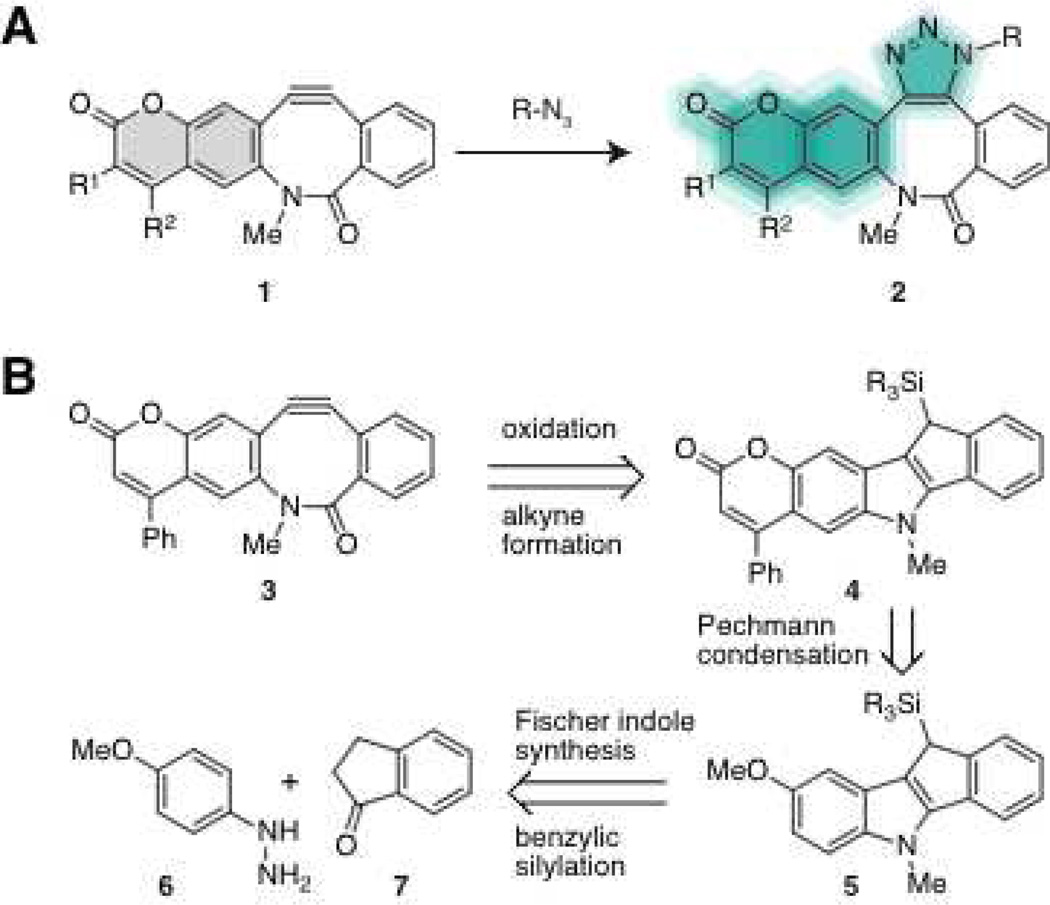
Here we describe the synthesis and photophysical characterization of a fluorogenic cyclooctyne reagent comprising previously-reported biarylazacyclooctynone (named BARAC)5d fused to a coumarin dye (1, Figure 1A). The parent compound BARAC reacts more rapidly with azides than any other reported cyclooctyne. We postulated that its modular synthesis using mild reactions would enable the incorporation of a fluorogenic moiety. Coumarin was an obvious first choice based on precedent from Zhou and Fahrni, who demonstrated that functionalization of the coumarin scaffold with a terminal alkyne can suppress fluorescence relative to the triazolefunctionalized congener.9b Their observation is consistent with the general consensus that an electron-donating functionality at the position meta to the oxygen atom in the lactone ring augments coumarin’s fluorescence quantum yield, and that triazoles are electron-rich.11 Additionally, coumarins are highly modular fluorophores and by simply changing the electronics of the functionality appended to the unsaturated lactone (R1 and R2, 1, Figure 1A) their brightness and color can be tuned.12 These considerations motivated the design of synthetic target 3 (Figure 1B) bearing a 4-phenylcoumarin moiety,13,14 which we call coumBARAC.
Figure 1.
A) A fluorogenic BARAC becomes fluorescent upon reaction with an azide to form a triazole. B) Retrosynthesis of a coumarin-fused BARAC.
In a retrosynthetic sense, we decided to install the coumarin’s lactone moiety as late in the synthesis as possible to take maximum advantage of the modular scaffolds that we were fusing. The late-stage synthetic steps would parallel our previous synthesis of BARAC and, as such, coumBARAC (3) was envisioned to derive from the fused indole coumarin 4 (Figure 1B). This coumarin could in turn come from a Pechmann condensation between a 5-methoxyindole, 5, and a beta-ketoester. As before, the indole would arise from a Fischer indole synthesis between 4-methoxyphenylhydrazine (6) and 1-indanone (7). In theory, the benzylic silylation could be accomplished at various stages of the synthesis.
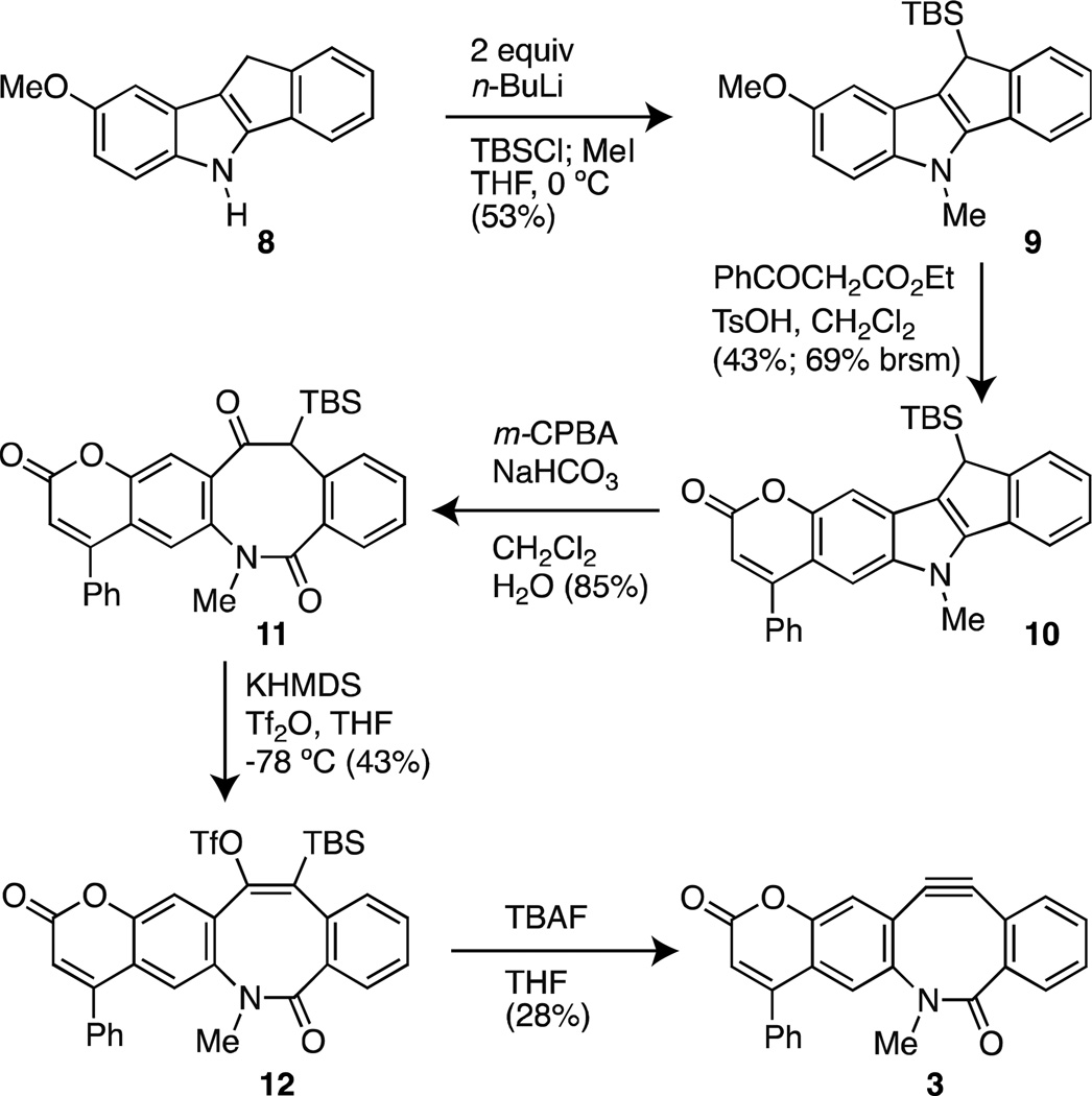
The synthesis of coumBARAC began with a Fischer indole synthesis to make known tetracyclic 5-methoxyindole 8 (Scheme 1).15 All attempts to silylate the N-methylated indole proved unsuccessful. This was a surprising result that we attribute to the reported capricious nature of N-alkyl-5-methoxyindoles.16 We circumvented this problem by double deprotonation of the free NH-indole to allow for benzylic silylation. By quenching the reaction with methyliodide we alkylated the deprotonated nitrogen atom to give 9 directly. The use of a t-butyldimethylsilyl group was advantageous for stability during subsequent reactions and purification schemes.
Scheme 1.
Synthesis of coumBARAC
After trying many traditional procedures for functionalization at the 6-position of both the 5-methoxy and 5-hydroxyindoles, we found that most conditions would leave the starting material untouched, or at times remove the silyl group. Friedel-Crafts acylation with acid chlorides under forcing conditions was possible, but only with many deleterious side reactions. With the 5-hydroxyindole, we observed products arising from reaction at the undesirable C4 position. Ultimately, we observed that methoxyindole 9 would condense with ethyl benzoyl acetate in the presence of triflic acid in dichloromethane. These non-traditional conditions were expected to furnish an alpha-beta unsaturated ester with the C5-methoxy group remaining intact,17 but, at a slow rate, they provided the highly fluorescent desired coumarin, 10, with few side products.
Oxidative cleavage of the indole C2–C3 bond in 10 with m-CPBA afforded keto-amide 11, which was only weakly fluorescent using standard hand-held UV-lamps set at 365 nm. This observation provided a qualitative indication that electronic perturbations at this position of the substituted coumarin would affect fluorescent output. Keto-amide 11 was taken on to silylenoltriflate 12 using KHMDS and triflic anhydride. Consistent with a previous account of strained alkyne formation from TBS-enoltriflates,18 12 was impervious to cesium fluoride and required TBAF to afford coumBARAC (3).
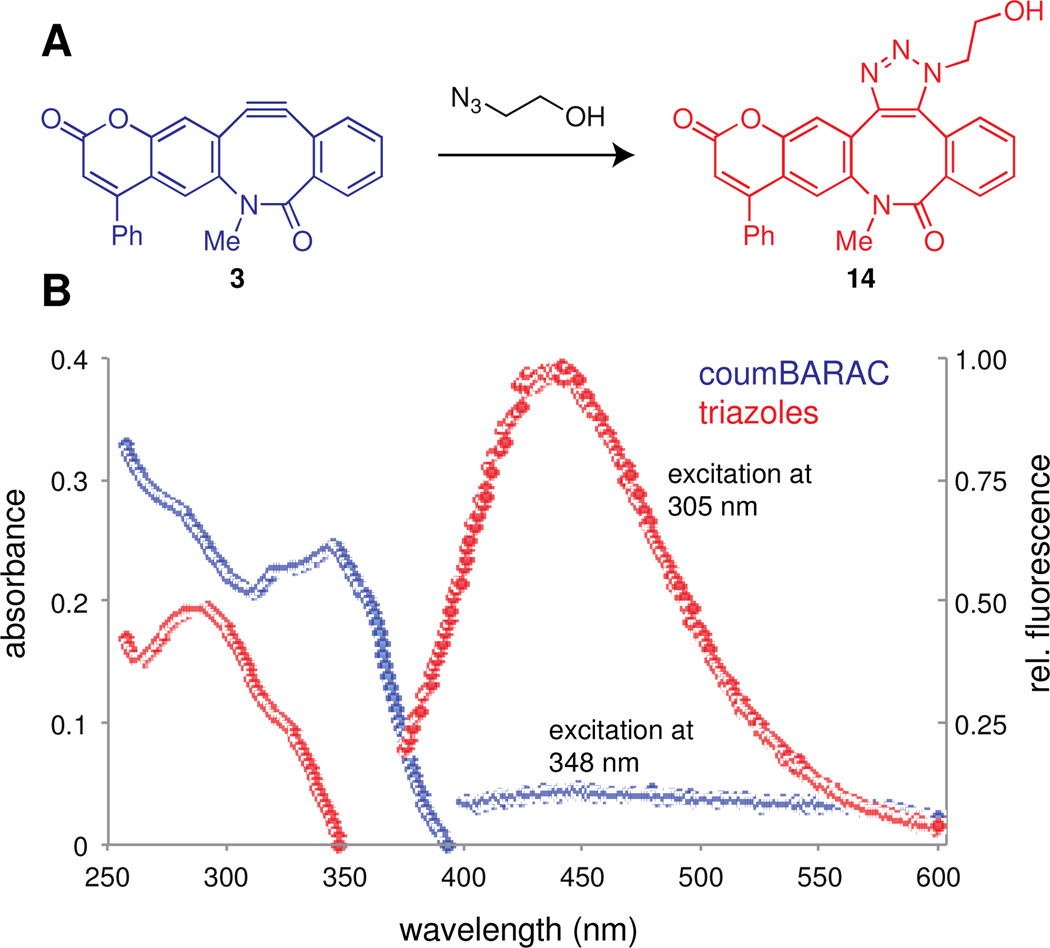
We were encouraged by the observation that coumBARAC (3) is only weakly fluorescent (Figure 2B). However, conversion of 3 to triazole 14 and its regioisomer (Figure 2A) by treatment with 2-azidoethanol19 was accompanied by a strong increase in fluorescence with a large Stokes shift into a standard range for coumarin emission (λem = 438 nm, Figure 2B). To quantitate the magnitude of this “turn-on” effect, we measured the fluorescence quantum yields of coumBARAC 3 (Φf ~ 0.003) and its triazole products 14 (Φf ~ 0.04).20 The observed ~10-fold increase exhibited by the triazoles versus coumBARAC is consistent with the magnitude of turn-on observed by Zhou and Fahrni.9b
Figure 2.
CoumBARAC is fluorogenic. A) CoumBARAC (blue) reacts with 2-azidoethanol to form a mixture of triazole products (one shown in red). B) The absorbance and emission spectra of 22 µM solutions coumBARAC (blue, λex = 348 nm) and triazoles (red, λex = 305 nm) in a solution of 1% DMSO in pH 7.4 phosphate buffer solution.
These results establish a platform for the design of fluorogenic cyclooctynes but improvements are warranted for applications in biological imaging. First, the calculated fluorescence quantum yield for triazoles 14 is quite low relative to standard fluorophores used in microscopy.21 We speculate that the low quantum yield reflects the inability of the aryl-triazole bond to freely rotate and optimize orbital overlap with the coumarin moiety. Also, the optimal excitation wavelength for the triazoles is relatively high-energy (~300 nm) and their excitation profile was sharp and only slightly overlapping with excitation lasers commonly used for imaging biological samples.22 Notably, the modular synthesis we report here should be useful for further functionalization of the coumarin scaffold toward achieving more convenient excitation (and emission) wavelegths. More broadly, this work provides a starting point en route to a new class of fluorogenic cyclooctynes for copper-free click chemistry.
Supplementary Material
Acknowledgment
The authors thank E. Sletten (UC Berkeley), K. Beatty (UC Berkeley), K. Dehnert (UC Berkeley), B. Beahm (UC Berkeley) and H. Aaron (UC Berkeley, imaging facility) for fruitful discussions and A. Lambert (UC Berkeley, C. Chang Laboratory) for technical assistance with fluorimetric measurements. J.C.J. was supported by a postdoctoral fellowship from the American Cancer Society. This work was supported by a grant to C.R.B. from the National Institutes of Health (GM58867).
Footnotes
Supporting Information Available Detailed experimental details pertaining to the synthesis and characterization of compounds 8–12 and 3 are available in a supporting information file.
References
- 1.For a recent review on fluorogenic probes designed for protein labeling, see; Sadhu KK, Mizukami S, Hori Y, Kikuchi K. ChemBioChem. 2011;12:1299–1308. doi: 10.1002/cbic.201100137. For a nice example of a pH sensitive probe, see; Hilderbrand SA, Kelly KA, Niedre M, Weissleder R. Bioconjugate Chem. 2008;19:1635–1639. doi: 10.1021/bc800188p.
- 2.Sletten EM, Bertozzi CR. Acc. Chem. Res. 2011;44:666–676. doi: 10.1021/ar200148z. [DOI] [PMC free article] [PubMed] [Google Scholar]
- 3.Saxon E, Bertozzi CR. Science. 2000;287:2007–2010. doi: 10.1126/science.287.5460.2007. [DOI] [PubMed] [Google Scholar]
- 4.Agard NJ, Prescher JA, Bertozzi CR. J. Am. Chem. Soc. 2004;126:15046–15047. doi: 10.1021/ja044996f. [DOI] [PubMed] [Google Scholar]
- 5.(a) Lemieux GA, de Graffenried CL, Bertozzi CR. J. Am. Chem. Soc. 2003;125:4708–4709. doi: 10.1021/ja029013y. [DOI] [PubMed] [Google Scholar]; (b) Hangauer MJ, Bertozzi CR. Angew. Chem. Int. Ed. 2008;47:2394–2397. doi: 10.1002/anie.200704847. [DOI] [PMC free article] [PubMed] [Google Scholar]
- 6.(a) Baskin JM, Prescher JA, Laughlin ST, Agard NJ, Chang PV, Miller IA, Lo A, Codelli JA, Bertozzi CR. Proc. Natl. Acad. Sci. U. S. A. 2007;104:16793–16797. doi: 10.1073/pnas.0707090104. [DOI] [PMC free article] [PubMed] [Google Scholar]; (b) Ning X, Guo J, Wolfert MA, Boons GJ. Angew. Chem. Int. Ed. 2008;47:2253–2255. doi: 10.1002/anie.200705456. [DOI] [PMC free article] [PubMed] [Google Scholar]; (c) Debets MF, van Berkel SS, Schoffelen S, Rutjes FPJT, van Hest JCM, van Delft FL. Chem. Commun. 2010;46:97–99. doi: 10.1039/b917797c. [DOI] [PubMed] [Google Scholar]; (d) Jewett JC, Sletten EM, Bertozzi CR. J. Am. Chem. Soc. 2010;132:3688–3690. doi: 10.1021/ja100014q. [DOI] [PMC free article] [PubMed] [Google Scholar]; (e) Dommerholt J, Schmidt S, Temming S, Hendriks LJA, Rutjes FPJT, van Hest JCM, Lefeber DJ, Friedl P, van Delft FL. Angew. Chem. Int. Ed. 2010;49:9422–9425. doi: 10.1002/anie.201003761. [DOI] [PMC free article] [PubMed] [Google Scholar]
- 7.Sletten EM, Bertozzi CR. Org. Lett. 2008;10:3097–3099. doi: 10.1021/ol801141k. [DOI] [PMC free article] [PubMed] [Google Scholar]
- 8.Stöckmann H, Neves AA, Stairs S, Ireland-Zecchini H, Brindle KM, Leeper FJ. Chem. Sci. 2011;2:932–936. doi: 10.1039/C0SC00631A. [DOI] [PMC free article] [PubMed] [Google Scholar]
- 9.For a recent review of fluorogenic click reactions see, Le Droumaguet C, Wang C, Wang Q. Chem. Soc. Rev. 2010;39:1233–1239. doi: 10.1039/b901975h. Also see, Zhou Z, Fahrni CJ. J. Am. Chem. Soc. 2004;126:8862–8863. doi: 10.1021/ja049684r.
- 10.Mbua NE, Guo J, Wolfert MA, Steet R, Boons GJ. ChemBioChem. 2011;12:1912–1921. doi: 10.1002/cbic.201100117. [DOI] [PMC free article] [PubMed] [Google Scholar]
- 11.Kleinpeter E, Wilde H, Hauptmann S. Magn. Reson. Chem. 1986;24:53–54. [Google Scholar]
- 12.Schiedel MS, Briehn CA, Bäuerle P. J. Organomet. Chem. 2002;653:200–208. [Google Scholar]
- 13.We found that the phenyl ring at the coumarin 4-position facilitated the synthesis because of its lack of acidic protons.
- 14.The 4-phenylcoumarin moiety has shown success in other fluorogenic applications, albeit with a different turn-on mechanism. Please see, Do JH, Kim HN, Yoon J, Kim JS, Kim HJ. Org. Lett. 2010;12:932–934. doi: 10.1021/ol902860f.
- 15.Brown DW, Graupner PR, Sainsbury M, Shertzer HG. Tetrahedron. 1991;47:4383–4408. [Google Scholar]
- 16.Sundberg RJ, Parton RL. J. Org. Chem. 1976;41:163–165. [Google Scholar]
- 17.Plazuk D, Zakrzewski J. J. Org. Chem. 2002;67:8672–8674. doi: 10.1021/jo025717e. [DOI] [PubMed] [Google Scholar]
- 18.Peña D, Cobas A, Pérez D, Guitián E. Synthesis. 2002;10:1454–1458. [Google Scholar]
- 19.The rate of reaction of coumBARAC with benzyl azide was comparable to that previously reported for the parent compound BARAC (ref 6d). In work not yet published, we found that the addition of electron-withdrawing or -donating substituents to BARAC's aryl rings has only a minor effect on reactivity.
- 20.We considered the possibility that the two regioisomeric triazoles formed by reaction of coumBARAC with 2-azidoethanol have different photophysical properties. To address this, we separated the ~1:1 mixture by column chromatography and analyzed their spectral properties independently. The triazoles had identical excitation/emission profiles, making the selective formation of one over the other irrelevant in the context of improving fluorescent properties.
- 21.The quantum yield was measured using quinine-sulfate as a standard.
- 22.The DAPI filter set is commonly the highest-energy light used to excite biological samples and the excitation is somewhat narrowly centered at 350 nm.
Associated Data
This section collects any data citations, data availability statements, or supplementary materials included in this article.


