Abstract
The majority of human pancreatic cancers have activating mutations in the KRAS proto-oncogene. These mutations result in increased activity of the NF-κB pathway and the subsequent constitutive production of proinflammatory cytokines. Here, we show that inhibitor of κB kinase 2 (Ikk2), a component of the canonical NF-κB signaling pathway, synergizes with basal Notch signaling to upregulate transcription of primary Notch target genes, resulting in suppression of antiinflammatory protein expression and promotion of pancreatic carcinogenesis in mice. We found that in the KrasG12DPdx1-cre mouse model of pancreatic cancer, genetic deletion of Ikk2 in initiated pre-malignant epithelial cells substantially delayed pancreatic oncogenesis and resulted in downregulation of the classical Notch target genes Hes1 and Hey1. Tnf-α stimulated canonical NF-κB signaling and, in collaboration with basal Notch signals, induced optimal expression of Notch targets. Mechanistically, Tnf-α stimulation resulted in phosphorylation of histone H3 at the Hes1 promoter, and this signal was lost with Ikk2 deletion. Hes1 suppresses expression of Pparg, which encodes the antiinflammatory nuclear receptor Pparγ. Thus, crosstalk between Tnf-α/Ikk2 and Notch sustains the intrinsic inflammatory profile of transformed cells. These findings reveal what we believe to be a novel interaction between oncogenic inflammation and a major cell fate pathway and show how these pathways can cooperate to promote cancer progression.
Introduction
Cancer-related inflammation has been shown to be critically linked with malignant disease — either by being the initiating, extrinsic cause or by supporting the intrinsic microenvironment during tumor progression (1). Most solid tumors are characterized by an intrinsic tumor-promoting inflammatory response (1). Activation of proto-oncogenes such as ras and/or inactivation of tumor suppressors orchestrates a proinflammatory transcriptional program and constitutive production of inflammatory cytokines and chemokines that shape a tumor-promoting microenvironment. Oncogenes and tumor suppressor genes are, however, difficult molecular targets in cancer therapy (2). In contrast, inflammatory cytokines and signaling pathways affected by the genetic changes occurring in malignant diseases are attractive druggable targets.
Activating mutations of the KRAS proto-oncogene are found in more than 90% of pancreatic ductal adenocarcinomas (PDACs), the most prevalent form of pancreatic cancer (3). Histological and molecular studies have demonstrated that disease progression occurs through a series of preinvasive lesions, pancreatic intraepithelial neoplasias (PanINs), that progress into invasive carcinoma (4). Mouse models with pancreas-specific activation of oncogenic Kras display the full spectrum of PanINs and recapitulate the features of human PDAC (5, 6). NF-κB, a major transcription factor for inflammatory responses, is found activated in Kras-transformed epithelial cells (7, 8). NF-κB activation is regulated through the inhibitor of κB kinase (Ikk) complex, which consists of two catalytic subunits, Ikk1 and Ikk2 and the regulatory protein Ikk3 (or Nemo) (reviewed in ref. 9). During canonical NF-κB signaling, inflammatory stimuli including cytokines such as Tnf-α generate signals that converge at the Ikk complex, phosphorylating Ikk2, which in turn phosphorylates the inhibitory molecule inhibitor of κB (IκB), resulting in its proteasomal degradation. This releases the p65/p50 NF-κB heterodimer, allowing its nuclear translocation and promoter binding for inflammatory gene transcription. A series of studies has indicated a requirement of Ikk2 and p65 in both murine and human Kras-induced transformation of lung epithelial cells and in models of inflammation-induced carcinogenesis (7, 8, 10, 11). However, the implication of the pathway in pancreatic cancer has so far been unexplored.
Interestingly, in many types of cancer, including pancreatic cancer, the NF-κB and Notch pathways are activated (12–15). Classical activation of Notch signaling is triggered by ligation of Notch receptors and ligands. This leads to proteolytic cleavage of Notch and the release of the Notch intracellular domain (NICD). NICD subsequently translocates to the nucleus and binds to the DNA-binding protein Rbp-j. This interaction results in assembly of a transcriptional activation complex that drives the expression of Notch target genes (16). Among the best-characterized direct Notch target genes are the Hes and Hey families of transcriptional repressors. These genes are found to be upregulated in early PanINs and throughout PDAC but not in normal pancreatic epithelium (5, 15). In the context of mutant Kras, Notch pathway activation has been shown to have a tumor-promoting role and has been implicated in mediating metaplasia of acinar to ductal epithelium, a critical process in pancreatic carcinogenesis (17–19).
In the present study we showed that genetic deletion of Ikk2 in Kras+/LSL-G12DPdx1-cre mice blocked the progression of PanIN lesions. We further demonstrated that Tnf-α stimulation of initiated pre-malignant epithelial cells via Ikk2 engaged with canonical Notch signaling to upregulate the expression of primary Notch target genes. The crosstalk between NF-κB and Notch downregulated Pparγ, a repressor of inflammatory gene expression and retained a constitutive production of proinflammatory mediators and cytokines by the transformed cells.
Results
Pancreas-specific deletion of Ikk2 blocks PanIN progression in KrasG12D mice.
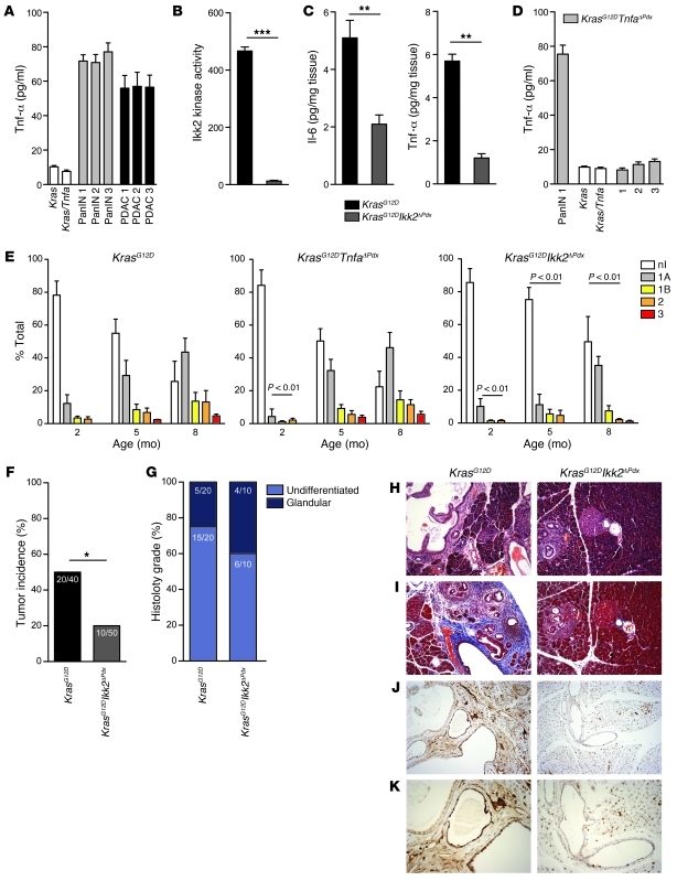
Kras+/LSL-G12DPdx1-cre (abbreviated as KrasG12D) mice express an endogenous oncogenic KrasG12D allele initially in pancreatic progenitors and later in the adult pancreas (5). We generated ductal epithelial cell lines from PanIN- and PDAC-bearing KrasG12D mice and identified constitutive secretion of Tnf-α (Figure 1A), similar to previous data indicating Tnf-α production by initiated pre-malignant ovarian epithelial cells (20). To determine the role of Ikk2/NF-κB signaling in formation and progression of PanINs, we generated Kras+/LSL-G12DIkk2fl/flPdx1-cre (KrasG12DIkk2ΔPdx) mice. In parallel, we assessed the contribution of malignant cell–derived Tnf-α using Kras+/LSL-G12DTnfafl/flPdx1-cre (KrasG12DTnfaΔPdx) mice.
Figure 1. Genetic deletion of Ikk2 inhibits PanIN progression.
(A) Tnf-α secretion by ductal cell lines derived from KrasG12D PanIN- or PDAC-bearing mice measured by ELISA. Control cells were generated from Kras and Kras/Tnfa cre-negative pancreases. (B) Cellular Ikk2 kinase activity in cell lines derived from KrasG12D and KrasG12DIkk2ΔPdx mice. (C) Il-6 and Tnf-α secretion in pancreatic tissue of KrasG12D and KrasG12DIkk2ΔPdx mice. n = 6; **P < 0.01, ***P < 0.01. (D) Tnf-α secretion by cell lines derived from KrasG12D (PanIN 1) or KrasG12DTnfaΔPdx PanIN- or PDAC-bearing mice. Cre-negative Kras and Kras/Tnfa control cells were included. Data in C are shown as mean + SD of n = 6 mice, and data in A, B, and D are mean + SD of triplicate experiments. (E) Quantification of the proportion of pancreas occupied by PanIN lesions. Frequency and grade of the lesions was quantified at 2, 5, and 8 months of age. Data are shown as mean + SD; P < 0.01. nl, no lesion. (F) Tumor incidence and (G) histology grade in KrasG12D and KrasG12DIkk2ΔPdx mice. *P < 0.05. (H–K) KrasG12D and KrasG12DIkk2ΔPdx 4-month old pancreases stained with (H) hematoxylin and eosin, (I) Masson’s trichrome (blue, collagen; red, muscle fibers and cytoplasm; black, nuclei) and (J and K) anti-PCNA. Original magnification, ×10 (H–J), ×20 (K).
The compound strains were generated by interbreeding C57BL/6 mice carrying floxed Ikk2 or Tnfa alleles with the Kras+/LSL-G12D and Pdx1-cre strains (Supplemental Figure 1A; supplemental material available online with this article; doi: 10.1172/JCI45797DS1). No gross pathology was observed in the pancreas of Ikk2ΔPdx or TnfaΔPdx mice (Supplemental Figure 1B). Activity of the Ikk complex was abolished in cells derived from KrasG12DIkk2ΔPdx mice, confirming excision of the Ikk2 locus (Figure 1B). Secretion of Tnf-α and Il-6 in the pancreas was also significantly decreased (P < 0.01, n = 6, Figure 1C). Similarly, cell lines derived from PanIN- and PDAC-bearing KrasG12DTnfaΔPdx mice secreted minimal levels of Tnf-α, confirming Tnfa inactivation (Figure 1D).
We assessed the development of PanIN lesions in cohorts (n = 12 per time point) of KrasG12D, KrasG12DIkk2ΔPdx, and KrasG12DTnfaΔPdx mice at 2, 5, and 8 months of age. Histological assessment for the proportion of pancreas occupied by PanINs was carried out as previously described (4). Ikk2 deletion in KrasG12DIkk2ΔPdx mice resulted in a profound decrease in the frequency of high-grade PanINs (PanINs 2 and 3) at all time points (P < 0.01; Figure 1E). Only low-grade PanINs were present in 5-month-old KrasG12DIkk2ΔPdx mice, while approximately 80% of the pancreatic parenchyma retained normal exocrine tissue. Even at 8 months of age, formation of grade 2 and 3 lesions was minimal, and the frequency of grade 1 PanINs was lower compared with both KrasG12D and KrasG12DTnfaΔPdx mice (P < 0.01; Figure 1E).
Two-month-old KrasG12DTnfaΔPdx mice exhibited a significant reduction in early PanIN lesions (P < 0.01, n = 12). However, by 5 months of age, PanINs had formed and progressed in a pattern similar to that in KrasG12D mice (Figure 1E). These results indicated that, in the context of mutant Kras, Ikk2 signaling was important for the development and progression of PanINs. Activation of the pathway by Tnf-α provided by the transformed epithelial cells was important early during the carcinogenic process. However, as the disease progressed, an influx of tumor-associated immune cells, primarily macrophages and neutrophils, compensated Tnf-α cytokine levels. To address the importance of the inflammatory infiltrate to compensate for the lack of inflammatory cytokines, we generated chimeras using Mx1-cre mice to target Tnf-α deletion in the leukocyte compartment (Supplemental Figure 2, A–D). Infiltration of these cells was minimal in KrasG12DIkk2ΔPdx pancreases, indicating that Ikk2 inactivation impaired their capacity to attract other cell types (Supplemental Figure 2, A and B).
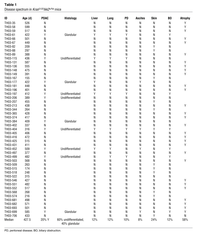
To assess whether Ikk2 depletion affected PDAC development, we followed cohorts of 50 KrasG12DIkk2ΔPdx and 40 KrasG12D mice for nearly 2 years (Table 1 and Supplemental Table 1). Mice were sacrificed when they developed signs of distress. 20% of KrasG12DIkk2ΔPdx mice had PDAC, while there was a 50% tumor incidence in KrasG12D mice (Figure 1F). Interestingly, deletion of Ikk2 changed the histopathological feature of the observed tumors, as shown by the ratio of undifferentiated to glandular morphology in these mice (Figure 1G) at end point (Table 1 and Supplemental Table 1).
Table 1 .
Disease spectrum in KrasG12DIkk2ΔPdx mice
Further histological analyses of KrasG12DIkk2ΔPdx pancreases showed a profound delay in stromal reaction (Figure 1, H and I). Proliferation of acinar cells was assessed by PCNA expression. As shown in Figure 1, J and K, there was a reduction in proliferating acinar cells in KrasG12DIkk2ΔPdx compared with KrasG12D pancreases. No difference was noted in the levels of apoptosis, measured by cleaved caspase-3 staining (data not shown). Collectively, these data indicated that PanIN progression and development of PDAC were dependent on epithelial Ikk2 depletion.
Notch target genes Hes1 and Hey1 are downregulated in KrasG12DIkk2ΔPdx PanINs.
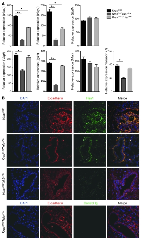
The Notch pathway, normally quiescent in the adult pancreas, is found to be reactivated in pancreatic cancer throughout PanIN and PDAC development (15, 17, 21). We assessed the regulation of Notch downstream targets as indicators of disease development (5). In accordance with previous studies, we found that the classical Notch target genes Hes1 and Hey1 were expressed in KrasG12D PanIN-bearing mice (5). However, there was a substantial decrease in their expression in age-matched KrasG12DTnfaΔPdx and KrasG12DIkk2ΔPdx mice (Figure 2A). The pancreases of KrasG12DIkk2ΔPdx showed decreased expression of Igfr1, Vegf, and tenascin C, all Notch target genes, while expression of Myc and the AP-1 family transcription factor Batf was not altered (Figure 2A). Inactivation of Tnfa had little impact on the expression levels of these genes. Immunofluorescence analysis revealed Hes1-positive staining in PanIN lesions of 3-month-old KrasG12D mice. Similar levels of Hes1 were found in KrasG12DTnfaΔPdx mice (Figure 2B). In contrast, Hes1 protein was minimal in age-matched KrasG12DIkk2ΔPdx animals (Figure 2B). These results indicated concurrent activity of the classical NF-κB and Notch pathways.
Figure 2. Molecular analysis of Notch and NF-κB target gene expression in KrasG12DTnfaΔPdx and KrasG12DIkk2ΔPdx pancreases.
(A) Relative mRNA expression of Hes1, Hey1, Batf, Vegf, Igfr1, Myc, and tenascin C in KrasG12DTnfaΔPdx and KrasG12DIkk2ΔPdx 3-month PanIN-bearing pancreases was measured by real-time PCR. Data are shown as mean + SD; n = 6. *P < 0.05, **P < 0.01. The experiment was done in duplicate. (B) Immunofluorescence staining for Hes1 and E-cadherin in PanIN-bearing pancreases from KrasG12DTnfaΔPdx, KrasG12DIkk2ΔPdx, and KrasG12D mice at 3 months of age. Original magnification, ×40. Blue, DAPI; red, E-cadherin; green, Hes1.
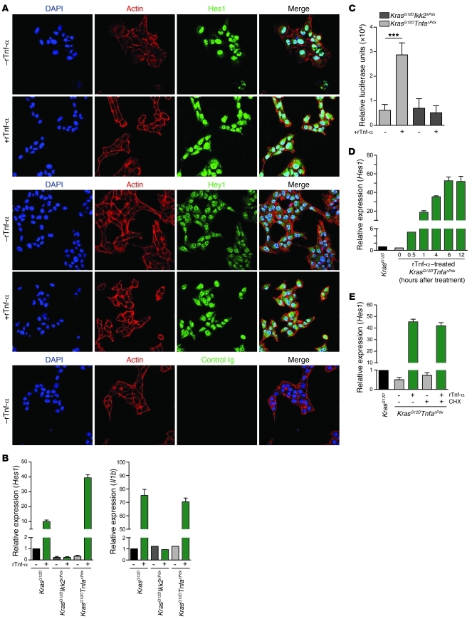
To further dissect the interaction of the Tnf-α/Ikk2 and Notch signaling pathways, we examined the response of cell lines derived from KrasG12D, KrasG12DTnfaΔPdx and KrasG12DIkk2ΔPdx mice to recombinant Tnf-α (rTnf-α) stimulation in vitro. Basal Notch activity in KrasG12D cell lines was demonstrated by nuclear localization of Hes1 and low levels of cytoplasmic staining (Figure 3A). Stimulation with rTnf-α increased expression of both nuclear and cytoplasmic Hes1 protein (Figure 3A). The expression of Hes1, Hey1, as well as Batf, Vegf, Igfr1, Myc, and tenascin C was increased in KrasG12D and KrasG12DTnfaΔPdx cells after rTnf-α stimulation. In contrast, rTnf-α failed to upregulate expression of these genes in KrasG12DIkk2ΔPdx cells (Figure 3B and Supplemental Figure 3A). We next transiently transfected the cell lines with a Hes1 luciferase reporter construct and stimulated them with 1 ng/ml rTnf-α. This resulted in enhanced transcriptional activity of the Hes1 promoter in KrasG12DTnfaΔPdx but not in KrasG12DIkk2ΔPdx cells (Figure 3C). These results showed that in initiated pre-malignant epithelial cells Ikk2 signaling enhanced the expression of Notch target genes.
Figure 3. Tnf-α–induced Notch and NF-κB target gene expression in PanIN cell lines.
(A) Expression of Hes1 and Hey1 in KrasG12D PanIN cell lines was examined by immunofluorescence staining; cells were left unstimulated or were stimulated with 10 ng/ml rTnf-α for 24 hours. Original magnification, ×40. Blue, DAPI; red, actin; green, Hes1. One representative experiment of 3 performed is shown. (B) Relative mRNA expression of Hes1 and Il1b in PanIN cell lines stimulated with 1 ng/ml rTnf-α for 6 hours. Relative expression was calculated by setting expression of untreated KrasG12D samples as 1. (C) Hes1 luciferase reporter assay in KrasG12DTnfaΔPdx and KrasG12DIkk2ΔPdx PanIN cell lines stimulated with 1 ng/ml rTnf-α for 6 hours. Results were normalized to firefly luciferase activity relative to internal control and are expressed as mean + SD from triplicate transfections. ***P < 0.01. One representative experiment of 3 performed is shown. (D) Kinetic analysis of Hes1 mRNA expression in KrasG12DTnfaΔPdx PanIN cell lines stimulated with 1 ng/ml rTnf-α. (E) KrasG12DTnfaΔPdx PanIN cells were treated with 1 ng/ml rTnf-α in the presence or absence of 15 μg/ml cycloheximide (CHX). Expression of Hes1 was quantified by real-time PCR. Relative expression was calculated by setting expression of untreated KrasG12D samples as 1. (B, D, and E) Data are shown as mean + SD of triplicate determinants, and 1 representative experiment of 3 is shown.
Activation of the NF-κB pathway is known to upregulate Notch receptors and ligands, both of which are found to be expressed on PanIN and PDAC cells (17, 22–26). However, an interaction downstream of the two pathways has not been described. We next assessed whether this enhanced expression of Notch target genes upon stimulation with rTnf-α was due to upregulation of Notch receptors and ligands, which would reinforce downstream signaling. We stimulated KrasG12DTnfaΔPdx cell lines with rTnf-α over a full 12-hour time course and assessed mRNA expression of Hes1 and Hey1. Upregulation of gene expression occurred within 30 minutes and reached a plateau between 6 and 12 hours after treatment (Figure 3D and Supplemental Figure 3B). This rapid upregulation of Hes1 and Hey1 was independent of new protein synthesis and suggested a direct interaction between the pathways (Figure 3E and Supplemental Figure 3C).
Tnf-α–induced Notch target gene expression requires canonical Notch signaling and Ikk2-mediated histone phosphorylation.
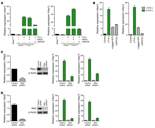
We next sought to determine whether Tnf-α–induced upregulation of Notch target genes required canonical Notch signaling. This is initiated by proteolytic cleavage of NICD following receptor-ligand interactions, mediated by the γ-secretase activity of a multiprotein complex (27). Pharmacological inhibition of γ-secretase using the synthetic inhibitor L685458 resulted in attenuation of Hes1 and Hey1 expression in rTnf-α–stimulated KrasG12DTnfaΔPdx PanIN cell lines (Figure 4A). While expression of both these genes was sensitive to L685458, transcription levels of Il1b, Mmp13, and Cox2, all NF-κB targets, remained unaffected (Supplemental Figure 4). We maximally engaged Notch receptors by stimulating KrasG12D cells with the classical Notch ligands Jagged-2 and Delta-like–1 (Dll1) and compared the levels of Hes1 and Hey1 expression with those after treatment with rTnf-α. rTnf-α induced higher Hes1 and Hey1 levels than ligand-mediated Notch activation of the pathway (Figure 4B).
Figure 4. Tnf-α–induced Notch target gene expression requires expression of Rbpj and Ikk2.
(A) Inhibition of Hes1 and Hey1 mRNA expression in Tnf-α–induced KrasG12DTnfaΔPdx PanIN cells treated with the γ-secretase inhibitor L685458 (5 μM). Cells were stimulated with 1 ng/ml rTnf-α. (B) KrasG12DTnfaΔPdx PanIN cells were treated with rTnf-α, 20 μg/ml Jagged-2/Fc, or cocultured with OP9-DL1 cells. Tnf-α was more efficient in inducing the expression of Hes1 and Hey1. The results were normalized to values obtained from KrasG12D cells. (C and D) KrasG12DTnfaΔPdx PanIN cell lines transfected with (C) Rbpj- or (D) Ikk2-specific siRNA. Forty-eight hours after transfection, cells were stimulated with 1 ng/ml rTnf-α for 6 hours, and expression of Hes1 was quantified by real-time PCR. Nontargeting siRNA and/or unstimulated controls were included. Results were normalized to uninfected and unstimulated KrasG12DTnfaΔPdx cells. All data are shown as mean + SD of triplicate determinants and are representative of 3 independent experiments.
We further examined the requirement of Notch signaling for Tnf-α–mediated upregulation of Hes1 and Hey1 using siRNA to knock down the expression of Rbpj, a nuclear transcription factor essential for Notch target gene expression. Transfection of KrasG12DTnfaΔPdx cell lines with Rbpj siRNA resulted in a 4-fold decrease in Hes1 and Hey1 transcripts, confirming the requirement of NICD–Rbp-j interaction for upregulation of target gene expression (Figure 4C and Supplemental Figure 5A). Expression of the NF-κB targets Il1b and Cox2 remained unaffected in Rbpj-knockdown cells (data not shown). Specific siRNA inhibition of Ikk2 also resulted in a downregulation of Hes1 and Hey1 expression following rTnf-α treatment (Figure 4D and Supplemental Figure 5B). This was consistent with our previous observation that KrasG12DIkk2ΔPdx cell lines lost the capacity to upregulate Hes1 and Hey1 upon rTnf-α stimulation. Similarly, knockdown of Nemo blocked Hes1 and Hey1 expression (Supplemental Figure 5C), while knockdown of Ikk1 (Supplemental Figure 5D) had no effect on Hes1 or Hey1 expression.
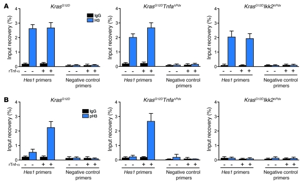
Hes1 expression is not known to be regulated by NF-κB (16). To investigate the pathways downstream of Ikk2 that lead to Hes1 activation, we examined phosphorylation of histone H3 at serine 10, a histone modification that is induced by Ikk2 and is linked with recruitment of RNA polymerase II and transcriptional activation (28–30). We carried out ChIP and real-time PCR assays and showed that rTnf-α stimulation induced phosphorylation of histone H3 at serine 10 at the Hes1 promoter (Figure 5). This inducible phosphorylation was abolished in KrasG12DIkk2ΔPdx cells (Figure 5). These results indicate a link between Tnf-α–stimulated Ikk2 signaling and the Hes1 locus, whereby Tnf-α enhanced the transcriptional activity of a classical Notch target gene via Ikk2 by inducing histone H3 phosphorylation.
Figure 5. Tnf-α–induced Notch target gene expression is dependent on Ikk2 and chromatin remodeling.
ChIP was performed on rTnf-α–treated KrasG12D, KrasG12DTnfaΔPdx, and KrasG12DIkk2ΔPdx samples with anti–histone H3 (A) or anti–phospho–histone H3 at serine 10 (pH3) (B). Rabbit IgG was used as control. Precipitated DNA was measured by real-time PCR using primers specific for Hes1. Results are shown as mean + SD of triplicate determinants and are representative of 3 independent experiments.
Tnf-α–induced crosstalk between NF-κB and Notch pathways leads to Hes1-mediated Pparγ inhibition.
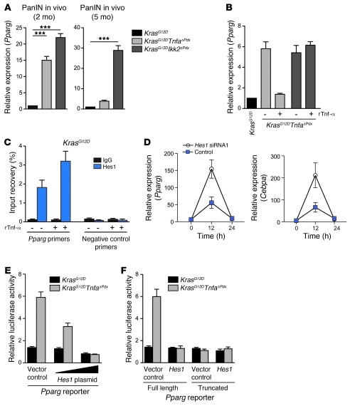
Hes1 is known to bind to the promoter region of the nuclear receptor Pparγ and suppress its expression (31). Pparγ represses inflammatory gene expression induced by other classes of transcription factors including NF-κB. We observed higher Pparg mRNA expression in 2-month-old KrasG12DTnfaΔPdx and KrasG12DIkk2ΔPdx compared with KrasG12D pancreases (Figure 6A). However, by 5 months of age, expression of Pparg in KrasG12DTnfaΔPdx was only marginally higher than in KrasG12D mice. In contrast, it remained elevated in KrasG12DIkk2ΔPdx pancreases (Figure 6A). Moreover, after rTnf-α stimulation, Pparg mRNA in KrasG12DTnfaΔPdx cells decreased to levels similar to those in KrasG12D cells (Figure 6B). Binding of Hes1 to the Pparg promoter in KrasG12D cells was confirmed by ChIP (Figure 6C). These data indicated that Tnf-α–induced Hes1 upregulation in initiated pre-malignant cells resulted in Pparg suppression.
Figure 6. Tnf-α/NF-κB and Notch crosstalk leads to Hes1-mediated Pparg inhibition.
(A) Pparg mRNA expression in 2- and 5-month-old KrasG12DTnfaΔPdx and KrasG12DIkk2ΔPdx pancreases. Data were normalized to KrasG12D pancreases. Data are shown as mean + SD; n = 6. ***P < 0.001. The experiment was performed in duplicate. (B) Tnf-α stimulation (1 ng/ml) induced downregulation of Pparg in KrasG12DTnfaΔPdx PanIN cell lines. (C) ChIP was performed on KrasG12D cells using anti-Hes1 or a control IgG. Precipitated DNA was amplified by real-time PCR using primers specific for Pparg. (D) siRNA knockdown of Hes1 upregulated Pparg and Cebpa expression in KrasG12D PanIN cells. (E) KrasG12D and KrasG12DTnfaΔPdx PanIN cells were cotransfected in duplicate with a Pparg reporter construct containing 1,500 bases of the proximal Pparg promoter (full length) and a Hes1 expression plasmid or empty vector control. Twenty-four hours after transfection, cells were analyzed for luciferase activity. (F) Transfection of KrasG12DTnfaΔPdx PanIN cells as described in E with a full-length Pparg reporter construct or a construct with a truncated Hes1-binding sequence. All data are shown as mean + SD from duplicate transfections and are representative of 3 independent experiments.
We further examined the interplay between Hes1 and Pparg using Hes1-specific siRNA to knock down Hes1 expression in KrasG12D PanIN cell lines. This resulted in robust upregulation of Pparg expression, which indicates Hes1-mediated inhibition of Pparg transcription (Figure 6D and Supplemental Figure 6, A and B). Similarly, Cebpa, a transcription factor whose expression requires Pparg, was also negatively regulated by Hes1 (Figure 6D and Supplemental Figure 6C).
Hes proteins suppress gene expression by a number of mechanisms that include binding to N boxes or suppressing E box–mediated transcription in promoters that contain tandem E boxes and Rbp-j sites (32–34). We investigated the mechanism by which Hes1 inhibits Pparg expression in our system by analyzing the effects of Hes1 on the activity of a Pparg promoter–driven reporter gene. We confirmed, in transient transfection assays, that Hes1 suppressed expression of a Pparg promoter–driven reporter gene, in a dose-dependent manner (Figure 6E), through sequences from –1,500 to –160 that contain 6 E-box elements (31). A truncated E box sequence abrogated the ability of Hes1 to inhibit Pparg promoter activity (Figure 6F).
Pharmacological intervention in Notch and Pparγ signaling modulates the inflammatory profile of malignant cells and inhibits PanIN growth.
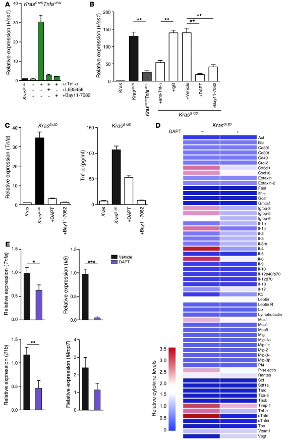
Pharmacological inhibition of NF-κB or Notch signaling by anti–Tnf-α, the NF-κB inhibitor Bay11-7082, or the γ-secretase inhibitor DAPT could block the expression of Hes1 in PanIN-bearing 5-month-old mice. As shown in Figure 7, each of these approaches inhibited Hes1 in PanIN-bearing pancreases and reduced Tnf-α cytokine levels in KrasG12D cells (P < 0.01; Figure 7, A–C).
Figure 7. Inhibition of Notch/NF-κB signaling attenuates the inflammatory profile of malignant cells.
(A) KrasG12DTnfaΔPdx cells were stimulated with 1 ng/ml rTnf-α for 6 hours in the presence or absence of L685458 or Bay11-7082. Hes1 mRNA expression was quantified by real-time PCR. Results were normalized to unstimulated KrasG12D cells. Data are shown as mean + SD of triplicate determinants and are representative of 3 independent experiments. (B) Hes1 mRNA expression in pancreases of 5-month-old untreated KrasG12D and KrasG12DIkk2ΔPdx mice and of KrasG12D mice treated with anti–Tnf-α, control IgG, DAPT, Bay11-7082, or the vehicle control. Results were normalized to Kras pancreases. Data are shown as mean + SD; n = 6. **P < 0.01. The experiment was done in duplicate. (C) KrasG12D PanIN cells were treated with DAPT or Bay11-7082. Tnfa mRNA expression and cytokine secretion are indicated. Data are shown as mean + SD of triplicate experiments and are representative of 3 independent experiments. (D) Cytokine and chemokine array on whole pancreases of DAPT or vehicle-treated 5-month-old KrasG12D mice. The data are represented graphically as normalized signal intensity. (E) Tnfa, Il6, Il1b, and Mmp7 expression in FACS-sorted EYFP-positive KrasG12D cells treated for 1 week with DAPT or vehicle. Data are shown as mean + SD; n = 12. *P < 0.05, **P < 0.01, ***P < 0.001. Analysis of Mmp7 expression did not reveal statistical significance.
We hypothesized that this interplay between NF-κB and Notch signaling and a coordinated downregulation of Pparg acted as a forward feedback loop that sustains expression of inflammatory cytokines and chemokines by the transformed cells. To address this hypothesis, we treated KrasG12D mice with DAPT, a γ-secretase inhibitor. Cytokine arrays on whole pancreases of untreated and DAPT-treated mice revealed downregulation of proinflammatory cytokines and chemokines (Figure 7D). To further strengthen the impact of Notch signaling on the inflammatory state of the transformed cells, we used KrasG12D mice carrying the Rosa26-LSL-Eyfp allele. In these mice, Eyfp expression was confined to the KrasG12D-expressing epithelial cell pool. Cohorts of n = 12 mice were treated with DAPT or vehicle, and the Eyfp-positive cells were isolated by FACS. Analysis of the sorted cells showed significant downregulation of Tnfa (P < 0.05), Il6 (P < 0.001), and Il1b expression (P < 0.01) in the DAPT-treated group (Figure 7E).
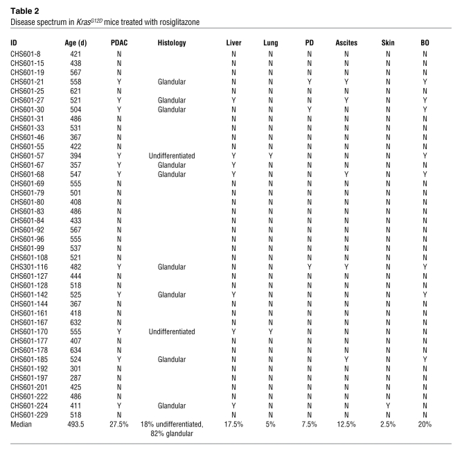
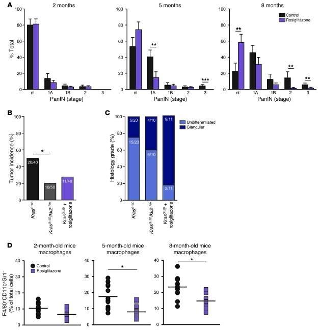
We finally asked whether treatment with rosiglitazone, a Pparγ agonist with antiinflammatory properties in vivo, would influence PanIN development in KrasG12D mice (35, 36). Mice were treated with 3 mg/kg/d rosiglitazone added to their daily diet, and cohorts of KrasG12D mice were followed for nearly 2 years (Table 2). Progression of PanINs was significantly delayed in rosiglitazone-treated mice compared with the untreated controls (P < 0.01, n = 12, Figure 8A). Tumor incidence was 2-fold lower (11 of 40) compared with that in untreated mice, greater than that observed in the Ikk2-depleted KrasG12DIkk2ΔPdx animals (10 of 50) (Figure 8, B and C). Analysis of the macrophage infiltrate showed a reduction in the frequency of these cells in rosiglitazone-treated animals (Figure 8D). In total, these data suggest that modulation of tumor-associated inflammatory networks can inhibit PanIN progression and restrain stromal inflammatory components.
Table 2 .
Disease spectrum in KrasG12D mice treated with rosiglitazone
Figure 8. Rosiglitazone treatment blocks PanIN progression in KrasG12D mice.
(A) Quantification of the percentage or pancreatic parenchyma occupied by PanINs in KrasG12D mice treated with rosiglitazone compared with untreated controls. Values are shown as mean + SD; n = 12. (B) Tumor incidence and (C) histology grade in rosiglitazone-treated KrasG12D mice compared with untreated KrasG12D and KrasG12DIkk2ΔPdx mice. (D) Percentage of F4/80+CD11b+Gr1– cells in the pancreas of 2-, 5-, and 8-month-old KrasG12D mice treated with rosiglitazone or untreated as measured by flow cytometry. Each data point represents an individual mouse. Mean values are depicted by the horizontal lines; n = 10. *P < 0.05, **P < 0.01, ***P < 0.001.
Discussion
The integrative interactions among proinflammatory cytokines, transcription factors, and oncogenic signaling pathways are currently the focus of extensive investigation. Here we demonstrated that in the context of Kras-driven pancreatic carcinogenesis, genetic inactivation of Ikk2 blocked the progression of PanIN lesions. Depletion of Ikk2 correlated with decreased expression of the classical Notch target genes Hes1 and Hey1. Our further work showed that Tnf-α–induced activation of the NF-κB pathway in initiated pre-malignant epithelial cells cooperated with basal Notch signals to enhance the expression of Notch target genes, in an Ikk2-dependent manner. The interplay between Ikk2 and Notch, via the expression of Hes1, repressed the antiinflammatory nuclear receptor Pparγ and created a forward feedback loop that retained the transformed cells in an inflammatory state.
Ikk2 is essential for canonical activation of NF-κB and has been shown to be required for carcinogenesis both in settings where NF-κB activation is driven by ras mutations and in inflammation-induced cancer models (7, 8, 10, 11). However, the role of the Ikk2/NF-κB axis is context and cell type dependent; in certain settings, such as those observed in hepatocarcinogenesis, Ikk2 depletion results in tumor promotion (37). Our data demonstrated that in the context of Kras-driven pancreatic carcinogenesis, genetic deletion of Ikk2 blocked the progression of malignant epithelial cell lesions.
Activation of NF-κB is known to regulate a number of cellular processes, including a malignant cell–intrinsic network of inflammatory cytokines and chemokines (38). These act in an autocrine and paracrine manner both on the malignant cells and on the surrounding stroma and induce the activity of a number of oncogenic transcription factors, including Stat3 and AP-1 as well as NF-κB itself (10, 11, 20, 39, 40). With deletion of Ikk2 in initiated pre-malignant epithelial cells, an array of inflammatory cytokines and chemokines at the tumor site was significantly downregulated, and recruitment of macrophages and neutrophils was profoundly decreased (Supplemental Figure 2 and data not shown). Cell-autonomous processes such as proliferation were also affected, and downregulation of Notch target genes was observed. Notch signaling has oncogenic properties in pancreatic carcinogenesis; however, a link between this pathway and canonical NF-κB had not been previously appreciated (17–19).
Tnf-α is a major inflammatory cytokine that activates the NF-κB pathway and is regulated in its expression by NF-κB. We demonstrated that Tnf-α stimulation of initiated pre-malignant epithelial cells resulted in upregulated expression of classical Notch targets. This occurred at the level of transcription by Ikk2-mediated phosphorylation of histone H3, a modification that is linked with transcriptional activation (28–30). Accordingly, Tnf-α–mediated upregulation of Hes1 and Hey1 was independent of de novo protein synthesis but required canonical Notch signaling. Our data suggest that activation of NF-κB signaling can synergize with basal Notch signals to induce maximal expression of Notch target genes.
Conversely, Vilimas et al. have demonstrated that in T cell acute lymphoblastic leukemia, constitutively active Notch results in activation of the NF-κB pathway (13). Work from the same group demonstrated that the Notch/Hes1 signaling sustained NF-κB pathway activation by repressing the deubiquitinase CYLD, a negative Ikk complex regulator (41). In conjunction with our data, these studies indicate a bidirectional interaction between the NF-κB and Notch pathways that can result in bidirectional expression of target genes and enhanced malignant cell growth.
Within the tumor microenvironment, Tnf-α stems from two sources: the tumor-infiltrating immune cells and the malignant cells. In accordance with previous studies, we found an influx of inflammatory cells, predominantly macrophages and neutrophils, in KrasG12D mice as disease progressed (42). These cells were a major source of the cytokine in aged mice. We also showed that Kras-induced PanIN and PDAC cells constitutively secreted low levels of Tnf-α. Crosstalk between NF-κB and Notch signaling can therefore be fueled both by a constitutive autonomous activation of NF-κB signaling due to mutant Kras and by inflammatory cytokines provided by the immune cells. Our data suggested that early in the carcinogenic process, Tnf-α secreted by the malignant cells is critical for their growth, while at later stages, influx of immune cells constitutes the major source of the cytokine.
Inflammation induced extrinsically by tissue damage (i.e., pancreatitis) or inflammation related to metabolic stress has been shown to accelerate PanIN development (43–45). The underlying mechanism is a deregulated regeneration process whereby constitutively active Notch permits PanIN formation. By identifying a direct link between NF-κB signaling and enhanced Notch activity, we provide evidence that a major proinflammatory and a developmental signaling pathway can cooperate in the context of mutant ras to promote carcinogenesis.
Repression of inflammatory genes by the nuclear receptor Pparγ has been highlighted as an important mechanism by which cells can regulate inflammatory responses and homeostasis (46). Our findings demonstrated that a Tnf-α/Hes1–driven mechanism of Pparγ inhibition operates in initiated pre-malignant pancreatic epithelial cells. Hes1 suppressed Pparg expression by targeting E box elements in the promoter of the gene. We propose that the coordinated activity of NF-κB and Notch along with a suppression of antiinflammatory transcription factors such as Pparγ leads to a sustained expression of inflammatory genes and transcription factors and a constitutive production of inflammatory mediators and chemokines by the transformed cells. Pharmacological inhibition of the Notch pathway in KrasG12D mice with a γ-secretase inhibitor resulted in attenuation of inflammatory gene expression by the transformed cells and downregulation of cytokine production in the pancreas. It has also been shown to significantly attenuate the development of PanINs (21).
Synthetic Pparγ ligands induce allosteric changes to the receptor and allow it to enter into a repression pathway (47). These agents are shown to have antiinflammatory activity in a variety of models of acute and chronic inflammation, as reviewed in ref. 35. By using rosiglitazone, a Pparγ ligand of the thiazolidinedione class, to treat KrasG12D mice, we demonstrated a marked decrease in the frequency of PanINs and their progression into invasive carcinoma. Nevertheless, Pparγ has a wide range of effects on metabolism (48). Thiazolidinedione drugs are best characterized by their insulin-sensitizing action and have been used in the treatment of diabetes. Notably, individuals with type 2 diabetes receiving metformin, a glucose-lowering drug, have a decreased risk of developing pancreatic cancer (49, 50).
Our findings and an increasing body of studies highlight the requirement for inflammatory signaling pathways in the development of pancreatic cancer and reveal key molecular targets to assist current treatments.
Methods
Mouse strains.
Kras+/LSL-G12D, Pdx1-cre (5), Tnfafl/fl (51), and Ikk2fl/fl strains (52) were interbred to obtain KrasG12D, KrasG12DTnfaΔPdx , and KrasG12DIkk2ΔPdx triple mutant mice on a mixed 129/SvJaa/C57BL/6 background (see Supplement Figure 1 for the breeding plan). Ikk2fl/fl mice were a gift from Toby Lawrence (Inflammation Biology Group, Centre d’Immunologie de Marseille-Luminy, CNRS-INSERM-Université de la Méditerranée, Marseille, France). Mx1-cre mice were obtained from The Jackson Laboratory (53) and crossed to Tnfafl/fl mice. The mice were genotyped at weaning by a commercial vendor (Transnetyx). Six-week-old KrasG12D and KrasG12DTnfaΔPdx female mice were lethally irradiated and underwent transplantation with bone marrow of female Tnfafl/fl or Tnfafl/flMx1-cre mice (n = 10 each group). Two-month old mice were injected 3 times with 5 μg/g body weight poly(I:C) to delete Tnfa. Deletion was examined by Tnf-α ELISA of peripheral leukocytes upon ex vivo LPS stimulation. For in vivo experiments, we used rosiglitazone (Cayman Chemical) incorporated into standard rodent chow (3 mg/kg/d; 8 weeks); the Notch antagonist N-[N-(3,5-difluorophenacetyl)-l-alanyl]-S-phenylglycine t-butyl ester (DAPT/γ-secretase inhibitor IX, Calbiochem) at 100 mg/kg/d i.p.; L685458 (Sigma-Aldrich) and Bay11-7082 (Alexis) at 10 mg/kg/d i.p. or vehicle; anti–murine Tnf-α (R&D Systems) at 10 mg/kg/d or respective IgG control antibody (R&D Systems). Five-month-old mice were treated for 1 week.
Cell lines and reagents.
Primary pancreatic ductal cell lines were derived from KrasG12D, KrasG12DTnfaΔPdx, and KrasG12DIkk2ΔPdx mice as previously described (54). PanIN cell lines were derived from PanIN lesions with no invasive cancer present within the pancreas of the mouse. OP9-DL1 cells, a bone marrow–derived stromal cell line that ectopically expresses the Notch ligand Dll1 (55), were cocultured with KrasG12DTnfaΔPdx pancreatic ductal cells. Cycloheximide and L685458 were purchased from Sigma-Aldrich. Recombinant mouse Tnf-α and Jagged-2/Fc chimeric protein were purchased from R&D Systems.
Histology and immunofluorescence.
Histological analysis of pancreases was carried out by standard procedures. Specimens were harvested from time-matched animals, fixed in buffered formalin, and embedded in paraffin. Tissue sections (5 μm) were stained with hematoxylin and eosin or used for immunostaining. PanIN lesions and PDACs were graded as previously described (56). Proliferating cells were assessed by immunohistochemistry using an anti-PCNA antibody (BD Biosciences). Trichrome (Masson’s) staining was carried out according to the manufacturer’s instructions (Sigma-Aldrich). For immunofluorescence staining, cells were stained using anti-Hes1 (Santa Cruz Biotechnology Inc.) or anti–E-cadherin (Invitrogen) primary antibodies. Cell nuclei were counterstained with DAPI, and cells were visualized under a LSM 510 confocal microscope.
Flow cytometry.
Pancreases were minced and digested with 2 mg/ml collagenase type IV (Sigma-Aldrich). Single-cell suspensions were prepared and cells were immunolabeled with fluorochrome-conjugated antibodies in PBS supplemented with 1% FBS. All antibodies were purchased from eBioscience: anti-F4/80–APC (clone BM8), anti-CD11b–PE (M1/70), anti-Ly6G–FITC (RB6-8C5). Flow cytometric data were subsequently acquired on a FACSCalibur flow cytometer (BD Biosciences) and analyzed using FlowJo software. For sorting of Eyfp-positive pancreatic epithelial cells, single-cell suspensions were further digested with 0.05% trypsin (Sigma-Aldrich), and EYFP-positive cells were collected using a FACSAria II sorter (BD Biosciences).
Real-time PCR analysis, protein expression, and kinase activity assay.
Total tissue RNA was prepared using an RNeasy kit (QIAGEN). Quantitative PCR was performed as described previously (57). We performed antibody-based multiplex cytokine arrays analyzing the abundance of 62 cytokines and chemokines (Millipore). Tnf-α levels in cell culture supernatants were determined using a commercially available ELISA kit (R&D Systems) according to the manufacturer’s instructions. Cells and tumors were lysed and analyzed by SDS-PAGE and immunoblotting (58) with antibodies to Hes1 and Rbp-j (both from Santa Cruz Biotechnology Inc.), Ikk2 (Cell Signaling Technology), and β-actin (Sigma-Aldrich). In vitro Ikk2 kinase activity was assessed using the Kinase Assay Kit (Cell Signaling Technology) according to the manufacturer’s instructions.
ChIP.
ChIP was performed with the EZ-ChIP Assay Kit (Millipore) in accordance with the manufacturer’s instructions. A total of 106 PanIN cells were used per condition. Antibodies against phospho–histone H3 at serine 10 (Cell Signaling Technology), anti–total histone H3 (Cell Signaling Technology), and control rabbit IgG (R&D Systems) were used.
siRNA transfection.
Rbpj-, Ikk2-, and Hes1-specific siRNAs and nontargeting control siRNAs were purchased from Dharmacon. siRNAs were transfected with Lipofectamine RNAiMAX reagent (Invitrogen).
Luciferase reporter assay.
For Hes1 reporter gene assays, primary KrasG12DTnfaΔPdx and KrasG12DIkk2ΔPdx cells were transfected in duplicate in 24-well plates with a Hes1 luciferase reporter construct containing the –194 to +160 promoter fragment of the Hes1 gene inserted upstream of the luciferase gene in pGL2 (gift from Sangram S. Sisodia, Department of Neurobiology, University of Chicago, Chicago, Illinois, USA) and an internal control plasmid encoding Renilla luciferase (Promega) with Lipofectamine Plus reagent from Invitrogen (59, 60). On the next day, cells were stimulated with rTnf-α for 6 hours, and cell lysates were prepared and analyzed for firefly and Renilla luciferase activity with the Dual-Luciferase Reporter Assay System (Promega). The Pparg reporter gene and Hes1 plasmids (gifts from Marc Montminy, Salk Institute for Biological Studies, La Jolla, California, USA) have been previously described (31). Cells were transfected with the Pparg reporter plasmids and an expression plasmid encoding Hes1 or a control vector as described for Hes1 reporter gene assays. Results are shown as firefly normalized to Renilla luciferase activity.
Statistics.
Results were tested for statistical significance using 1- or 2-way ANOVA and Bonferonni’s multiple comparisons test on GraphPad Prism version 4.0c software. Tumor incidence was analyzed by Fisher’s exact test. P values of 0.05 or less were considered statistically significant.
Study approval.
Mice were maintained under specific pathogen–free conditions in the Biological Services Unit, Barts Cancer Institute, Queen Mary University of London, and used according to the established institutional guidelines under the authority of a UK Home Office project license (Guidance on Operation of Animals [Scientific Procedures] Act 1986; all animal studies were approved by the UK Home Office).
Supplementary Material
Acknowledgments
This work is supported by the Medical Research Council (grant G0601867) and Cancer Research UK (grant C18270/A11251). F.R. Balkwill acknowledges the support of the Higher Education Funding Council for England. D.A. Tuveson acknowledges the support of the University of Cambridge, Cancer Research UK, and Hutchison Whampoa Limited. We thank Simon Hallam (Barts Cancer Institute, United Kingdom) for critical review of the manuscript.
Footnotes
Conflict of interest: The authors have declared that no conflict of interest exists.
Citation for this article: J Clin Invest. 2011;121(12):4685–4699. doi:10.1172/JCI45797.
References
- 1.Mantovani A, Allavena P, Sica A, Balkwill F. Cancer-related inflammation. Nature. 2008;454(7203):436–444. doi: 10.1038/nature07205. [DOI] [PubMed] [Google Scholar]
- 2.Downward J. Targeting RAS signalling pathways in cancer therapy. Nat Rev Cancer. 2003;3(1):11–22. doi: 10.1038/nrc969. [DOI] [PubMed] [Google Scholar]
- 3.Jaffee EM, Hruban RH, Canto M, Kern SE. Focus on pancreas cancer. Cancer Cell. 2002;2(1):25–28. doi: 10.1016/S1535-6108(02)00093-4. [DOI] [PubMed] [Google Scholar]
- 4.Hruban RH, et al. Pancreatic intraepithelial neoplasia: a new nomenclature and classification system for pancreatic duct lesions. Am J Surg Pathol. 2001;25(5):579–586. doi: 10.1097/00000478-200105000-00003. [DOI] [PubMed] [Google Scholar]
- 5.Hingorani SR, et al. Preinvasive and invasive ductal pancreatic cancer and its early detection in the mouse. Cancer Cell. 2003;4(6):437–450. doi: 10.1016/S1535-6108(03)00309-X. [DOI] [PubMed] [Google Scholar]
- 6.Guerra C, et al. Tumor induction by an endogenous K-ras oncogene is highly dependent on cellular context. Cancer Cell. 2003;4(2):111–120. doi: 10.1016/S1535-6108(03)00191-0. [DOI] [PubMed] [Google Scholar]
- 7.Meylan E, et al. Requirement for NF-kappaB signalling in a mouse model of lung adenocarcinoma. Nature. 2009;462(7269):104–107. doi: 10.1038/nature08462. [DOI] [PMC free article] [PubMed] [Google Scholar]
- 8.Bassères DS, Ebbs A, Levantini E, Baldwin AS. Requirement of the NF-kappaB subunit p65/RelA for K-Ras-induced lung tumorigenesis. Cancer Res. 2010;70(9):3537–3546. doi: 10.1158/0008-5472.CAN-09-4290. [DOI] [PMC free article] [PubMed] [Google Scholar]
- 9.Hagemann T, Biswas SK, Lawrence T, Sica A, Lewis CE. Regulation of macrophage function in tumors: the multifaceted role of NF-kappaB. Blood. 2009;113(14):3139–3146. doi: 10.1182/blood-2008-12-172825. [DOI] [PMC free article] [PubMed] [Google Scholar]
- 10.Greten FR, et al. IKKbeta links inflammation and tumorigenesis in a mouse model of colitis-associated cancer. Cell. 2004;118(3):285–296. doi: 10.1016/j.cell.2004.07.013. [DOI] [PubMed] [Google Scholar]
- 11.Yang J, et al. Conditional ablation of Ikkb inhibits melanoma tumor development in mice. J Clin Invest. 2010;120(7):2563–2574. doi: 10.1172/JCI42358. [DOI] [PMC free article] [PubMed] [Google Scholar]
- 12.Lu Z, et al. miR-301a as an NF-kappaB activator in pancreatic cancer cells. Embo J. 2010;30(1):57–67. doi: 10.1038/emboj.2010.296. [DOI] [PMC free article] [PubMed] [Google Scholar]
- 13.Vilimas T, et al. Targeting the NF-kappaB signaling pathway in Notch1-induced T-cell leukemia. Nat Med. 2007;13(1):70–77. doi: 10.1038/nm1524. [DOI] [PubMed] [Google Scholar]
- 14.Wu WK, et al. Dysregulation of cellular signaling in gastric cancer. Cancer Lett. 2010;295(2):144–153. doi: 10.1016/j.canlet.2010.04.025. [DOI] [PubMed] [Google Scholar]
- 15.Miyamoto Y, et al. Notch mediates TGF alpha-induced changes in epithelial differentiation during pancreatic tumorigenesis. Cancer Cell. 2003;3(6):565–576. doi: 10.1016/S1535-6108(03)00140-5. [DOI] [PubMed] [Google Scholar]
- 16.Bray SJ. Notch signalling: a simple pathway becomes complex. Nat Rev Mol Cell Biol. 2006;7(9):678–689. doi: 10.1038/nrm2009. [DOI] [PubMed] [Google Scholar]
- 17.Mazur PK, et al. Notch2 is required for progression of pancreatic intraepithelial neoplasia and development of pancreatic ductal adenocarcinoma. Proc Natl Acad Sci U S A. 2010;107(30):13438–13443. doi: 10.1073/pnas.1002423107. [DOI] [PMC free article] [PubMed] [Google Scholar]
- 18.De La O JP, et al. Notch and Kras reprogram pancreatic acinar cells to ductal intraepithelial neoplasia. Proc Natl Acad Sci U S A. 2008;105(48):18907–18912. doi: 10.1073/pnas.0810111105. [DOI] [PMC free article] [PubMed] [Google Scholar]
- 19.Morris JP, 4th, Cano DA, Sekine S, Wang SC, Hebrok M. β-catenin blocks Kras-dependent reprogramming of acini into pancreatic cancer precursor lesions in mice. J Clin Invest. 2010;120(2):508–520. doi: 10.1172/JCI40045. [DOI] [PMC free article] [PubMed] [Google Scholar]
- 20.Kulbe H, et al. The inflammatory cytokine tumor necrosis factor-alpha generates an autocrine tumor-promoting network in epithelial ovarian cancer cells. Cancer Res. 2007;67(2):585–592. doi: 10.1158/0008-5472.CAN-06-2941. [DOI] [PMC free article] [PubMed] [Google Scholar]
- 21.Plentz R, et al. Inhibition of gamma-secretase activity inhibits tumor progression in a mouse model of pancreatic ductal adenocarcinoma. Gastroenterology. 2009;136(5):1741–1749. doi: 10.1053/j.gastro.2009.01.008. [DOI] [PMC free article] [PubMed] [Google Scholar]
- 22.Bash J, et al. Rel/NF-kappaB can trigger the Notch signaling pathway by inducing the expression of Jagged1, a ligand for Notch receptors. Embo J. 1999;18(10):2803–2811. doi: 10.1093/emboj/18.10.2803. [DOI] [PMC free article] [PubMed] [Google Scholar]
- 23.Amsen D, Blander JM, Lee GR, Tanigaki K, Honjo T, Flavell RA. Instruction of distinct CD4 T helper cell fates by different notch ligands on antigen-presenting cells. Cell. 2004;117(4):515–526. doi: 10.1016/S0092-8674(04)00451-9. [DOI] [PubMed] [Google Scholar]
- 24.Fung E, et al. Delta-like 4 induces notch signaling in macrophages: implications for inflammation. Circulation. 2007;115(23):2948–2956. doi: 10.1161/CIRCULATIONAHA.106.675462. [DOI] [PubMed] [Google Scholar]
- 25.Monsalve E, et al. Notch-1 up-regulation and signaling following macrophage activation modulates gene expression patterns known to affect antigen-presenting capacity and cytotoxic activity. J Immunol. 2006;176(9):5362–5373. doi: 10.4049/jimmunol.176.9.5362. [DOI] [PubMed] [Google Scholar]
- 26.Palaga T, et al. Notch signaling is activated by TLR stimulation and regulates macrophage functions. Eur J Immunol. 2008;38(1):174–183. doi: 10.1002/eji.200636999. [DOI] [PubMed] [Google Scholar]
- 27.Gordon WR, Arnett KL, Blacklow SC. The molecular logic of Notch signaling — a structural and biochemical perspective. J Cell Sci. 2008;121(pt 19):3109–3119. doi: 10.1242/jcs.035683. [DOI] [PMC free article] [PubMed] [Google Scholar]
- 28.Anest V, Hanson JL, Cogswell PC, Steinbrecher KA, Strahl BD, Baldwin AS. A nucleosomal function for IkappaB kinase-alpha in NF-kappaB-dependent gene expression. Nature. 2003;423(6940):659–663. doi: 10.1038/nature01648. [DOI] [PubMed] [Google Scholar]
- 29.Saccani S, Natoli G. Dynamic changes in histone H3 Lys 9 methylation occurring at tightly regulated inducible inflammatory genes. Genes Dev. 2002;16(17):2219–2224. doi: 10.1101/gad.232502. [DOI] [PMC free article] [PubMed] [Google Scholar]
- 30.Yamamoto Y, Verma UN, Prajapati S, Kwak YT, Gaynor RB. Histone H3 phosphorylation by IKK-alpha is critical for cytokine-induced gene expression. Nature. 2003;423(6940):655–659. doi: 10.1038/nature01576. [DOI] [PubMed] [Google Scholar]
- 31.Herzig S, Hedrick S, Morantte I, Koo SH, Galimi F, Montminy M. CREB controls hepatic lipid metabolism through nuclear hormone receptor PPAR-gamma. Nature. 2003;426(6963):190–193. doi: 10.1038/nature02110. [DOI] [PubMed] [Google Scholar]
- 32.Fischer A, Gessler M. Delta-Notch — and then? Protein interactions and proposed modes of repression by Hes and Hey bHLH factors. Nucleic Acids Res. 2007;35(14):4583–4596. doi: 10.1093/nar/gkm477. [DOI] [PMC free article] [PubMed] [Google Scholar]
- 33.Iso T, Kedes L, Hamamori Y. HES and HERP families: multiple effectors of the Notch signaling pathway. J Cell Physiol. 2003;194(3):237–255. doi: 10.1002/jcp.10208. [DOI] [PubMed] [Google Scholar]
- 34.Tanigaki K, Honjo T. Regulation of lymphocyte development by Notch signaling. Nat Immunol. 2007;8(5):451–456. doi: 10.1038/ni1453. [DOI] [PubMed] [Google Scholar]
- 35.Straus DS, Glass CK. Anti-inflammatory actions of PPAR ligands: new insights on cellular and molecular mechanisms. Trends Immunol. 2007;28(12):551–558. doi: 10.1016/j.it.2007.09.003. [DOI] [PubMed] [Google Scholar]
- 36.Straus DS, et al. 15-deoxy-delta 12,14-prostaglandin J2 inhibits multiple steps in the NF-kappa B signaling pathway. Proc Natl Acad Sci U S A. 2000;97(9):4844–4849. doi: 10.1073/pnas.97.9.4844. [DOI] [PMC free article] [PubMed] [Google Scholar]
- 37.Maeda S, Kamata H, Luo JL, Leffert H, Karin M. IKKbeta couples hepatocyte death to cytokine-driven compensatory proliferation that promotes chemical hepatocarcinogenesis. Cell. 2005;121(7):977–990. doi: 10.1016/j.cell.2005.04.014. [DOI] [PubMed] [Google Scholar]
- 38.Grivennikov SI, Greten FR, Karin M. Immunity, inflammation, and cancer. Cell. 2010;140(6):883–899. doi: 10.1016/j.cell.2010.01.025. [DOI] [PMC free article] [PubMed] [Google Scholar]
- 39.Grivennikov SI, Karin M. Dangerous liaisons: STAT3 and NF-kappaB collaboration and crosstalk in cancer. Cytokine Growth Factor Rev. 2010;21(1):11–19. doi: 10.1016/j.cytogfr.2009.11.005. [DOI] [PMC free article] [PubMed] [Google Scholar]
- 40.Moore RJ, et al. Mice deficient in tumor necrosis factor-alpha are resistant to skin carcinogenesis. Nat Med. 1999;5(7):828–831. doi: 10.1038/10552. [DOI] [PubMed] [Google Scholar]
- 41.Espinosa L, et al. The Notch/Hes1 pathway sustains NF-kappaB activation through CYLD repression in T cell leukemia. Cancer Cell. 2010;18(3):268–281. doi: 10.1016/j.ccr.2010.08.006. [DOI] [PMC free article] [PubMed] [Google Scholar]
- 42.Clark CE, Hingorani SR, Mick R, Combs C, Tuveson DA, Vonderheide RH. Dynamics of the immune reaction to pancreatic cancer from inception to invasion. Cancer Res. 2007;67(19):9518–9527. doi: 10.1158/0008-5472.CAN-07-0175. [DOI] [PubMed] [Google Scholar]
- 43.Guerra C, et al. Chronic pancreatitis is essential for induction of pancreatic ductal adenocarcinoma by K-Ras oncogenes in adult mice. Cancer Cell. 2007;11(3):291–302. doi: 10.1016/j.ccr.2007.01.012. [DOI] [PubMed] [Google Scholar]
- 44.Khasawneh J, et al. Inflammation and mitochondrial fatty acid beta-oxidation link obesity to early tumor promotion. Proc Natl Acad Sci U S A. 2009;106(9):3354–3359. doi: 10.1073/pnas.0802864106. [DOI] [PMC free article] [PubMed] [Google Scholar]
- 45.Siveke JT, Einwächter H, Sipos B, Lubeseder-Martellato C, Klöppel G, Schmid RM. Concomitant pancreatic activation of Kras(G12D) and Tgfa results in cystic papillary neoplasms reminiscent of human IPMN. Cancer Cell. 2007;12(3):266–279. doi: 10.1016/j.ccr.2007.08.002. [DOI] [PubMed] [Google Scholar]
- 46.Glass CK, Saijo K. Nuclear receptor transrepression pathways that regulate inflammation in macrophages and T cells. Nat Rev Immunol. 2010;10(5):365–376. doi: 10.1038/nri2748. [DOI] [PubMed] [Google Scholar]
- 47.Zoete V, Grosdidier A, Michielin O. Peroxisome proliferator-activated receptor structures: ligand specificity, molecular switch and interactions with regulators. Biochim Biophys Acta. 2007;1771(8):915–925. doi: 10.1016/j.bbalip.2007.01.007. [DOI] [PubMed] [Google Scholar]
- 48.Kota BP, Huang TH, Roufogalis BD. An overview on biological mechanisms of PPARs. Pharmacol Res. 2005;51(2):85–94. doi: 10.1016/j.phrs.2004.07.012. [DOI] [PubMed] [Google Scholar]
- 49.Currie CJ, Poole CD, Gale EA. The influence of glucose-lowering therapies on cancer risk in type 2 diabetes. Diabetologia. 2009;52(9):1766–1777. doi: 10.1007/s00125-009-1440-6. [DOI] [PubMed] [Google Scholar]
- 50.Evans JM, Donnelly LA, Emslie-Smith AM, Alessi DR, Morris AD. Metformin and reduced risk of cancer in diabetic patients. Bmj. 2005;330(7503):1304–1305. doi: 10.1136/bmj.38415.708634.F7. [DOI] [PMC free article] [PubMed] [Google Scholar]
- 51.Grivennikov SI, et al. Distinct and nonredundant in vivo functions of TNF produced by T cells and macrophages/neutrophils: protective and deleterious effects. Immunity. 2005;22(1):93–104. doi: 10.1016/j.immuni.2004.11.016. [DOI] [PubMed] [Google Scholar]
- 52.Fong CH, et al. An antiinflammatory role for IKKbeta through the inhibition of “classical” macrophage activation. J Exp Med. 2008;205(6):1269–1276. doi: 10.1084/jem.20080124. [DOI] [PMC free article] [PubMed] [Google Scholar]
- 53.Kühn R, Schwenk F, Aguet M, Rajewsky K. Inducible gene targeting in mice. Science. 1995;269(5229):1427–1429. doi: 10.1126/science.7660125. [DOI] [PubMed] [Google Scholar]
- 54.Schreiber FS, et al. Successful growth and characterization of mouse pancreatic ductal cells: functional properties of the Ki-RAS(G12V) oncogene. Gastroenterology. 2004;127(1):250–260. doi: 10.1053/j.gastro.2004.03.058. [DOI] [PubMed] [Google Scholar]
- 55.Taghon TN, David ES, Zúñiga-Pflücker JC, Rothenberg EV. Delayed, asynchronous, and reversible T-lineage specification induced by Notch/Delta signaling. Genes Dev. 2005;19(8):965–978. doi: 10.1101/gad.1298305. [DOI] [PMC free article] [PubMed] [Google Scholar]
- 56.Hruban RH, et al. Pathology of genetically engineered mouse models of pancreatic exocrine cancer: consensus report and recommendations. Cancer Res. 2006;66(1):95–106. doi: 10.1158/0008-5472.CAN-05-2168. [DOI] [PubMed] [Google Scholar]
- 57.Charles KA, et al. The tumor-promoting actions of TNF-α involve TNFR1 and IL-17 in ovarian cancer in mice and humans. J Clin Invest. 2009;119(10):3011–3023. doi: 10.1172/JCI39065. [DOI] [PMC free article] [PubMed] [Google Scholar]
- 58.Hagemann T, et al. “Re-educating” tumor-associated macrophages by targeting NF-kappaB. J Exp Med. 2008;205(6):1261–1268. doi: 10.1084/jem.20080108. [DOI] [PMC free article] [PubMed] [Google Scholar]
- 59.Jarriault S, Brou C, Logeat F, Schroeter EH, Kopan R, Israel A. Signalling downstream of activated mammalian Notch. Nature. 1995;377(6547):355–358. doi: 10.1038/377355a0. [DOI] [PubMed] [Google Scholar]
- 60.Berechid BE, Thinakaran G, Wong PC, Sisodia SS, Nye JS. Lack of requirement for presenilin1 in Notch1 signaling. Curr Biol. 1999;9(24):1493–1496. doi: 10.1016/S0960-9822(00)80121-9. [DOI] [PubMed] [Google Scholar]
Associated Data
This section collects any data citations, data availability statements, or supplementary materials included in this article.


