Abstract
We have successfully expressed and characterized mechanosensitive channel of small conductance (MscS) from Escherichia coli in oocytes of the African clawed frog, Xenopus laevis. MscS expressed in oocytes has the same single-channel conductance and voltage dependence as the channel in its native environment. Two hallmarks of MscS activity, the presence of conducting substates at high potentials and reversible adaptation to a sustained stimulus, are also exhibited by oocyte-expressed MscS. In addition to its ease of use, the oocyte system allows the user to work with relatively large patches, which could be an advantage for the visualization of membrane deformation. Furthermore, MscS can now be compared directly to its eukaryotic homologues or to other mechanosensitive channels that are not easily studied in E. coli.
INTRODUCTION
Ion channels gated by mechanical stimuli are found in bacteria, animals, and plants, where they are proposed to mediate the perception of sound, touch, pain, gravity, and osmotic stress (Monshausen and Gilroy, 2009; Arnadóttir and Chalfie, 2010; Kung et al., 2010). The best-studied prokaryotic mechanosensitive ion channels are mechanosensitive channel of small conductance (MscS) and mechanosensitive channel of large conductance (MscL) from Escherichia coli. MscS and MscL are often described as “osmotic safety valves,” as they are redundantly required for cell survival of extreme hypo-osmotic shock (Levina et al., 1999). MscS forms a homoheptameric channel that is gated directly by membrane stretch (Bass et al., 2002; Okada et al., 2002; Sukharev, 2002; Wang et al., 2008).
Electrophysiological studies of MscS have traditionally been performed in one of two systems. MscS activity was first described in giant E. coli spheroplasts, which are produced from multiple cells by enzymatic inhibition of septation and subsequent digestion of the cell wall (Ruthe and Adler, 1985; Martinac et al., 1987). The cloning of MscS (Levina et al., 1999) made the reconstitution of liposomes with purified channel protein possible (Okada et al., 2002; Sukharev, 2002; Vásquez et al., 2007). Giant spheroplasts are the system of choice when a native lipid environment is critical, whereas reconstituted liposomes allow modulation of membrane lipid composition and the complete absence of any cell-related structures.
In general, the electrophysiological characteristics of MscS in giant spheroplasts are the same as in reconstituted patches. For the most part, the channel is nonselective, exhibiting only a slight preference for anions (Li et al., 2002; Sukharev, 2002; Sotomayor et al., 2007). MscS single-channel conductance is ∼1.2 nS in giant spheroplasts (measured in 200 mM KCl; Akitake et al., 2005; Edwards et al., 2005; Sotomayor et al., 2007) and ∼0.5 nS in liposomes (measured in 100 mM salt; Sukharev, 2002; Vásquez et al., 2007). At negative pipette potentials, the single-channel conductance of MscS is approximately half of that measured at positive potentials (Sukharev, 2002). Although gating of MscS by membrane stretch is voltage independent, at higher membrane potentials the channel produces multiple subconducting states that somewhat complicate its characterization (Li et al., 2002; Sukharev, 2002; Shapovalov and Lester, 2004; Akitake et al., 2005; Sotomayor et al., 2007; Edwards et al., 2008). In excised patches or reconstituted liposomes, or in the cell-attached configuration, MscS is subject to desensitization, the reversible loss of response to a sustained stimulus (Levina et al., 1999; Li et al., 2002; Sukharev, 2002; Schumann et al., 2004; Akitake et al., 2005; Grajkowski et al., 2005; Sotomayor et al., 2007; Edwards et al., 2008; Koprowski et al., 2011), although the physiological significance of this phenomenon has been questioned (Belyy et al., 2010b; Booth et al., 2011). An asymmetric response to tension during the opening and closing transitions (hysteresis) also appears to be an intrinsic feature of MscS and has been attributed to the hydration characteristics of the channel pore (Sukharev, 2002, 2007; Anishkin et al., 2010).
We have tested a third system for the study of MscS that exploits the promiscuous transcription and translation apparatus of the oocytes of the African clawed frog Xenopus laevis (Gurdon et al., 1971; Stuhmer and Parekh, 1995). Xenopus oocytes have proven to be a very effective tool for the study of ion channels from a variety of eukaryotic systems, including mammals, insects, and plants (Miller and Zhou, 2000; Sigel and Minier, 2005). Several prokaryotic ligand-gated ion channels have been successfully expressed and characterized by two-electrode voltage clamp in Xenopus oocytes (Bocquet et al., 2007; Choi et al., 2010; Hilf et al., 2010; Weng et al., 2010). Furthermore, oocytes have been used for the expression and study of mammalian mechanosensitive channels, including TREK-1, TRPC1, and TRPV4 (Patel et al., 1998; Maroto et al., 2005; Loukin et al., 2010). Here, we describe the expression of MscS–green fluorescent protein (GFP) in Xenopus and use single-channel patch-clamp electrophysiology to show that the mechanosensitive behavior of untagged MscS in oocyte membranes is comparable to that of MscS in E. coli membranes and in reconstituted liposomes. We anticipate that this system will provide a useful tool for future studies of MscS structure and function.
MATERIALS AND METHODS
Molecular biology
To obtain pOO2-MscS, MscS was amplified from pFLAG-CTC-MscS (Haswell and Meyerowitz, 2006) using the primers 5′-GCTCTAGAATGGAAGATTTGAATGTTGTCGATAGC-3′ and 5′-GGGGTACCTTACGCAGCTTTGTCTTCTTTCAC-3′, and introduced into the pOO2 vector (a pBF-derived oocyte expression plasmid; Ludewig et al., 2002) between the XbaI and KpnI sites. To obtain pOO2-MscS-GFP, GFP was subcloned into pOO2 between the EcoRI and BglII sites. Next, MscS was amplified from pB10b-MscS using the primers 5′-ATAAGCTTATGGAAGATTTGAATGTTGTC-3′ and 5′-ATGAATTCCGCAGCTTTGTCTTCTTTCAC-3′, and introduced into pOO2-GFP between the HindIII and EcoRI sites, resulting in a construct without the β-globin 5′UTR upstream of the MscS ATG. The sequences were verified and plasmid DNA isolated using the QIAprep Miniprep kit (QIAGEN). Capped cRNA was transcribed in vitro by SP6 polymerase using the mMessenger mMachine kit (Invitrogen) and stored at −80°C at ∼1,000 ng/µl until use.
Spheroplast preparation
Cells from the wild-type E. coli strain Frag-1 were used for making spheroplasts, as described in Martinac et al. (1987). Isolated spheroplasts were stored at −80°C, and thawed spheroplast preparations were discarded after 1 d.
Oocyte preparation
Xenopus oocytes (Dumont stage V or VI) were collected and isolated as described elsewhere (Yang and Sachs, 1990; Stuhmer and Parekh, 1995) and incubated in complete ND96 buffer (96 mM NaCl, 2 mM KCl, 1.8 mM CaCl2, 1 mM MgCl2, and 5 mM HEPES, pH 7.4) supplemented with 50 mg/l gentamicin at 18°C overnight. The next day, cells were injected with cRNA, typically 50 nl of ∼1,000 ng/µl of RNA prep per cell. Oocytes injected with pOO2-MscS or pOO2-MscS-GFP RNAs were patched 3–7 d after injection. Vitelline membranes were removed from the cells before patching using a pair of dull forceps in complete ND96 buffer.
Confocal microscopy
2–4 d after injection with pOO2-MscS-GFP cRNA, de-vitellinized oocytes were placed onto cavity slides and covered with thin coverslips. A Fluoview-1000 confocal with BX-61 microscope and FV10-ASW application software suite (all from Olympus) were used for imaging and image acquisition and handling.
Electrophysiology
De-vitellinized oocytes were patched in symmetric complete ND96 buffer using pipette bubble number (BN) 6.5–7, unless otherwise specified. Giant spheroplasts were patched in symmetric potassium (200 mM KCl, 90 mM MgCl2, 5 mM CaCl2, and 5 mM HEPES, pH 7.4); the bath solution also included 300 mM sucrose. Pipette BN was 4.5–5. All the traces presented in this report except for that shown in Fig. 1 A were obtained from inside-out (excised) patches. Pressure ramps and steps were generated by means of a high speed pressure system (HSPS-1; ALA Scientific Instruments). Recordings were made and digitized with the Axopatch 200B patch-clamp amplifier and the Digidata 1440A digitizer (Molecular Devices). Data were collected at 20 kHz and filtered at 5 kHz. Data were acquired and analyzed with the pClamp10 software suite (Molecular Devices). For pipette fabrication, patch glass (Kimax 51; Kimble Products) and a puller (P-97; Sutter Instrument) were used. A micromanipulator system was used for membrane patching (PatchStar 700; Scientifica). All measurements were performed at +30-mV command potential or as specified in the text.
Figure 1.
Suitability of Xenopus oocytes for the expression of bacterial mechanosensitive channel MscS. (A) Inactivation of endogenous mechanosensitive channels upon patch excision. Traces from the same water-injected oocyte are shown in cell-attached configuration (top) and excised-patch (bottom). Pipettes with BN 7 at a potential of +40 mV were used. (B) Confocal scan showing a portion of an oocyte expressing MscS-GFP 4 d after injection. Bright field and GFP signal (in green) are superimposed. (C) MscS activation by negative and positive pressure in the pipette, as recorded from the same excised patch in symmetric ND96 buffer. Pipette potential was +30 mV with BN 6.
RESULTS
To begin to evaluate Xenopus oocytes as an appropriate system for the analysis of MscS, we first considered possible interference by endogenous mechanosensitive channels, which have been characterized by several authors (Methfessel et al., 1986; Sobczak et al., 2010; Terhag et al., 2010). In the cell-attached configuration, these channels have an amplitude of ∼5 pA at ∼50-mV pipette potential and are activated at relatively low membrane tensions (Yang and Sachs, 1990). In our hands, endogenous mechanosensitive channel activity either was not detected in the oocyte membranes at all (in 30–40% of patches), or became inactive upon patch excision in ND96 (Fig. 1 A). In all subsequent experiments, water-injected oocytes were routinely tested for endogenous activity; in rare cases when the latter was observed after patch excision (2/17 batches), the entire batch of oocytes was discarded.
Oocytes injected with MscS-GFP cRNA exhibited strong GFP signal at the periphery of the cell within 48 h of incubation, indicating efficient translation of the injected cRNA (Fig. 1 B). Preliminary observations indicated that an untagged version of MscS had reproducibly higher activity than MscS-GFP under identical experimental conditions (n = 21 oocytes for MscS-GFP; n = 40 oocytes for untagged MscS); therefore, we used untagged MscS for electrophysiological characterization in oocytes. Oocytes expressing untagged MscS reproducibly exhibited channel response to membrane stretch generated both by negative and positive pipette pressures (Fig. 1 C). As has been observed with other mechanosensitive channels (Suchyna and Sachs, 2007; Suchyna et al., 2009), we saw a characteristic difference in the number of channels per patch at positive versus negative pressure, with fewer channels activated at subsaturating pressure. Thus, untagged MscS can be easily expressed in Xenopus oocytes and is capable of forming a functional tension-gated channel within the context of a metazoan membrane.
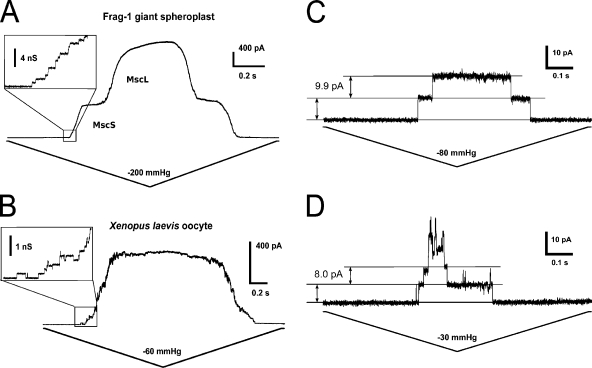
We next compared number and conductance of MscS channels expressed in Xenopus ooctyes to that of endogenous MscS in giant E. coli spheroplasts (Fig. 2 A). In oocytes, the number of channels per patch ranged from the hundreds to only a few (Fig. 2, B and C), depending on the patch size (pipette BN 5–7) and time of incubation after cRNA injection. These expression levels were similar to that of native MscS in wild-type E. coli Frag-1 spheroplasts, although lower than the number of channels previously observed in spheroplasts derived from E. coli–overexpressing MscS (Levina et al., 1999; Akitake et al., 2005).
Figure 2.
MscS conductance in oocytes is comparable to that in E. coli. (A) MscS and MscL channel activities in spheroplasts, recorded in 200 mM KCl plus 90 mM MgCl2. (B) MscS channel activity in oocytes, recorded in 96 mM NaCl plus 2 mM KCl (ND96). (C) Opening and closing of single MscS channels in response to stretch in an excised Xenopus oocyte patch. Pipette, BN 5. (D) Single-channel conductance measured in 98 mM TEA-Cl. Pipette, BN 7. Pipette potential was +40 mV in A and B, +30 mV in C and D.
We consistently observed the expected tension-sensitive channels of ∼1.2-nS conductance when wild-type E. coli spheroplasts were patched under the standard conditions for MscS (n = 6 patches). The buffer used for oocyte readings had fourfold lower ionic strength than the buffer used for spheroplasts; taking into account MscS’s preference for anions over cations, we expected a nearly fourfold decrease in current from MscS channels expressed in oocytes, or a single-channel conductance of ∼300 pS. Our experimental values are in good agreement with this prediction, as MscS in oocytes exhibited a single-channel conductance of ∼330 pS at +30 mV (Fig. 2 C).
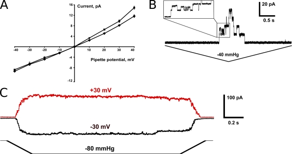
Although MscS is largely voltage independent, slight inward rectification has been previously reported in both spheroplasts and in liposomes (Li et al., 2002; Sukharev, 2002; Sotomayor et al., 2007; Edwards et al., 2008), and has been attributed to the production of conducting substates (see below). We therefore established the current–voltage relationship for MscS in oocytes under symmetric salt, as shown in Fig. 3 A. At positive pipette potentials, the single-channel conductance for MscS derived from the slope of the current–voltage curve was 351 ± 3 pS, whereas at negative pipette potentials, the slope of the current–voltage curve was 218 ± 2 pS. The ratio of the conductance of MscS in oocytes at positive to negative pipette potentials is 1.6, close to the value of 2.0 obtained with purified MscS reconstituted into liposomes (Sukharev, 2002). Thus, the data shown in Figs. 2, B and C, and 3 A demonstrate that the conductance of MscS in oocytes is similar to that of MscS in giant spheroplasts and in liposomes.
Figure 3.
MscS exhibits low voltage dependence when expressed in oocytes. (A) The current–voltage relationship for MscS in symmetric ND96 (squares; n = 7 patches) and in TEA-Cl (diamonds; n = 4 patches). Error bars indicate standard deviation. (B) Example of conductive substates as recorded at +40-mV pipette potential. (C) Nearly symmetric activation curves recorded at opposite potentials in the same excised patch.
We also tested the effect of replacing the KCl in pipette and bath solutions with TEA-Cl, a specific blocker of K+ channels (Armstrong, 1971; Stanfield, 1983). The single-channel conductance of MscS was almost unchanged in Na+-, K+-free symmetric buffer (98 mM TEA-Cl, 1.8 mM CaCl2, 1 mM MgCl2, and 5 mM HEPES, pH 7.4) at negative pipette potentials (233 ± 3 pS), whereas at positive potentials, it decreased slightly to 280 ± 4 pS (Figs. 2 D and 3 A). These results imply that the TEA+ ion, with a predicted diameter of ∼8 Å, similar to that of a hydrated potassium ion (Bezanilla and Armstrong, 1972), can pass relatively efficiently through the MscS channel pore. This interpretation is consistent with molecular dynamics simulations and a recent crystal structure of MscSA106V that indicate a pore diameter of at least 10–15 Å in the open state (Anishkin et al., 2008; Vásquez et al., 2008; Wang et al., 2008).
A characteristic of MscS activity in both spheroplasts and liposomes is the appearance of conducting substates at high potentials, although the relationship of these substates to the normal gating cycle of MscS is unclear (Li et al., 2002; Sukharev, 2002; Shapovalov and Lester, 2004; Akitake et al., 2005; Sotomayor et al., 2007; Edwards et al., 2008). MscS expressed in Xenopus oocytes also exhibited conducting substates at high potentials (Fig. 3 B); in fact, their appearance prevented us from plotting a current–voltage relationship at pipette voltages over +40 mV and below −40 mV. We occasionally observed “flickering” at negative pipette potentials, which may be a result of an increased rate of switching between subconducting states (Akitake et al., 2005). Importantly, the number of channels activated in excised patches under negative and positive potentials was the same (Fig. 3 C), as reported previously for MscS expressed in E. coli spheroplasts (Akitake et al., 2005).
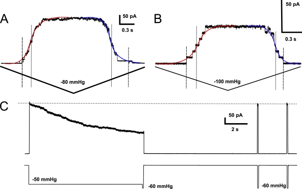
To compare the activation dynamics of MscS expressed in oocytes with that in giant spheroplasts, we calculated the midpoint gating pressure (Fig. 4 A). The previously reported midpoint pressure for MscS was 120–150 mmHg when measured with a pipette of BN 4 (Akitake et al., 2005; Belyy et al., 2010b). These pipettes should have inner tip diameters of <0.5 µm, whereas the pipettes of BN 7 used in our oocyte patches should have diameters of ∼2 µm (Schnorf et al., 1994). According to the law of Laplace (γ = Pr/2), and assuming that the curvature radius of the membrane patch changes proportionally to the diameter of the pipette, we expected to see approximately fourfold less pressure (P) required to produce the same linear tension (γ) in oocytes than in spheroplasts, or 30–50 mmHg. Opening and closing curves fitted with a Boltzmann distribution function (Fig. 4 A, red and blue, respectively) indicated an average activation midpoint pressure of −27.0 ± 5.5 mmHg and inactivation midpoint pressure of −32.6 ± 6.4 mmHg (n = 7 patches) when measured with a pipette of BN 7 (Fig. 4 A). This result is therefore consistent with the previously reported midpoint pressure for MscS. The average activation threshold pressure (defined as the pressure at which the first stable channel opening is detected) in these experiments was −19.1 ± 5.1 mmHg, and the average inactivation threshold pressure (closure of the last channel) was −15.8 ± 5.3 mmHg. The average activation/inactivation midpoint ratio was 0.89 ± 0.14, whereas the average activation/inactivation threshold ratio was 1.48 ± 0.50 (n = 7 patches). This mild hysteresis, wherein MscS channels required lower tension to open than to close, and opened at a faster rate, is similar to that reported previously (Sukharev et al., 2007; Anishkin et al., 2010; Belyy et al., 2010a).
Figure 4.
Activation and inactivation behavior of MscS expressed in oocytes. (A) Representative trace from a short triangle stimulation ramp; pipette, BN 7. (B) Hysteresis of MscS is more prominent in small patches (pipette, BN 5). In A and B, slope fits for channel opening are shown in red and for channel closing in blue; dashed and dotted lines indicate threshold and midpoint tensions, respectively. Pipette potential was +40 mV. (C) Slow inactivation at subsaturating pressure with subsequent recovery during saturating tension pulses.
We observed more prominent hysteresis in about half of recordings made with smaller (BN 5) pipettes. Contrary to the results reported above with large patches, in these recordings MscS channels required a lower tension to open than to close, and opened at a slower rate. In the example shown in Fig. 4 B, the threshold and midpoint pressures for channel opening were −39.7 and −50.1 mmHg, respectively, whereas for channel closing they were −27.4 and −34.8 mmHg. This phenomenon could be caused by differences in membrane relaxation mechanisms or in the contributions made by cooperative gating in patches of different sizes.
Another hallmark of MscS activity is the loss of channel response after sustained channel stimulation or inactivation (Levina et al., 1999; Akitake et al., 2005; Grajkowski et al., 2005). In oocytes, MscS inactivation was not detected during short periods of stimulation with 1–2-s pressure ramps (Fig. 4 A). However, at longer periods of constant tension and especially when smaller pipettes were used, slow and reversible inactivation (with current decaying over a 10-s interval) was observed (Fig. 4 C). This behavior appears to be reversible and much slower than that reported for MscS expressed in spheroplasts, where the current decayed in <1 s (Akitake et al., 2007). This difference in the rate of inactivation of MscS expressed in ooctyes versus spheroplasts might be explained by the larger pipette diameters used for oocyte patches, as patch geometry has been shown to affect MscS adaptation in excised patches (Belyy et al., 2010b). Alternatively, the different lipid composition of Xenopus and E. coli membranes might lead to variable bilayer relaxation behaviors.
DISCUSSION
A common concern regarding the use of Xenopus oocytes for the heterologous expression of ion channels is the presence of endogenous channels, which may complicate single-channel studies by providing unwanted background signal (Sobczak et al., 2010; Terhag et al., 2010). An important consideration for our studies was potential interference by endogenous mechanosensitive channels, which have been reported in both excised and cell-attached patches of Xenopus ootyes (Methfessel et al., 1986; Taglietti and Toselli, 1988; Yang and Sachs, 1990; Lane et al., 1991). However, as shown in Fig. 1 B, although endogenous mechanosensitive channels are frequently present in cell-attached patches, they are not active in excised patches under our conditions. Although we cannot completely rule out a minor effect of endogenous channels on our recordings, their contribution to the final conductance measured under tension appears negligible; in traces at relatively high tensions, with all MscS single-channel events resolved, no other channels or current drift was observed (see Figs. 2 C and 4 C). Other drawbacks to oocyte expression include those inherent to the system, such as variability in oocyte quality and expression capacity that depend on the genetic background of the frog, its health, and even the season (Stuhmer and Parekh, 1995; Terhag et al., 2010). Although we show here that the general features of MscS channel activity are unchanged in oocytes (Figs. 2–4), it remains possible that eukaryotic posttranslational modifications or the lipid environment of the oocyte membrane could more subtly alter its properties.
A Xenopus oocyte system for the analysis of heterologously expressed ion channels presents several advantages for the study of MscS. The large size of an oocyte (∼1 mm in diameter, compared with ∼6 µm in diameter for giant E. coli spheroplasts) allows for the use of much larger patch pipettes, which facilitates the detection of rare single-channel events. The chance of detecting poorly expressed or unstable channels can also be improved by simply increasing the incubation time after injection, as oocytes are stable in vitro for up to 2 wk (Stuhmer and Parekh, 1995). The ease with which oocytes can be injected and the high efficiency of cRNA expression may facilitate the study of heteromeric channels formed between mutant versions of MscS (or between MscS and its five E. coli homologues; Booth et al., 2007) when coinjected, and will allow for the incorporation of unnatural amino acids (Nowak et al., 1998; Torrice et al., 2009). It may also be possible to make use of high-throughput technologies to quickly analyze MscS variants or the effect of drugs on MscS function (Dunlop et al., 2008; Papke and Smith-Maxwell, 2009).
A particularly useful application of MscS expression in oocytes may be in the measurement of membrane tension, a parameter of interest to those who study the biophysics of mechanosensitive channel gating (for example, see Phillips et al., 2009). When MscS is analyzed in spheroplast patches, the threshold tension required to open MscS is often reported relative to that required to open MscL (Blount et al., 1996; Li et al., 2002; Edwards et al., 2005, 2008; Nomura et al., 2006). A more direct approach was used with MscS-reconstituted liposomes, wherein the tension applied to the membrane was calculated from visual observations of membrane curvature under stretch in the pipette (Sukharev, 2002). More recently, spheroplast radius was measured and compared with channel open probability in whole cell mode (Belyy et al., 2010b). However, these direct approaches to measuring membrane tension are limited by the requirement for MscS protein expression and purification, or by the relatively small size of E. coli spheroplasts. In comparison, we expect that large numbers of MscS mutants may be relatively easily assayed and membrane curvature accurately measured in the large patches typically possible with Xenopus oocytes (for example, see Hamill and McBride, 1992).
Expressing MscS in Xenopus oocytes may also be useful for direct comparison to MscS homologues from eukaryotes (Pivetti et al., 2003; Haswell, 2007; Balleza and Gómez-Lagunas, 2009). Although an MscS homologue from the unicellular alga Chlamydomonas was expressed and characterized in E. coli spheroplasts (Nakayama et al., 2007), attempts to do so with MscS-like proteins from higher plants have been unsuccessful (unpublished data). If MscS-like proteins are functional in oocytes (as are many plant channels and transporters), their characteristics could be directly compared with those of MscS. Furthermore, comparisons between MscS and evolutionarily unrelated mechanosensitive channels from animals are also now possible, and MscS should serve as a valuable internal standard for the mechanosensitivity of eukaryotic channels coexpressed in the same patch. Although we did not include MscL in our studies, our results suggest that other prokaryotic mechanosensitive channels may also be functional in oocytes. In summary, we anticipate that Xenopus oocytes provide a system for the expression and electrophysiological study of the prokaryotic mechanosensitive channel MscS that will complement the approaches currently in use.
Acknowledgments
The authors thank Daniel Schachtman for his expert advice and guidance in setting up a Xenopus system in our laboratory and for providing the pOO2 plasmid, and Dr. Ellen Martin-Tryon for generating pOO2-MscS-GFP.
This work was supported by an American Recovery and Reinvestment Act supplement to National Institutes of Health (R01GM084211-01).
Kenton J. Swartz served as editor.
Footnotes
Abbreviations used in this paper:
- BN
- bubble number
- GFP
- green fluorescent protein
- MscL
- mechanosensitive channel of large conductance
- MscS
- mechanosensitive channel of small conductance
References
- Akitake B., Anishkin A., Sukharev S. 2005. The “dashpot” mechanism of stretch-dependent gating in MscS. J. Gen. Physiol. 125:143–154 10.1085/jgp.200409198 [DOI] [PMC free article] [PubMed] [Google Scholar]
- Akitake B., Anishkin A., Liu N., Sukharev S. 2007. Straightening and sequential buckling of the pore-lining helices define the gating cycle of MscS. Nat. Struct. Mol. Biol. 14:1141–1149 10.1038/nsmb1341 [DOI] [PubMed] [Google Scholar]
- Anishkin A., Kamaraju K., Sukharev S. 2008. Mechanosensitive channel MscS in the open state: modeling of the transition, explicit simulations, and experimental measurements of conductance. J. Gen. Physiol. 132:67–83 10.1085/jgp.200810000 [DOI] [PMC free article] [PubMed] [Google Scholar]
- Anishkin A., Akitake B., Kamaraju K., Chiang C.S., Sukharev S. 2010. Hydration properties of mechanosensitive channel pores define the energetics of gating. J. Phys. Condens. Matter. 22:454120 10.1088/0953-8984/22/45/454120 [DOI] [PubMed] [Google Scholar]
- Armstrong C.M. 1971. Interaction of tetraethylammonium ion derivatives with the potassium channels of giant axons. J. Gen. Physiol. 58:413–437 10.1085/jgp.58.4.413 [DOI] [PMC free article] [PubMed] [Google Scholar]
- Arnadóttir J., Chalfie M. 2010. Eukaryotic mechanosensitive channels. Annu Rev Biophys. 39:111–137 10.1146/annurev.biophys.37.032807.125836 [DOI] [PubMed] [Google Scholar]
- Balleza D., Gómez-Lagunas F. 2009. Conserved motifs in mechanosensitive channels MscL and MscS. Eur. Biophys. J. 38:1013–1027 10.1007/s00249-009-0460-y [DOI] [PubMed] [Google Scholar]
- Bass R.B., Strop P., Barclay M., Rees D.C. 2002. Crystal structure of Escherichia coli MscS, a voltage-modulated and mechanosensitive channel. Science. 298:1582–1587 10.1126/science.1077945 [DOI] [PubMed] [Google Scholar]
- Belyy V., Anishkin A., Kamaraju K., Liu N., Sukharev S. 2010a. The tension-transmitting ‘clutch’ in the mechanosensitive channel MscS. Nat. Struct. Mol. Biol. 17:451–458 10.1038/nsmb.1775 [DOI] [PubMed] [Google Scholar]
- Belyy V., Kamaraju K., Akitake B., Anishkin A., Sukharev S. 2010b. Adaptive behavior of bacterial mechanosensitive channels is coupled to membrane mechanics. J. Gen. Physiol. 135:641–652 10.1085/jgp.200910371 [DOI] [PMC free article] [PubMed] [Google Scholar]
- Bezanilla F., Armstrong C.M. 1972. Negative conductance caused by entry of sodium and cesium ions into the potassium channels of squid axons. J. Gen. Physiol. 60:588–608 10.1085/jgp.60.5.588 [DOI] [PMC free article] [PubMed] [Google Scholar]
- Blount P., Sukharev S.I., Schroeder M.J., Nagle S.K., Kung C. 1996. Single residue substitutions that change the gating properties of a mechanosensitive channel in Escherichia coli. Proc. Natl. Acad. Sci. USA. 93:11652–11657 10.1073/pnas.93.21.11652 [DOI] [PMC free article] [PubMed] [Google Scholar]
- Bocquet N., Prado de Carvalho L., Cartaud J., Neyton J., Le Poupon C., Taly A., Grutter T., Changeux J.P., Corringer P.J. 2007. A prokaryotic proton-gated ion channel from the nicotinic acetylcholine receptor family. Nature. 445:116–119 10.1038/nature05371 [DOI] [PubMed] [Google Scholar]
- Booth I.R., Edwards M., Miller S., Li C., Black S., Bartlett W., Schumann U. 2007. Structure-function relations of MscS. Mechanosensitive Ion Channels, Part A. Hamill O.P., editor Academic Press, New York: 269–292 [Google Scholar]
- Booth I.R., Rasmussen T., Edwards M.D., Black S., Rasmussen A., Bartlett W., Miller S. 2011. Sensing bilayer tension: bacterial mechanosensitive channels and their gating mechanisms. Biochem. Soc. Trans. 39:733–740 [DOI] [PubMed] [Google Scholar]
- Choi S.B., Kim J.U., Joo H., Min C.K. 2010. Identification and characterization of a novel bacterial ATP-sensitive K+ channel. J. Microbiol. 48:325–330 10.1007/s12275-010-9231-9 [DOI] [PubMed] [Google Scholar]
- Dunlop J., Bowlby M., Peri R., Vasilyev D., Arias R. 2008. High-throughput electrophysiology: an emerging paradigm for ion-channel screening and physiology. Nat. Rev. Drug Discov. 7:358–368 10.1038/nrd2552 [DOI] [PubMed] [Google Scholar]
- Edwards M.D., Li Y., Kim S., Miller S., Bartlett W., Black S., Dennison S., Iscla I., Blount P., Bowie J.U., Booth I.R. 2005. Pivotal role of the glycine-rich TM3 helix in gating the MscS mechanosensitive channel. Nat. Struct. Mol. Biol. 12:113–119 10.1038/nsmb895 [DOI] [PubMed] [Google Scholar]
- Edwards M.D., Bartlett W., Booth I.R. 2008. Pore mutations of the Escherichia coli MscS channel affect desensitization but not ionic preference. Biophys. J. 94:3003–3013 10.1529/biophysj.107.123448 [DOI] [PMC free article] [PubMed] [Google Scholar]
- Grajkowski W., Kubalski A., Koprowski P. 2005. Surface changes of the mechanosensitive channel MscS upon its activation, inactivation, and closing. Biophys. J. 88:3050–3059 10.1529/biophysj.104.053546 [DOI] [PMC free article] [PubMed] [Google Scholar]
- Gurdon J.B., Lane C.D., Woodland H.R., Marbaix G. 1971. Use of frog eggs and oocytes for the study of messenger RNA and its translation in living cells. Nature. 233:177–182 10.1038/233177a0 [DOI] [PubMed] [Google Scholar]
- Hamill O.P., McBride D.W., Jr 1992. Rapid adaptation of single mechanosensitive channels in Xenopus oocytes. Proc. Natl. Acad. Sci. USA. 89:7462–7466 10.1073/pnas.89.16.7462 [DOI] [PMC free article] [PubMed] [Google Scholar]
- Haswell E.S. 2007. MscS-like proteins in plants. Mechanosensitive Ion Channels, Part A. Hamill O.P., editor Academic Press, New York: 329–353 [Google Scholar]
- Haswell E.S., Meyerowitz E.M. 2006. MscS-like proteins control plastid size and shape in Arabidopsis thaliana. Curr. Biol. 16:1–11 10.1016/j.cub.2005.11.044 [DOI] [PubMed] [Google Scholar]
- Hilf R.J., Bertozzi C., Zimmermann I., Reiter A., Trauner D., Dutzler R. 2010. Structural basis of open channel block in a prokaryotic pentameric ligand-gated ion channel. Nat. Struct. Mol. Biol. 17:1330–1336 10.1038/nsmb.1933 [DOI] [PubMed] [Google Scholar]
- Koprowski P., Grajkowski W., Isacoff E.Y., Kubalski A. 2011. Genetic screen for potassium leaky small mechanosensitive channels (MscS) in Escherichia coli: recognition of cytoplasmic β domain as a new gating element. J. Biol. Chem. 286:877–888 10.1074/jbc.M110.176131 [DOI] [PMC free article] [PubMed] [Google Scholar]
- Kung C., Martinac B., Sukharev S. 2010. Mechanosensitive channels in microbes. Annu. Rev. Microbiol. 64:313–329 10.1146/annurev.micro.112408.134106 [DOI] [PubMed] [Google Scholar]
- Lane J.W., McBride D.W., Jr, Hamill O.P. 1991. Amiloride block of the mechanosensitive cation channel in Xenopus oocytes. J. Physiol. 441:347–366 [DOI] [PMC free article] [PubMed] [Google Scholar]
- Levina N., Tötemeyer S., Stokes N.R., Louis P., Jones M.A., Booth I.R. 1999. Protection of Escherichia coli cells against extreme turgor by activation of MscS and MscL mechanosensitive channels: identification of genes required for MscS activity. EMBO J. 18:1730–1737 10.1093/emboj/18.7.1730 [DOI] [PMC free article] [PubMed] [Google Scholar]
- Li Y., Moe P.C., Chandrasekaran S., Booth I.R., Blount P. 2002. Ionic regulation of MscK, a mechanosensitive channel from Escherichia coli. EMBO J. 21:5323–5330 10.1093/emboj/cdf537 [DOI] [PMC free article] [PubMed] [Google Scholar]
- Loukin S., Zhou X., Su Z., Saimi Y., Kung C. 2010. Wild-type and brachyolmia-causing mutant TRPV4 channels respond directly to stretch force. J. Biol. Chem. 285:27176–27181 10.1074/jbc.M110.143370 [DOI] [PMC free article] [PubMed] [Google Scholar]
- Ludewig U., von Wirén N., Frommer W.B. 2002. Uniport of NH4+ by the root hair plasma membrane ammonium transporter LeAMT1;1. J. Biol. Chem. 277:13548–13555 10.1074/jbc.M200739200 [DOI] [PubMed] [Google Scholar]
- Maroto R., Raso A., Wood T.G., Kurosky A., Martinac B., Hamill O.P. 2005. TRPC1 forms the stretch-activated cation channel in vertebrate cells. Nat. Cell Biol. 7:179–185 10.1038/ncb1218 [DOI] [PubMed] [Google Scholar]
- Martinac B., Buechner M., Delcour A.H., Adler J., Kung C. 1987. Pressure-sensitive ion channel in Escherichia coli. Proc. Natl. Acad. Sci. USA. 84:2297–2301 10.1073/pnas.84.8.2297 [DOI] [PMC free article] [PubMed] [Google Scholar]
- Methfessel C., Witzemann V., Takahashi T., Mishina M., Numa S., Sakmann B. 1986. Patch clamp measurements on Xenopus laevis oocytes: currents through endogenous channels and implanted acetylcholine receptor and sodium channels. Pflugers Arch. 407:577–588 10.1007/BF00582635 [DOI] [PubMed] [Google Scholar]
- Miller A.J., Zhou J.J. 2000. Xenopus oocytes as an expression system for plant transporters. Biochim. Biophys. Acta. 1465:343–358 10.1016/S0005-2736(00)00148-6 [DOI] [PubMed] [Google Scholar]
- Monshausen G.B., Gilroy S. 2009. Feeling green: mechanosensing in plants. Trends Cell Biol. 19:228–235 10.1016/j.tcb.2009.02.005 [DOI] [PubMed] [Google Scholar]
- Nakayama Y., Fujiu K., Sokabe M., Yoshimura K. 2007. Molecular and electrophysiological characterization of a mechanosensitive channel expressed in the chloroplasts of Chlamydomonas. Proc. Natl. Acad. Sci. USA. 104:5883–5888 10.1073/pnas.0609996104 [DOI] [PMC free article] [PubMed] [Google Scholar]
- Nomura T., Sokabe M., Yoshimura K. 2006. Lipid-protein interaction of the MscS mechanosensitive channel examined by scanning mutagenesis. Biophys. J. 91:2874–2881 10.1529/biophysj.106.084541 [DOI] [PMC free article] [PubMed] [Google Scholar]
- Nowak M.W., Gallivan J.P., Silverman S.K., Labarca C.G., Dougherty D.A., Lester H.A. 1998. In vivo incorporation of unnatural amino acids into ion channels in Xenopus oocyte expression system. Methods Enzymol. 293:504–529 10.1016/S0076-6879(98)93031-2 [DOI] [PubMed] [Google Scholar]
- Okada K., Moe P.C., Blount P. 2002. Functional design of bacterial mechanosensitive channels. Comparisons and contrasts illuminated by random mutagenesis. J. Biol. Chem. 277:27682–27688 10.1074/jbc.M202497200 [DOI] [PubMed] [Google Scholar]
- Papke R.L., Smith-Maxwell C. 2009. High throughput electrophysiology with Xenopus oocytes. Comb. Chem. High Throughput Screen. 12:38–50 10.2174/138620709787047975 [DOI] [PMC free article] [PubMed] [Google Scholar]
- Patel A.J., Honoré E., Maingret F., Lesage F., Fink M., Duprat F., Lazdunski M. 1998. A mammalian two pore domain mechano-gated S-like K+ channel. EMBO J. 17:4283–4290 10.1093/emboj/17.15.4283 [DOI] [PMC free article] [PubMed] [Google Scholar]
- Phillips R., Ursell T., Wiggins P., Sens P. 2009. Emerging roles for lipids in shaping membrane-protein function. Nature. 459:379–385 10.1038/nature08147 [DOI] [PMC free article] [PubMed] [Google Scholar]
- Pivetti C.D., Yen M.R., Miller S., Busch W., Tseng Y.H., Booth I.R., Saier M.H., Jr 2003. Two families of mechanosensitive channel proteins. Microbiol. Mol. Biol. Rev. 67:66–85 10.1128/MMBR.67.1.66-85.2003 [DOI] [PMC free article] [PubMed] [Google Scholar]
- Ruthe H.J., Adler J. 1985. Fusion of bacterial spheroplasts by electric fields. Biochim. Biophys. Acta. 819:105–113 10.1016/0005-2736(85)90200-7 [DOI] [PubMed] [Google Scholar]
- Schnorf M., Potrykus I., Neuhaus G. 1994. Microinjection technique: routine system for characterization of microcapillaries by bubble pressure measurement. Exp. Cell Res. 210:260–267 10.1006/excr.1994.1038 [DOI] [PubMed] [Google Scholar]
- Schumann U., Edwards M.D., Li C., Booth I.R. 2004. The conserved carboxy-terminus of the MscS mechanosensitive channel is not essential but increases stability and activity. FEBS Lett. 572:233–237 10.1016/j.febslet.2004.07.045 [DOI] [PubMed] [Google Scholar]
- Shapovalov G., Lester H.A. 2004. Gating transitions in bacterial ion channels measured at 3 µs resolution. J. Gen. Physiol. 124:151–161 10.1085/jgp.200409087 [DOI] [PMC free article] [PubMed] [Google Scholar]
- Sigel E., Minier F. 2005. The Xenopus oocyte: system for the study of functional expression and modulation of proteins. Mol. Nutr. Food Res. 49:228–234 10.1002/mnfr.200400104 [DOI] [PubMed] [Google Scholar]
- Sobczak K., Bangel-Ruland N., Leier G., Weber W.M. 2010. Endogenous transport systems in the Xenopus laevis oocyte plasma membrane. Methods. 51:183–189 10.1016/j.ymeth.2009.12.001 [DOI] [PubMed] [Google Scholar]
- Sotomayor M., Vásquez V., Perozo E., Schulten K. 2007. Ion conduction through MscS as determined by electrophysiology and simulation. Biophys. J. 92:886–902 10.1529/biophysj.106.095232 [DOI] [PMC free article] [PubMed] [Google Scholar]
- Stanfield P.R. 1983. Tetraethylammonium ions and the potassium permeability of excitable cells. Rev. Physiol. Biochem. Pharmacol. 97:1–67 10.1007/BFb0035345 [DOI] [PubMed] [Google Scholar]
- Stuhmer W., Parekh A. 1995. Electrophysiological recordings from Xenopus oocytes. Single-Channel Recording. Sakmann B., Neher E., Springer, New York: 341–356 [Google Scholar]
- Suchyna T.M., Sachs F. 2007. Mechanosensitive channel properties and membrane mechanics in mouse dystrophic myotubes. J. Physiol. 581:369–387 10.1113/jphysiol.2006.125021 [DOI] [PMC free article] [PubMed] [Google Scholar]
- Suchyna T.M., Markin V.S., Sachs F. 2009. Biophysics and structure of the patch and the gigaseal. Biophys. J. 97:738–747 10.1016/j.bpj.2009.05.018 [DOI] [PMC free article] [PubMed] [Google Scholar]
- Sukharev S. 2002. Purification of the small mechanosensitive channel of Escherichia coli (MscS): the subunit structure, conduction, and gating characteristics in liposomes. Biophys. J. 83:290–298 10.1016/S0006-3495(02)75169-2 [DOI] [PMC free article] [PubMed] [Google Scholar]
- Sukharev S., Akitake B., Anishkin A. 2007. The bacterial mechanosensitive channel MscS: emerging principles of gating and modulation. Mechanosensitive Ion Channels. Hamill O., editor Academic Press, New York: 236–263 [Google Scholar]
- Taglietti V., Toselli M. 1988. A study of stretch-activated channels in the membrane of frog oocytes: interactions with Ca2+ ions. J. Physiol. 407:311–328 [DOI] [PMC free article] [PubMed] [Google Scholar]
- Terhag J., Cavara N.A., Hollmann M. 2010. Cave Canalem: how endogenous ion channels may interfere with heterologous expression in Xenopus oocytes. Methods. 51:66–74 10.1016/j.ymeth.2010.01.034 [DOI] [PubMed] [Google Scholar]
- Torrice M.M., Bower K.S., Lester H.A., Dougherty D.A. 2009. Probing the role of the cation-pi interaction in the binding sites of GPCRs using unnatural amino acids. Proc. Natl. Acad. Sci. USA. 106:11919–11924 10.1073/pnas.0903260106 [DOI] [PMC free article] [PubMed] [Google Scholar]
- Vásquez V., Cortes D.M., Furukawa H., Perozo E. 2007. An optimized purification and reconstitution method for the MscS channel: strategies for spectroscopical analysis. Biochemistry. 46:6766–6773 10.1021/bi700322k [DOI] [PubMed] [Google Scholar]
- Vásquez V., Sotomayor M., Cordero-Morales J., Schulten K., Perozo E. 2008. A structural mechanism for MscS gating in lipid bilayers. Science. 321:1210–1214 10.1126/science.1159674 [DOI] [PMC free article] [PubMed] [Google Scholar]
- Wang W., Black S.S., Edwards M.D., Miller S., Morrison E.L., Bartlett W., Dong C., Naismith J.H., Booth I.R. 2008. The structure of an open form of an E. coli mechanosensitive channel at 3.45 A resolution. Science. 321:1179–1183 10.1126/science.1159262 [DOI] [PMC free article] [PubMed] [Google Scholar]
- Weng Y., Yang L., Corringer P.J., Sonner J.M. 2010. Anesthetic sensitivity of the Gloeobacter violaceus proton-gated ion channel. Anesth. Analg. 110:59–63 10.1213/ANE.0b013e3181c4bc69 [DOI] [PMC free article] [PubMed] [Google Scholar]
- Yang X.C., Sachs F. 1990. Characterization of stretch-activated ion channels in Xenopus oocytes. J. Physiol. 431:103–122 [DOI] [PMC free article] [PubMed] [Google Scholar]


