INTRODUCTION
Brassinosteroids (BRs) comprise a class of over 40 polyhydroxylated sterol derivatives that appear to be ubiquitously distributed throughout the plant kingdom. Among plant hormones, BRs are structurally the most similar to animal steroid hormones, which have well-known functions in regulating embryonic and post-embryonic development and adult homeostasis. Like their animal counterparts, BRs regulate the expression of numerous genes, impact the activity of complex metabolic pathways, contribute to the regulation of cell division and differentiation, and help control overall developmental programs leading to morphogenesis. They are also involved in regulating processes more specific to plant growth including photomorphogenesis and skotomorphogenesis, and cell expansion in the presence of a potentially growth-limiting cell wall.
Numerous books (Cutler et al., 1991; Khripach et al., 1999; Sakurai et al., 1999) and recent reviews covering all currently known aspects of BR biology and chemistry are available, including biosynthesis (Fujioka and Sakurai, 1997; Sakurai, 1999; Yokota, 1997), physiological effects (Altmann, 1999; Clouse, 1997; Clouse and Sasse, 1998; Sasse, 1999; Sasse, 1997), molecular genetics (Clouse and Feldmann, 1999), signal transduction (Schumacher and Chory, 2000), natural occurrence (Fujioka, 1999) and research history (Steffens, 1991; Yokota, 1999).
Short History of BR Research
The discovery of plant growth-promoting compounds that were subsequently shown to be steroids, was accomplished independently by research at the United States Department of Agriculture (USDA) and at Nagoya University in Japan (Yokota, 1999). In an effort to isolate new plant hormones, Mitchell and his USDA colleagues examined organic extracts of pollen from numerous species over a thirty-year period. The most active growth-promoting extracts were isolated from Brassica napus pollen, and were hence named “brassins”. Brassins had a pronounced effect on cell elongation and division in the bean second-internode bioassay, and were found to increase yields when sprayed on young seedlings of radishes, leafy vegetables and potatoes. Based on this preliminary data, Mitchell et al. (Mitchell et al., 1970) somewhat prematurely, but perhaps prophetically, attributed hormonal status to the brassins “because they are specific translocatable organic compounds isolated from a plant and have induced measurable growth control when applied in minute amounts to another plant.” However, they incorrectly predicted that the active component of brassins was a fatty acid ester.
The true chemical nature of the brassin active component was discovered after a major coordinated effort involving several USDA laboratories, a truckload of 227 kg of bee-collected B. napus pollen, pilot-plant solvent extractions, and extensive column chromatography (Steffens, 1991). The net result was 4 mg of a pure substance that was identified by single crystal X-ray analysis to be a steroidal lactone, which was named brassinolide (Grove et al., 1979). Within two years brasssinolide (BL) and its stereo isomer, 24-epiBL, had been chemically synthesized, eliminating the need for such massive plant extraction procedures. With ample synthetic compound in hand, research in the 1980's focused on determination of BR physiological effects in a wide variety of biological systems and on testing greenhouse and field applications for enhanced crop yield (Cutler et al., 1991).
The first report of the effects of BR on Arabidopsis growth appeared in 1991 (Clouse and Zurek, 1991) followed shortly thereafter by a description of a screen for BR-insensitive mutants in Arabidopsis and demonstration that BRs regulated gene expression in that species (Clouse et al., 1993). The early 1990's also saw significant progress by several Japanese groups in unraveling the biosynthetic pathway to BRs from common membrane sterols (Fujioka and Sakurai, 1997). While most chemists and biologists involved in BR research believed these compounds were indeed a new class of plant hormone (Sasse, 1991), unequivocal proof of their indispensable role in plant growth and development was not available until 1996, when a series of four independent reports described the identification and properties of one BR-insensitive (Clouse et al., 1996; Kauschmann et al., 1996) and three BR-deficient mutants in Arabidopsis (Kauschmann et al., 1996; Li et al., 1996; Szekeres et al., 1996). The mutants exhibited an extreme dwarf phenotype, which could be rescued to wildtype by BR treatment of the deficient mutants. Moreover, it was demonstrated that two of the deficient mutants resulted from lesions in genes encoding steroid biosynthetic enzymes. Thus, convincing genetic and biochemical evidence was provided that BRs were essential for normal plant growth and development. BRs were then accepted by most scientists as a new class of plant hormone and the number of researchers studying their activity began to increase.
Chemical Structure
BL is a polyhydroxylated derivative of 5α-cholestan, namely (22R,23R,24S)-2α,3α,22,23-tetrahydroxy-24-methyl-B-homo-7-oxa-5α-cholestan-6-one (Figure 1). Thus, plants possess a growth-promoting steroid with structural similarity to cholesterol-derived animal steroid hormones such as androgens, estrogens and corticosteroids from vertebrates, and ecdysteroids from insects and crustacea. The BR family consists of BL and about 40 other free BRs plus 4 conjugates (Fujioka, 1999). These differ from BL by variations at C-2 and C-3 in the A ring; the presence of a lactone, ketone, or de-oxo function at C-6 in the B ring; the stereochemistry of the hydroxyl groups in the side chain, and the presence or absence of a methyl (methylene) or ethyl (ethylene) group at C-24. The conjugates are glycosylated, meristylated and laurylated derivatives of the hydroxyls in ring A or in the side chain. Many of the known BRs are biosynthetic precursors or metabolic products of BL, although castasterone, the immediate precursor of BL, is believed to have independent biological activity in some plants. The optimal structure for highest BR activity normally is that found in BL, consisting of a lactone function at C-6/C-7, cis-vicinal hydroxyls at C-2 and C-3, R configuration of the hydroxyls at C-22/C-23 and a methyl substitution at C-24 (Mandava, 1988).
Figure 1.

Structure of naturally occurring brassinosteroids. The structure of brassinolide is presented with possible variations in the A and B rings and the side chain shown in boxes. Compounds to the right have been identified in plants and numbers represent sturcture of A-Ring: B-Ring: Side Chain. The C and D rings remain the same for all compounds.
Natural Occurrence and Distribution in the Plant Kingdom
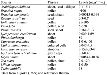
BRs have been found in all cases in which a rigorous examination of a plant species by GC-MS or LC has been undertaken; including at least 37 angiosperms (28 dicots and 9 monocots), 5 gymnosperms, 1 alga and 1 pteridophyte (Fujioka, 1999). Endogenous levels of BRs vary across plant organ type, tissue age and species, with pollen and immature seeds containing the highest levels. Young, growing shoots contain higher BR levels than mature tissue, which is not surprising in view of the greater physiological response of immature tissue to BRs. Table 1 summarizes endogenous BR levels detected in a range of species and tissue types.
Table 1.
Distribution and Endogenous Levels of BRs in Selected Plantsa
Overview of Physiological Effects
Early studies of BR activity in plants depended on exogenous BR application followed by recording the observable response. From these experiments it became clear that BRs had a dramatic positive effect on stem elongation, including promotion of epicotyl, hypocotyl and peduncle elongation in dicots, and enhanced growth of coleoptiles and mesocotyls of dicots (Mandava, 1988). Exogenous BRs also stimulated tracheary element differentiation in Helianthus tuberosus and Zinnia elegans, the two primary model systems for xylogenesis (Clouse and Zurek, 1991; Fukuda, 1997). In many test systems BRs increased rates of cell division, particularly under conditions of limiting auxin and cytokinin. BRs were also shown to accelerate senescence, cause hyperpolarization of membranes, stimulate ATPase activity, and alter the orientation of cortical microtubules. Besides direct effects on growth regulation, BRs also were shown to mediate abiotic and biotic stresses, including salt and drought stress, temperature extremes and pathogen attack (Clouse and Sasse, 1998).
Many of the observed physiological responses elicited by exogenous application of BRs were later shown to occur in planta by examination of the phenotypes of BR-deficient and -insensitive mutants in Arabdiopsis, tomato, and pea. Arabidopsis BR mutants show a characteristic phenotype in the light (Fig. 2), including dwarf stature, dark green, rounded leaves, prolonged life-span, reduced fertility, and altered vascular development. In the dark, they exhibit some of the features of light-grown plants including shortened hypocotyl and open cotyledons. In BR-deficient mutants, all of these phenotypic alterations are rescued to wildtype by exogenous application of BL (Altmann, 1999; Clouse and Feldmann, 1999).
Figure 2.
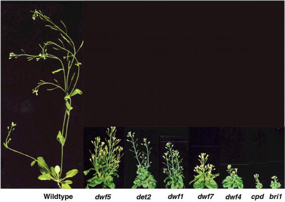
Phenotype of BR dwarf mutants. Comparison of several 5-week-old BR-deficient or – insensitive (bri1) mutants of Arabidopsis with a wild-type Arabidopsis plant of the same age. See the text for a description of each mutant. Adapted from a photo courtesy of S. Choe and K. Feldmann.
Biosynthesis
A clear understanding of how endogenous BR levels are regulated via synthesis and metabolism is a required component of any molecular model of BR action. The basic features of BL biosynthesis were uncovered utilizing cell suspension cultures of Catharanthus roseus, which were fed deuterated and tritiated putative intermediates in BL biosynthesis followed by analysis with sensitive techniques of gas chromatography-mass spectrometry to monitor conversion of the labeled compounds (Fujioka and Sakurai, 1997; Sakurai, 1999). Based on side chain structure and stereochemistry, wide distribution in the plant kingdom, and the relative biological activities in bioassays, it was predicted that the plant sterol campesterol would be converted to BL via teasterone, typhasterol and castasterone (Yokota et al., 1991). This skeletal pathway, in addition to several intermediate steps, was confirmed in C. roseus cells and seedlings. Moreover, it was found that C-6 oxidation could occur before (Early C-6 oxidation pathway) or after (Late C-6 oxidation pathway) hydroxylation of the side chain.
While campesterol has been shown to be the BL progenitor, other BRs are likely to also be derived from common plant sterols with appropriate side chain structure such as sitosterol, isofucosterol, 24-methylenecholesterol, and 24-epicampesterol (Yokota, 1997). Plant sterols are synthesized by the isoprenoid biosynthetic pathway via acetyl-CoA, mevalonate, isopentenyl pyrophosphate, geranyl pyrophosphate and farnesyl pyrophosphate (Fig. 3). Squalene is produced by condensation of two farnesyl pyrophosphate molecules, which is then converted via squalene-2,3-epoxide to cycloartenol, the parent compound of all plant sterols (Anderson and Beardall, 1991). The conversion of squalene-2,3-epoxide to cycloartenol is unique to plants. In animals and fungi squalene-2,3-epoxide is converted to lanosterol, the precursor of cholesterol and the animal steroid hormones (Benveniste, 1986). As described in detail below, several mutants have been isolated and characterized in detail that are blocked at various steps in the biosynthesis of plant sterols, and in their conversion to BRs. Interestingly, most of the sterol and BR-deficient mutants were isolated in screens for other physiological processes, such as dwarfism, constitutive photomorphogenesis in darkness, or defects in embryogenesis, and were only later shown to be involved in sterol or steroid biosynthesis when the sequence of the cloned genes suggested such a role.
Figure 3.
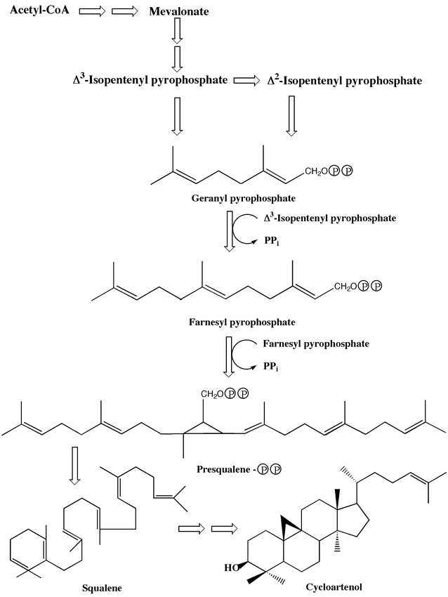
Biosynthetic pathway of the plant sterol precursor, cycloartenol, from mevalonate.
MODULATION OF ENDOGENOUS BR LEVELS IN ARABIDOPSIS
Biosynthesis of Sterol Precursors
The biosynthetic pathway to BL can be divided into general sterol synthesis (cycloartenol to campesterol), and the BR-specific pathway from campesterol to BL. Besides their role as BR precursors, plant sterols such as campesterol are integral membrane components which serve to regulate the fluidity and permeability of membranes and directly affect the activity of membrane associated proteins, including enzymes and signal transduction components (Hartmann, 1998). Moreover, recent evidence from animal studies suggests that in addition to their role as steroid hormone precursors, sterols can themselves serve as ligands for nuclear receptors and interact with other transcription complexes to directly modulate signal transduction pathways (Edwards and Ericsson, 1999). In view of the multiple and critical roles sterols play, it is not surprising that mutations in genes encoding sterol biosynthetic enzymes can have a dramatic affect on eukaryotic development. Mutations in genes encoding five biosynthetic enzymes in the pathway leading to plant sterols have now been identified, in addition to five mutants affecting the specific pathway to BRs. Comparison of the phenotype of these mutants and their response to BR application, is increasing our understanding of the role plant sterols and their hormone derivatives play in controlling both embryonic and post-embryonic plant development.
Arabidopsis mutants with blocks very early in the sterol biosynthetic pathway have some of the characteristics of BR-deficient mutants in adult plants, but show unique defects in embryogenesis not previously reported in BR mutants or in sterol biosynthetic mutants later in the pathway (Fig. 4). The fackel mutant was identified in a systematic screen for mutations affecting body organization in Arabidopsis seedlings (Mayer et al., 1991), while the extra-long-lifespan mutant was uncovered in a genetic screen for constitutive cytokinin response mutants. Subsequent analysis revealed that extra-long-lifespan and fackel are allelic (Jang et al., 2000). Molecular characterization of the fackel mutants has provided convincing evidence that the FACKEL gene encodes a sterol C-14 reductase required for the conversion of 4α-methyl-5α-ergosta-8,14,24(28)-trien-3β-ol to 4α-methylfecosterol (Jang et al., 2000; Schrick et al., 2000). The FACKEL gene shares significant sequence similarity to human, rat, chicken and fungal C-14 sterol reductases, and a FACKEL cDNA rescues the defective sterol C-14 reductase in the yeast mutant erg24. Furthermore, the endogenous levels of various BRs, campesterol and sitosterol were severely reduced in the mutant, while the substrate sterol C-14 reductase accumulated ten-fold. Interestingly, fackel accumulates three unusual 8,14-diene sterols, similar to plant cell suspension cultures treated with the C-14 reductase inhibitor, 15-azasterol. The smt1 mutant of Arabidopsis shares many of the phenotypic properties of fackel and has been shown to disrupt the step leading from cycloartenol to 24-methylene cycloartenol (Diener et al., 2000). The SMT1 gene encodes an enzyme capable of C-24 alkylation of the sterol side chain in the presence of S-adenosylmethionine and recombinant SMT1 catalyzes the conversion of cycloartenol to 24-methylene cycloartenol in vitro. Cholesterol accumulates in the smt1 mutant at the expense of sitosterol, although other sterol levels are relatively normal.
Figure 4.
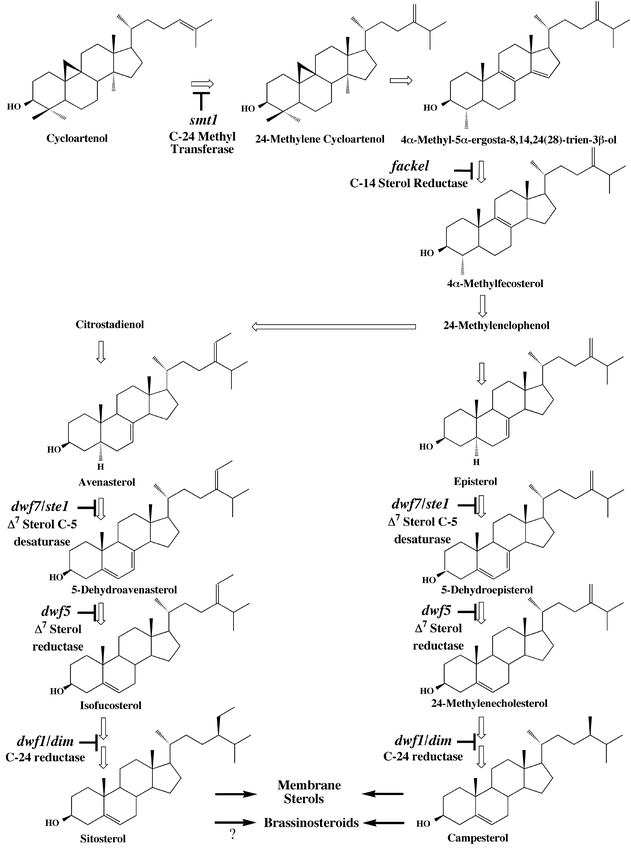
Biosynthetic pathways of sterols from cycloartenol. Steps in which mutants in Arabidopsis are available are indicated.
Embryogenesis is severely disrupted in fackel and smt1 mutants. Initially, a lack of asymmetrical cell division in the globular stage embryo is observed, and while wild-type embryos progress to the heart stage, fackel embryos remain globular and disorganized (Fig. 4). Multiple shoot apical meristems are initiated in the mutant and developing seedlings often have more than two cotyledons. Furthermore, the cotyledons appear to be directly attached to the root, with little development of the hypocotyl (Fig. 4). Determination of mRNA levels by in situ hybridization showed prominent expression of the FACKEL and SMT1 genes during wild-type embryo development (Diener et al., 2000; Schrick et al., 2000). Finally, it should be noted that while adult fackel plants exhibit many of the features of BR-deficient mutants (and are themselves BR-deficient), they are not rescued by BR treatment, even though they retain some sensitivity to BRs (Jang et al., 2000). An intriguing explanation for the lack of BR rescue of these early sterol mutants suggests that sterols themselves may serve as signaling molecules regulating embryogenesis and these sterol signals are disrupted in the fackel and smt1 mutants. Arguments in support of this hypothesis have been summarized in a recent review (Clouse, 2000).
The next step in the sterol biosynthetic pathway for which mutants are available involves the introduction of a C-5 double bond in the B ring of episterol (Fig. 4). A screen of ethane-methylsulfonate (EMS) mutagenized Arabidopsis seedlings by gas chromatography uncovered a mutant, ste1 (Gachotte et al., 1995), that accumulated Δ7 sterols with a decrease in the corresponding Δ5 sterols. Transgenic plants expressing the yeast ERG3 gene, a Δ7-C-5-desaturase, regained the normal sterol profile. The ste1 mutant was apparently a weak or leaky allele, since it showed no visible phenotype. Subsequent cloning and sequencing of the STE1 cDNA and ste1 allele showed that the mutation resulted from a Thr to Ile substitution at amino acid 114 (Husselstein et al., 1999). Null alleles of ste1, dwarf7-1 (dwf7-1) and dwf7-2, were discovered in screens for dwarfs in a population of T-DNA mutagenized and EMS mutagenized Arabidopsis plants respectively. These mutants had many of the phenotypic features of BR-deficient mutants (although not as severe), which were rescued by BR treatment (Choe et al., 1999b). The STE1/DWF7 gene is 38% identical (50% similar) to the yeast ERG3 gene and 35% identical (47% similar) to a human ortholog. All of these Δ7-C-5-desaturases have conserved transmembrane domains and histidine clusters required for activity and both dwf7-1 and dwf7-2 are nonsense mutations resulting in premature stop codons that eliminate some of these essential domains. An apparent DWF7/STE1 homolog with 80% amino acid identity has been cloned in Arabidopsis, suggesting that duplicate genes regulate this step. Such redundancy could account for the less severe phenotype of the dwf7 mutants when compared to mutants in the BR-specific pathway. Measurement of endogenous sterol and BR levels in dwf7-1 by GC-SIM showed severe reduction in 24-methylenecholesterol, campesterol, campestanol and all downstream BRs. These results were confirmed by feeding experiments with 13C-mevalonic acid, which showed that dwf7 accumulated 13C-episterol while 13C-5-dehydroepisterol was not detected. Thus, a very through genetic and biochemical analysis of these mutants has rigorously identified dwf7/ste1 alleles as lesions in the gene encoding the Δ7-C-5-desaturase of Arabidopsis sterol biosynthesis (Choe et al., 1999b).
The next step in the pathway, conversion of 5-dehydroepisterol to 24-methylenecholesterol by a Δ7-sterol reductase (Fig. 4), has also been thoroughly characterized. The dwf5 mutant of Arabidopsis was assigned to this step by similar methods to those described above for dwf7, including sequence analysis of the Arabidopsis homolog of Δ7-sterol reductase in six loss-of-function dwf5 alleles to pinpoint the mutations, measurement of endogenous BR and sterol levels, feeding experiments with labeled intermediates, and rescue of the mutant to wildtype by applying exogenous BRs. Moreover, the dwf5 mutant was also complemented by transformation with the Arabidopsis Δ7-sterol reductase gene. The phenotype of dwf5 is similar to dwf7 and other BR-deficient mutants, but has some unique characteristics. The dwf5-1 allele has increased fertility compared to other BR and sterol-deficient mutants, but had abnormal seeds with reduced germination rates. Furthermore, dwf5-1 did not show the prolonged life cycle typical of BR mutants (Choe et al., 2000).
The completion of the sterol biosynthetic pathway involves the isomerization and reduction of 24-methylenecholesterol to campesterol (and isofucosterol to sitosterol by a parallel reaction). Numerous allelic dwarf mutants have been associated with this conversion. The first mutant, dwarf1 (dwf1) was originally isolated from the first population of T-DNA lines generated by seed infection (Feldmann et al., 1989). An allele of dwf1, dimunito (dim), was independently isolated from another T-DNA screen (Takahashi et al., 1995). However, it was not known that dwf1/dim were BR-deficient mutants until a third allele, cabbage1 (cbb1), was shown to be rescued by BR treatment (Kauschmann et al., 1996). Another eight alleles of dwf1 were identified in various screens and sequence analysis showed that DWF 1 encoded an oxidoreductase with a flavin adenine dinucleotide-binding domain, and a putative membrane anchoring region (Choe et al., 1999a). A majority of the alleles contained lesions in this domain, suggesting a critical role for FAD-binding in DWF1 function. Subsequent work with a green fluorescent protein-DWF1 fusion confirmed that DWF1 is an integral membrane protein, as expected for most sterol and steroid biosynthetic enzymes (Klahre et al., 1998). Two independent groups confirmed that the DWF1 protein is responsible for both steps in the conversion of 24-methylenecholesterol to campesterol, and isofucosterol to sitosterol (Choe et al., 1999a; Klahre et al., 1998).
Biosynthesis of BL from Campesterol
The conversion of the membrane sterol campesterol to BL occurs via a series of reductions, hydroxylations, epimerizations and oxidations that have been extensively studied in several species. The first four reactions lead to campestanol via reduction of the C-5 double bound in campesterol (Fig. 5). One of these steps has been characterized in detail. The dwarf mutant de-etiolated2 (det2) was identified in a screen for plants that grew in the dark as if they were in the light (Chory et al., 1991). Map-based cloning of DET2 revealed significant sequence identity in the predicted protein (38–42%) to mammalian 5α-steroid reductases that catalyze the NADPH-dependent reduction of testosterone to dihydrotestosterone (Li et al., 1996). The mammalian reductase and the DET2 protein share 80% of conserved residues including a glutamate that is required for mammalian enzyme activity and which is mutated to a lysine in the det2-1 and det2-6 alleles. Moreover, recombinant DET2 protein expressed in human embryonic kidney cells reduced several 3-oxo, Δ4,5 mammalian steroids, such as testosterone and progesterone, while the mutant det2 protein failed to do so (Li et al., 1997). The DET2 enzyme activity was inhibited by 4-MA, a competitive inhibitor of mammalian 5α-reductases. Overexpression of human 5α-reductases in transgenic det2 plants resulted in wild-type phenotype without BR application, and the rescue was inhibited by 4-MA.
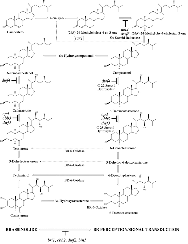
Figure 5.The biosynthetic pathway from campesterol to brassinolide showing known mutants in Arabidopsis that block the conversion of intermediates at the steps indicated, or prevent BR perception and/or signal transduction. The pathway on the left is termed Early C-6 Oxidation and that on the right, Late C-6 Oxidation. BR-6-Oxidase has been shown to convert 6-Deoxocastasterone and 6-Deoxotyphasterol to the corresponding 6-oxo compounds in Arabidopsis, while the conversion of 3-Dehydro-6-deoxoteasterone and 6-Deoxoteasterone has been demonstrated so far in recombinant yeast cells only.
BRs rescue det2 to wildtype in the light and the dark (Li et al., 1996) and endogenous levels of campestanol are reduced about 90% in det2 compared to wildtype (Fujioka et al., 1997). While det2 has the typical BR-mutant phenotype, it is not as severe as cpd and bri1 (see below), and this may be due to the residual campestanol present in det2. This, in turn, could result from a second reductase catalyzing the same reaction, similar to the case of dwf7. A putative DET2 homolog is predicted from Southern blot analysis. Measurements of endogenous BRs and feeding wildtype and det2 seedlings with deuterated substrates showed that DET2 catalyzes the step involving conversion of (24R)-24-methylcholest-4-en-3-one to (24R)-24-methyl-5α-choestan-3-one (Noguchi et al., 1999b).
Another mutant, sax1, may also affect steps in the conversion of campesterol to campestanol. sax1 was identified in a screen for mutants that were hypersensitive to auxin and abscisic acid (Ephritikhine et al., 1999a). While sax1 shares the dwarf phenotype of BR-deficient mutants and is partially rescued by BR treatment, it does not exhibit the typical de-etiolated phenotype in the dark. sax1 appears to block BL synthesis in a step preceeding det2, and feeding experiments with synthetic intermediates (Ephritikhine et al., 1999b) suggest that SAX1 acts in a putative side pathway involving conversion of campersterol to 6-deoxocathasterone via 22-hydroxylated intermediates (Fig. 5). However, this pathway has not been demonstrated to occur in planta and the SAX1 gene has not been cloned. Thus the role of SAX1 in BR biosynthesis is not as clear as in the BR-deficient mutants that have been more fully characterized.
From campestanol, the BL biosynthetic pathway diverges into the Early and Late C-6 oxidation branches. BRs lacking a ketone or lactone in the B ring occur widely, although they have low biological activity in bioassays (Yokota, 1997). The endogenous occurrence of 6-deoxocastasterone, 6-deoxotyphasterol, 3-dehyrdo-6-deoxoteasterone and 6-deoxoteasterone has been demonstrated in numerous plants where castasterone and BL are also present including bean, wheat, rice, rye, Arizona cypress, C. roseus and Arabidopsis (Choi et al., 1997; Fujioka et al., 1996; Fujioka and Sakurai, 1997; Griffiths et al., 1995). The co-occurrence of 6-deoxo and 6-oxo forms of BL precursors suggests that a late C-6 oxidation pathway, in which ketone formation at C-6 follows A ring hydroxyl and side chain modification, operates simultaneously with early C-6 oxidation in a wide array of plants. Feeding a range of deuterium-labeled substrates to seedlings followed by GC-MS has clearly demonstrated that both of these pathways are fully functional in Arabidopsis (Noguchi et al., 2000).
The conversion of campestanol to 6-deoxocathasterone (Late C-6 oxidation) and of 6-oxocampestanol to cathasterone (Early C-6 oxidation) are both accomplished by the product of the DWF4 gene, which encodes a cytochrome P450 with sequence similarity to mammalian steroid hydroxylases (Choe et al., 1998). The phenotype of dwf4 resembles that of other BR-deficient mutants and feeding experiments showed that only 22α-hydroxylated intermediates in BL biosynthesis, and synthetic compounds such as 22-hydroxycampesterol, could rescue dwf4 to wildtype suggesting that DWF4 functions as a C-22 steroid hydroxylase. The next step in both branches of the pathway also involves side chain hydroxylation and is catalyzed by the product of the CPD gene, which encodes a cytochrome P450 with 43% identity (66% similarity) to DWF4. The cpd mutant is an extreme dwarf which is rescued only by 23α-hydroxylated BRs, indicating that CPD acts as a C-23 steroid hydroxylase (Szekeres et al., 1996).
Mutants blocking steps after CPD in the Arabidopsis BL biosynthetic pathway have not been clearly identified, although it is possible that dwf8 affects the hydroxylation of typhasterol to castasterone (Clouse and Feldmann, 1999). However, in tomato, the DWARF gene encodes a cytochrome P450 monooxygenase that has been shown to catalyze the oxidation of 6-deoxocastasterone to castasterone via 6α-hydroxycastasterone (Bishop et al., 1999). Using the tomato DWARF sequence for database searches, several ESTs corresponding to putative homologs of the tomato gene were identified and RT-PCR was employed to obtain a full-length cDNA, which was named AtBR6ox. The 466 amino acid protein shared 68% identity (81% similarity) to the tomato DWARF cytochrome P450, and was indeed more closely related to the tomato gene than other Arabidopsis P450s in the pathway such as DWF4 and CPD. AtBR6ox was expressed in yeast, and by feeding deuterated substrates followed by GC-MS, it was shown that recombinant AtBR6ox is a steroid-6-oxidase that can convert not only 6-deoxocastasterone to castasterone, but also 6-deoxotyphasterol to typhasterol; 3-dehydro-6-deoxoteasterone to 3-dehydroteasterone; and 6-deoxoteasterone to teasterone (Shimada et al., 2001). The conversion of 6-deoxocastesterone to castasterone and 6-deoxotyphasterol to typasterol has been confirmed in Arabidopsis seedlings (Noguchi et al., 2000). Thus, BL biosynthesis in Arabidopsis appears to be more of a metabolic grid than two independent linear pathways of Early and Late C-6 oxidation. Double mutant studies showing additive phenotypes between mutants at different steps of the pathway supports the grid hypothesis vs. the independent linear pathways (Clouse and Feldmann, 1999).
Regulation of BL Biosynthesis
Modulation of the activity and/or levels of biosynthetic enzymes in the BL pathway is an obvious way in which BR concentrations could be increased or decreased in the plant cell. Such regulation could occur at the level of transcription, mRNA stability, translation, enzyme catalytic activity and accessibility to substrates. Possible regulatory factors affecting these steps might include environmental signals and developmental cues such as light and other hormones. Homeostasis of BL could be achieved by feedback inhibition by the pathway end product. Such feedback inhibition in fact has been demonstrated in several instances. Transcription of the CPD gene is specifically down-regulated by BL in both dark and light, and sensitivity of the response to cycloheximide suggests a requirement for de novo synthesis of a regulatory factor (Mathur et al., 1998). DWF4 transcript levels are also regulated by endogenous BRs. DWF4 expression is very low in wildtype but increases dramatically in the dwf1 and cpd mutants. An equivalent increase is also seen in the BR-insensitive mutant, bri1, indicating that active BR signaling is required for repression of DWF4 expression in wildtype. The general importance of BR signaling in BL homeostasis is confirmed by the observation that bri1 mutants accumulate much higher levels of BRs than wildtype (Noguchi et al., 1999a) and also show increased metabolic flow through the pathway (Noguchi et al., 2000).
Regulation of transcript and protein levels of enzymes involved in rate-limiting steps of BL biosynthesis would yield the greatest impact on overall BL levels. If one assumes that levels of intermediates should be elevated immediately prior to a control point, then both the reactions catalyzed by DWF4 and CPD are candidates for rate-limiting steps in BL biosynthesis. In Arabidopsis, the substrate of DWF4, campestanol, is present in 500-fold excess over the product, 6-deoxocathasterone, which in turn serves as the CPD substrate and occurs at 20-fold higher levels than the product, 6-deoxoteasterone. Another potential rate-limiting step is the conversion of 6-deoxocastasterone to castasterone, since the substrate is in ten-fold excess over the product (Nomura et al., 2001). In 26-day-old light-grown Arabidopsis plants, 6-deoxocastasterone occurs at the highest level of any BR measured (Nomura et al., 2001). Indeed, all of the 6-deoxo intermediates occur at much higher levels than the corresponding 6-oxo intermediates, suggesting that at least in the light, Late C-6 oxidation is the prominent pathway. Based on feeding experiments with det2 and dwf4, it has been proposed that 6-deoxo intermediates of the late C-6 oxidation pathway are more active in the light, while 6-oxo intermediates of the early C-6 oxidation pathway are more active in the dark (Choe et al., 1998; Fujioka et al., 1997). What biological significance this has is uncertain, particularly since endogenous BR levels in dark-grown seedlings has not yet been reported. Moreover, this is not a general phenomenon, since 6-deoxo BRs were equally effective in the light and the dark in rescuing the BR-deficient dumpy mutant of tomato (Koka et al., 2000).
The recent discovery of a specific BR-biosynthesis inhibitor provides a new tool for dissecting BR physiology and regulating endogenous BR levels at different stages in the plant growth cycle (Asami and Yoshida, 1999). Like the gibberellin biosynthesis inhibitors uniconazole and paclobutrazol, the BR biosynthesis inhibitor brassinazole is a triazole derivative which inhibits cytochrome P450 enzymes. Treatment of Arabidopsis plants with brassinazole results in a similar morphology to BR-deficient mutants, and these developmental defects are rescued by BR but not GA treatment (Asami et al., 2000; Nagata et al., 2000). A modified form of brassinazole, termed brassinazole 2001, exhibits even higher specificity in bioassays than the parent compound (Sekimata et al., 2001). These new reagents augment studies using BR-deficient mutants and provide a valuable tool for clarifying BR molecular mechanisms.
Metabolism
Most of the studies on BR metabolism have been in species other than Arabidopsis (Adam and Schneider, 1999). In cell suspension cultures of Lycopersicon esculentum, 25-hydroxy-24-epibrassinolide and 25-β-D-glucosyloxy-24-epibrassinolide constituted the major radioactive BRs seven days after feeding with [3H]-24-epibrassinolide (Schneider et al., 1994). Further studies with [3H]-24-epicastasterone showed that tomato cells metabolized this BR not only to the 25-hydroxy form but also to 26-hydroxy-24-epicastasterone and 26-β-D-glucopyranosyloxy-24-epicastasterone, and the same system was used to show that direct glucosylation could occur at either of the hydroxyls in the A ring after epimerization (Hai et al., 1996). The use of specific inhibitors suggested that one cytochrome P-450 and another hydroxylase were involved in C25 and C26 hydroxylation (Winter et al., 1997). Besides glucosylated forms, BR conjugation with a variety of fatty acid esters has been reported. Teasterone 3-laurate and teasterone 3-myristate were identified in lily pollen and studies of endogenous BR levels during pollen development suggested that conjugated teasterone may be a storage form which releases teasterone during pollen maturation to allow the biosynthesis of BL (Asakawa et al., 1996).
An important role for BR metabolism in regulating Arabidopsis growth and development has been suggested by the work of Neff et al. (Neff et al., 1999). The bas1 mutant was identified in an activation-tagging screen for the ability to suppress the long hypocotyl phenotype caused by mutations in the phytochrome B photoreceptor. The bas1 mutant is BR-deficient and accumulates 26-OH-BL, a proposed inactive metabolite of BL. The molecular basis of this event is the overexpression of the BAS1 gene, which encodes a cytochrome P450 enzyme capable of hydroxylating BR to 26-OH-BL. Thus, BAS1 may be an important regulatory protein in controlling endogenous levels of active BL in Arabidopsis.
MODES OF BR ACTION IN ARABIDOPSIS
Cell Elongation
Cell expansion, critical for growth and differentiation in all plant organs, is controlled by coordinated alterations in wall mechanical properties, cell hydraulics, biochemical processes and gene expression (Cosgrove, 1997). The primary cell walls in dicotyledenous and non-Poaceae monoctoyledenous plants are thought to consist of cellulose microfibrils tethered into a network by non-covalent attachment to hemicelluloses (primarily xyloglucans) which, in turn, are embedded in a pectic gel matrix (Carpita and Gibeaut, 1993). In order for turgor-driven cell expansion to proceed, the cell wall must transiently yield by slippage or breakage of the hemicellulose tethers, accompanied by incorporation of new wall polymers to prevent thinning and weakening of the walls. Regulation of the synthesis and activity of wall-modifying enzymes such as xyloglucan endotransglycosylases (XETs), glucanases, expansins, sucrose synthase and cellulose synthase, becomes an obvious target for hormones involved in cell elongation. In support of this model, BR regulation of genes encoding XETs and expansins has been demonstrated in soybean, tomato and Arabidopsis (Clouse, 1997) and BRs have been shown by biophysical measurements to promote wall loosening in soybean epicotyls (Zurek et al., 1994) and hypocotyls of Brassica chinensis and Cucurbita maxima (Tominaga et al., 1994; Wang et al., 1993).
In elongating soybean epicotyls, BR application resulted in increased plastic extensibility of the walls within two hours with a concomitant increase in the mRNA level of a gene named BRU1 (Zurek et al., 1994). The regulation of BRU1 expression under these conditions is specific to BR and occurs at the post-transcriptional level (Zurek and Clouse, 1994). Recombinant BRU1 protein exhibits xyloglucan-specific transglycosylation in vitro, and shares significant sequence identity with numerous XETs from several plant species. In situ hybridization in apical epicotyl cross-sections reveals that BRU1 is more highly expressed in inner vs. outer stem tissue, particularly in phloem cells, in parenchyma cells surrounding the xylem elements and in the starch sheath layer (Oh et al., 1998). Besides soybean, BR-regulated XETs have also been identified in tomato (Catala et al., 1997; Koka et al., 2000) and Arabidopsis.
The Arabidopsis TCH4 gene was shown to encode an XET with sequence similarity to BRU1, and TCH4 expression was increased within 30 minutes of BR treatment, with a maximum at two hours (Xu et al., 1995). Environmental stimuli such as touch, darkness and temperature also affect TCH4 expression. TCH4 is strongly expressed in expanding tissues, particularly in dark-grown hypocotyls, and in organs that undergo cell wall modification such as vascular elements. A promoter:GUS (β-glucuronidase) fusion of the Arabidopsis TCH4 gene lacking any 5′ untranslated sequence, results in a similar pattern of BR-regulated GUS expression as that observed for TCH4 expression in BR-treated non-transgenic plants, suggesting that TCH4 is transcriptionally regulated by BR (Xu et al., 1995). XETs are encoded by a large multi-gene family in Arabidopsis, only a subset of which are BR regulated (Xu et al., 1996).
As discussed above, the dwarf stature of BR-deficient mutants and the specific ability of BRs to rescue the dwarf phenotype, provides convincing genetic evidence that BRs are essential for normal plant growth. Examination of cell files in wild-type Arabidopsis and cbb, dwf4, cpd and dim mutant plants by light and electron microscopy has provided direct physical evidence that longitudinal cell expansion is greatly reduced in BR mutants (Azpiroz et al., 1998; Kauschmann et al., 1996; Szekeres et al., 1996; Takahashi et al., 1995). As expected, the expression of genes associated with cell elongation, such as TCH4, is also reduced in BR mutants (Kauschmann et al., 1996). The arrangement of cortical microtubules is known to be important in determining the orientation of cell expansion and BRs have been shown to affect re-configuration of microtubules to transverse orientation to allow longitudinal growth (Mayumi and Shibaoka, 1995). Interestingly, in the dim mutant there is reduced expression of a specific tubulin gene thought to be involved in cell elongation (Takahashi et al., 1995). However, it has also been shown using immunofluorescence of α-tubulin in a BR-deficient mutant that BRs can promote microtubule organization and cell elongation directly without increases in tubulin gene expression (Catterou et al., 2001). Besides alterations in cell wall properties, BRs may also affect transport of water via aquaporins and the activity of a vacuolar H+-ATPase, both of which are associated with cell elongation (Morillon et al., 2001; Schumacher et al., 1999).
Cell Division
Early work using the bean second internode bioassay suggested that BRs affected cell division as well as elongation (Steffens, 1991). BRs have been shown to stimulate cell division (in the presence of auxin and cytokinin) in cultured parenchyma cells of Helianthus tuberosus (Clouse and Zurek, 1991), and in protoplasts of Chinese cabbage and petunia (Nakajima et al., 1996; Oh and Clouse, 1998). Preliminary results from our laboratory have shown that BL affects the kinetics of the cell cycle in synchronized cell cultures of tobacco and also regulates the expression of genes associated with the S phase, including histone H2B and High Mobility Group-1 protein (Jiang, J and Clouse, S D, unpublished). A promotive role for BRs in Arabidopsis cell division has been implicated by the recent finding that 24-epiBL treatment of det2 cell suspension cultures increases transcript levels of the gene encoding cyclinD3 (CycD3), a protein involved in the regulation of G1/S transition in the cell cycle. CycD3 is also regulated by cytokinins and it is interesting to note that 24-epiBL could effectively substitute for zeatin in the growth of Arabidosis callus and cell suspension cultures (Hu et al., 2000).
Cell Differentiation
Besides the well-known role of auxins and cytokinins in vascular differentiation, a good deal of accumulated evidence supports the role of BRs in this process. Nanomolar levels of BL stimulate tracheid formation in H. tuberosus explants and isolated mesophyll cells of Zinnia elegans, the two primary model systems for study of xylogenesis (Clouse and Zurek, 1991; Iwasaki and Shibaoka, 1991). In the Zinnia system, BRs also regulate the expression of several genes associated with xylem formation (Fukuda, 1997). High levels of BRU1 expression in paratracheary parenchyma cells surrounding vessel elements in soybean epicotyls also suggests a role for BRs, and XETs, in xylem formation (Oh et al., 1998), and it is relevant that BRs have been identified in cambial scrapings of Pinus silvestris (Kim et al., 1990). In Arabidopsis, it is again the microscopic analysis of BR mutants that has shown a role for BRs in vascular differentiation in this species. The BR-deficient mutant cpd exhibits unequal division of the cambium, producing extranumerary phloem cell files at the expense of xylem cells (Szekeres et al., 1996). The sterol and BR-deficient mutant dwf7 shows the same increase in phloem vs. xylem cells and the number of vascular bundles is reduced from eight in the wildtype to six in the mutant. Furthermore, the spacing between vascular bundles is irregular and two vascular bundles can be joined without a separating layer of parenchyma cells (Choe et al., 1999b).
Reproductive Biology and Senescence
Reduced fertility or male sterility is a common characteristic of most BR-deficient and insensitive mutants. Pollen is a rich source of endogenous BRs and in vitro studies in Prunus avium have suggested that pollen tube elongation could depend in part on BRs (Hewitt et al., 1985). Also, pollination is often the initial step for the genesis of haploid plants, and in both Arabidopsis and Brassica juncea, treatment with BL induced the formation of haploid seeds which developed into stable plants (Kitani, 1994). In support of the view that BRs are important elements in male fertility, it was suggested that the male sterility observed in the cpd mutant was due to the inability of pollen to elongate during germination (Szekeres et al., 1996). However, in dwf4 the pollen appears to be viable and sterility is due to the reduced length of stamen filaments which results in the deposition of pollen on the ovary wall rather than the stigmatic surface (Choe et al., 1998). Interestingly, dwf5-1, the only BR mutant with wild-type fertility, is also the only mutant with stamens longer than the gynoecium (Choe et al., 2000). Despite its increased fertility, dwf5-1 has seeds that do not develop normally and that require exogenous BR application for full germination. While no other BR mutant requires BR in the germination medium, a possible endogenous role for BRs in Arabidopsis seed germination has been recently proposed. ABA and GA play antagonistic roles in establishing and breaking dormancy during seed development and germination. It was found that BRs can rescue the germination defect in GA biosynthetic and insensitive mutants, and that the BR mutants det2 and bri1 are more susceptible to inhibition of germination by ABA than wildtype. Thus it was concluded that BR signaling may be required to reverse ABA-induced dormancy and to sitmulate germination (Steber and McCourt, 2001).
Besides reduced fertility, most of the BR mutants also exhibit an extended life span and delayed senescence. While a typical wild-type Arabidopsis plant senesces after appoximately 60 days, BR mutants can remain green and initiating new flowers well after 100 days. The extent of the delayed senescence is correlated with reduced fertility, with the sterile mutants such as bri1 having the most delayed development. It has been proposed that the inability to produce signals for the onset of senescence in the infertile mutants leads to the observed extended life span (Choe et al., 1999a), and indeed the fertile dwf5-1 mutant does not show delayed senescence (Choe et al., 2000). Senescence of leaf and cotyledon tissue has often been shown to be retarded in vitro by administration of cytokinins, while 24-epiBL accelerated senescence in such systems (Ding and Zhao, 1995; He et al., 1996; Zhao et al., 1990). Delayed senescence in Arabidopsis BR mutants would tend to support the role of BRs in accelerating senescence in normal plants, however it is not clear whether BRs play a critical function in the intrinsic program of senescence in vegetative tissue. Examination of the effect of BR application on senescence-associated mutants of Arabidopsis and study of the expression of senescence-associated genes in BR mutants should help clarify this question.
Light, BR and Arabidopsis Development
Light quality, duration and intensity profoundly influence plant development throughout the life cycle and the etiolated dicot seedling is a particularly striking example that demonstrates how absence of light affects morphogenesis. Dark-grown seedlings exhibit greatly expanded hypocotyls or epicotyls, have a pronounced apical hook and contain undifferentiated chloroplast precursors. Upon exposure to light, stem elongation slows dramatically, the apical hook opens and true leaves with mature chloroplasts develop (Chory et al., 1996). While light is an essential signal influencing transition from the etiolated to de-etiolated state, plant hormones also play a critical role in regulating cell expansion, but the molecular mechanisms integrating these signals is not clear. Several de-etiolated mutants isolated in screens for plants that grow in darkness as if they were in the light, were subsequently revealed to be mutations in genes encoding BR biosynthetic enzymes (Li et al., 1996; Szekeres et al., 1996).
Numerous BR-deficient mutants in Arabidopsis, pea and tomato show defects in cell expansion in the dark. Depending on the severity of the mutant allele and the species, some of these mutants also show other characteristics of light-grown plants in the dark, such as expanded cotyledons, lack of an apical hook and aberrant expression of light-regulated genes (Clouse and Feldmann, 1999). While some of these de-etiolated characteristics have been attributed simply to the short stature of Arabidopsis BR mutants and their growth on agar plates (Azpiroz et al., 1998), it is also possible that light signal transduction pathways might impact the de novo synthesis or activity of BR biosynthetic enzymes, the metabolism of BRs, or BR signal transduction. As mentioned above, Neff et al. showed that the Arabidopsis bas1 mutant, which inactivates BL by overexpresses a C-26 BL hydroxylase, is capable of suppressing the long hypocotyl phenotype of phytochrome B mutants, suggesting a possible link between BR metabolism and phytochrome signaling (Neff et al., 1999). In a recent development in pea, Kang et al. have shown that the dark-inducible, light-repressible small G protein, Pra2, interacts with and activates a cytochrome P450 C-2 hydroxylase involved in brassinolide biosynthesis (Kang et al., 2001). Thus, a novel link between light signal transduction and the endogenous levels of BR has been established. Arabidopsis contains a large multigene family of small G proteins, and it would be interesting to study possible interactions of these proteins with other cytochrome P450s involved in BL biosynthesis such as CPD and DWF.
BR SIGNAL TRANSDUCTION
Screening for BR-Insensitive Mutants
The majority of work on BR signal transduction over the past five years has focused on a single mutant, brassinosteroid insensitive 1 (bri1) which affects a gene encoding a plasma membrane-bound leucine-rich repeat receptor kinase (Clouse et al., 1996; Kauschmann et al., 1996; Li and Chory, 1997). Based on the observation that most plant hormones at the appropriate concentration can inhibit primary root elongation in Arabidopsis, bri1 was first identified in a screen of 70,000 EMS-mutagenized seedlings by its ability to elongate roots in the presence of 0.1 μM 24-epiBL (Clouse et al., 1993; Clouse et al., 1996). The bri1 phenotype is among the most severe of BR mutants and exhibits extreme dwarfism, dark green downward curling leaves, male sterility, delayed development, and reduced apical dominance, particularly in older plants (Fig. 6). The insensitivity of bri1 to inhibition of root elongation extends over a wide range of BR concentrations and is highly specific. The bri1 mutant retains sensitivity to auxins, cytokinins, GA, and ethylene, and shows hypersensitivity to ABA (Clouse et al., 1996). Numerous other BR-insensitive mutants have been identified in a variety of independent screens, but so far genetic analysis has shown all of them to be allelic to bri1 (Kauschmann et al., 199a; Li and Chory, 1997; Noguchi et al., 1999a), which maps to the bottom of chromosome 4, near the CAPS marker DHS and SSLP marker nga1107. Table 2 shows the known alleles of bri1 and the location of the mutation within the BRI1 gene.
Figure 6.
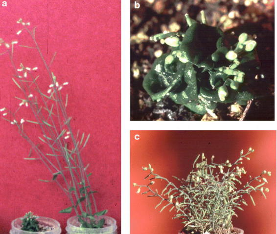
Phenotype of the bri1 mutant. Panel A shows 2-month-old mutant (left) and wild type (right) plants grown in 50 ml centrifuge tubes in a 23 C growth chamber (16 hr light / 8 hr dark). Panels B and shows close-up view of a 2-month-old bri1 mutant plant. Panel C shows the same plant after 4 months. All bars represent 1 cm. Adapted from Clouse et al. (1996).
Table 2.
bri1 mutant alleles

BRI1 is a LRR-Receptor Kinase Involved in BR Perception
BRI1 was identified by positional cloning and verified by sequencing numerous mutant alleles (Li and Chory, 1997). The predicted protein is an 1196 amino acid leucine-rich repeat (LRR) receptor kinase that contains the three major domains common to all animal and plant receptor kinases; the extracellular ligand-binding domain, the transmembrane domain and the cytoplasmic kinase domain. The extracellular domain begins with an N-terminal signal peptide, followed by a leucine zipper motif and 25 tandem copies of a 24-amino acid LRR with 13 potential N-glycosylation sites flanked by conservatively spaced cysteines. Both leucine zippers and LRRs are involved in protein-protein interactions and many receptor kinases dimerize in response to ligand binding (Heldin, 1995; Kobe and Deisenhofer, 1994). The molecular structure of the BRI1 extracellular domain suggests that it is involved in the formation of homo or heterodimers, but this has not been verified experimentally. A unique segment of the BRI1 extracellular domain that has been shown by mutant analysis to be critical for proper protein function, is a 70 amino acid island between LRR 21 and 22 (Li and Chory, 1997). Following the LRR region there is a predicted transmembrane domain spanning amino acids 793–814 followed by a cytoplasmic juxtamembrane region from amino acids 815–882. Based on sequence alignments with many known kinases (Hanks et al., 1988), the region from Phe-883 to Phe-1155 is predicted to comprise the active BRI1 kinase domain, and all invariant amino acid residues and the twelve conserved kinase sub-domains are clearly present. Lastly, amino acids 1156–1196 represent the carboxy-terminal segment of the cytoplasmic domain.
Hundreds of putative receptor kinases exist in Arabidopsis and several have been shown to function in diverse physiological processes including growth and development, embryogenesis, fertilization, abscission, disease resistance and light-mediated responses (Lease et al., 1998). Such functional diversity is expected to be generated by a wide range of ligands that bind to cognate extracellular domains of the receptor kinases. Mechanisms for signal transduction pathway-specific cytoplasmic components to bind to intracellular domains of receptor kinases are also required. A paradigm of animal receptor kinase action has been established by extensive study of several model systems. For example, binding of vertebrate epidermal growth factor to its cognate receptor kinase results in receptor dimerization and autophosphorylation on tyrosine residues in the kinase domain (Heldin, 1995). The activated kinase phosphorylates an intracellular transcription factor, Stat3, which is then translocated to the nucleus where it transcriptionally activates specific epidermal growth factor responsive genes (Park et al., 1996). Ligand-dependent dimerization followed by autophosphorylation is likely to be a conserved mechanism in plants, although conclusive confirmation of all steps in this pathway, including identification of all dimerization-dependent autophosphorylation sites and kinase substrates, has not yet been accomplished in plants. BRI1 shares significant sequence identity with many plant receptor kinases, particularly those in the LRR family of extracellular domains including CLAVATA1 and ERECTA, both involved in regulating different aspects of Arabidopsis development (Clark et al., 1997; Torii et al., 1996).
Recently, Joanne Chory's group has found that BRI1 is localized in the plasma membrane and is ubiquitously expressed in all organs of young, growing Arabidopsis plants (Friedrichsen et al., 2000). Furthermore, using a chimeric construct of the extracellular domain of BRI1 and the kinase domain of the rice receptor kinase XA21, they showed that the extracellular domain of BRI1 perceived BRs (He et al., 2000). In a direct confirmation that BRI1 is either the BR receptor or part of a BR receptor complex, the same group reported evidence that transgenic plants overexpressing a BRI1/green fluorescent protein had increased binding of tritiated BR in their plasma membranes compared to wild-type plants (Wang et al., 2001). The BR-binding activity could be precipitated by antibodies to green fluorescent protein, could be competitively inhibited by active BRs, and was abolished by mutations in the extracellular domain but not the kinase domain of BRI1. Since LRRs are generally not known for binding small molecules such as steroids, it is possible that BL first binds to a steroid-binding protein, and this complex then associates with the extracellular domain of BRI1. In animals, high affinity binding proteins for sex steroids have been identified that are not in the classical superfamily of steroid receptors. These include the membrane progesterone receptors (Falkenstein et al., 1996; Gerdes et al., 1998) and the soluble sex hormone binding globulins (Forest and Pugeat, 1986). A search of the Arabidopsis EST database reveals several clones that have significant homology to these mammalian sequences (213E3T7, K3H2TP, 240O1T7,E2A2T7, 110K7T7). It would be of great interest to determine if recombinant proteins derived from these sequences could bind labeled BR directly. Activation tagging has identified a putative secreted serine carboxypeptidase, BRS1, that may act to process a protein involved in early events of BR signaling. The authors propose the putative steroid-binding proteins other than BRI1 may be the targets of this peptidase (Li et al., 2001).
Biochemical Studies on the BRI1 Kinase Domain
Recent studies on the kinase domain of BRI1 (BRI1-KD) have confirmed that it functions as an active kinase in vitro and specific Ser and Thr residues within the KD have been shown to be autophosphorylated (Friedrichsen et al., 2000; Oh et al., 2000). Phosphoamino acid analyses reveal that plant receptor-like kinases autophosphorylate on Ser/Thr residues (as opposed to Tyr in most animal receptor kinases) and BRI1 is no exception (Fig. 7). The traditional method of determining protein phosphorylation sites is by tryptic digest of proteins, separation of the resulting peptides by HPLC and finally, modified Edman degragdation to determine specific Ser, Thr or Tyr residues that are phosphorylated (Hunter and Sefton, 1991). This method is useful, but suffers from several limitations including including expense, time required and the necessity for relatively large amounts (typically greater than picomole quantities) of protein. The application of Matrix-Assisted Laser Desorption/Ionization Mass Spectrometry (MALDI/MS) to phosphopeptide analysis, increases sensitivity and efficiency with which multiple phosphorylation sites can be analyzed (Liao et al., 1994). A MALDI/MS analysis of BRI1-KD identified at least twelve sites of in vitro autophosphorylation in the BRI-KD, five uniquely and seven with some remaining ambiguity because of multiple Thr or Ser residues within the peptide (Oh et al., 2000).
Figure 7.

Autophosphorylation and phosphoamino acid analysis of recombinant BRI1-KD. A) Affinity purified calmodulin-binding peptide-BRI1 kinase domain recombinant protein (CBP-BRI1-KD, Lane 1) or the mutant CBP-BRI1-K911E (Lane 2) was incubated with 20 μCi [γ-32P]-ATP in kinase buffer for 1 h at ambient temperature, followed by PAGE. Lanes 3 and 4 represent the Coomassie Blue stained gel corresponding to the autoradiograph in Lanes 1 and 2. Molecular mass of CBP-BRI1-KD was determined by MALDI/MS. B) CBP-BRI1-KD was autophosphorylated and transferred to PVDF membrane as described above. The membrane was digested with HCl and subjected to phosphoamino acid analysis by TLE with pSer, pThr and pTyr standards. CBP-BRI1-KD autophosphorylated primarily on Ser residues with a minor Thr component and no detectable phospho-Tyr residues. Adapted from Oh et al., (2000).
Some of the Ser and Thr residues of BRI1-KD autophosphorylated in vitro are conserved at a corresponding positions in related plant Ser/Thr kinases. The region of greatest Ser/Thr conservation occurs in the peptide 1038-DTHLSVSTLAGTPGYVPPEYYQSFR-1062, which lies in the highly conserved activation loop of kinase subdomain VIII (Lease et al., 1998). It is interesting to note that several bri1 mutant alleles are affected in this region (Table 2). Outside of subdomain VIII, the only other strongly conserved sites for Ser or Thr in related kinases were in the positions equivalent to T-872 in the juxtamembrane region and T-982 in kinase subdomain VIa of the BRI1-KD (Oh et al., 2000). Autophosphorylation in subdomain VIII likely leads to kinase activation, while multiple autophosphorylation sites in the juxtamembrane and carboxy terminal regions, if reflected in vivo, might indicate multiple interacting cytoplasmic partners for BRI1, each with a specific phosphorylated target sequence within these regions of BRI1.
Downstream Components of BR Signal Transduction
Identification of cytoplasmic binding partners and kinase domain substrates for BRI is critical for understanding downstream signaling events. A variety of molecular genetic and biochemical approaches can be employed to identify putative in vivo binding partners including yeast two-hybrid analysis (Bower et al., 1996; Gu et al., 1998), interaction cloning (Stone et al., 1994), immunoprecipitation and purification of receptor-protein complexes (Trotochaud et al., 1999), and use of synthetic peptides to understand binding motifs and substrate recognition consensus sequences (Kuriyan and Cowburn, 1997). Reports of yeast two-hybrid screens with the BRI1-KD have appeared in meeting abstracts and hopefully research reports detailing these studies will be published soon. A study of BRI1-KD phosphorylation recognition sequences in synthetic peptides has been reported (Oh et al., 2000). A similar configuration of basic and hydrophobic amino acids at P-3 through P-6 (relative to the phosphorylated Ser) as that found for target sequences of SNF1-related kinases (McMichael et al., 1993; Toroser and Huber, 1998) seems to be optimum for BRI-KD phosphorylation. The optimum sequence tested was GRMKKIASVEMMKK, and using analogs of this peptide it was found that the positioning of residues both N- and C-terminal of the phosphorylated Ser was critical for optimal activity of BRI1-KD. Positive residues at P-3, P-4, P+5, and P+6 were essential with a preference for Lys over Arg. A moderate reduction of activity was observed after substitution of the hydrophobic group at P-5 and P+4, with a more dramatic effect at P+3.
Using the preliminary consensus sequence [RK]-[RK]-X(2)-S-X(2)-[LMVIFY]-X-[RK]-[RK] (corresponding to the most important residues identified to date) to search the Arabidopsis non-redundant protein database resulted in 109 hits. A variety of interesting proteins were found, some of which had obvious connections to signal transduction pathways, and others, such as phytochrome D, are intriguing given the connection of BRs and photomorphogenesis. Presently, there is no direct evidence that these are true substrates in vivo, but the BRI1-KD substrate recognition sequence may provide a valuable molecular tool for further analysis. Interestingly, one protein containing elements of the BRI1-KD recognition sequence was originally identified by subtractive hybridization in a screen for BR-regulated genes (Jiang and Clouse, 2001). This WD-domain protein has extensive sequence similarity to mammalian TRIP-1 (TGF-β Receptor Interacting Protein), which is a kinase domain substrate of the TGF-β Type II receptor (Chen et al., 1995). Unlike most animal receptor kinases that autophosphorylate on Tyr, the TGF-β family of receptors are Ser/Thr receptor kinases. Subsequent experiments showed that TRIP-1 functioned as a modulator of TGF-β receptor signaling in vivo (Choy and Derynck, 1998). Suprisingly, TRIP-1 also has a dual function as a subunit of the eukaryotic translation initiation factor, eIF3. At 600 kDa, eIF3 is the largest of the eukaryotic translation initiation factors and is composed of 10 or more subunits, with TRIP-1 comprising the p36 subunit. eIF3 promotes dissociation of 80S ribosomes and is required for ribosomal binding of mRNA to the 40S subunit, thus playing an essential role in the initiation of eukaryotic protein synthesis. The Arabidopsis TRIP-1 homolog has been shown to be a functional component of the plant eIF3 complex (Burks et al., 2000).
Enhanced expression of TRIP-1 by BR, as has been observed in bean, tobacco and Arabidopsis (Jiang and Clouse, 2001), might lead to increased protein translation and serve as a general mechanism of BR-promoted growth. Besides alteration in transcript levels, phosphorylation of eIF3 subunits might be another way in which signal transduction pathways controlling development could impinge directly on translation initiation. TRIP-1 in vertebrates is known to be a substrate of the TGF-β ser/thr receptor kinase and it is conceivable that BRI1, or one of the many other plant receptor kinases involved in developmental control might phosphorylate TRIP-1, enhancing its competence to initiate assembly of the eIF3 complex. We have recently found that recombinant Arabidopsis TRIP-1 is strongly phosphorylated by BRI-KD in vitro, predominately on Thr residues (W. K. Ray and S.D. Clouse, unpublished). A synthetic peptide representing the TRIP-1 sequence most resembling the BRI-KD consensus described above, is also phosphorylated by BRI-KD in vitro. We are currently employing immunoprecipitation followed by liquid chromatography/tandem mass spectrometry (LC/MS/MS) to determine if TRIP-1 is phosphorylated by BRI1 in vivo.
To assess the effects of loss of function of TRIP-1 on Arabidopsis development, we constructed numerous independent transgenic lines expressing antisense AtTRIP-1 RNA (α-TRIP-1). The majority of the independent antisense lines examined (11 out of 16) germinated three to four days later than the wild-type control and then produced tiny seedlings with bright red cotyledons. All of these seedlings died before appearance of the first true leaves. Five out of the 16 lines survived beyond the seedling stage and all five lines showed dramatic alterations in several developmental pathways that shared some of the characteristics of BR-deficient and – insensitive mutants, including dwarfism, altered leaf morphology, delayed development, reduced apical dominance and reduced fertility (Fig. 8a-c). Several of the lines also exhibited unique developmental defects not seen in BR mutants, including inappropriate initiation of multiple rosettes, multiple floral meristems before bolting, and higher numbers of floral buds differentiating from floral meristems than wildtype. Like BR mutants, α-TRIP-D also showed an abnormally long life span with a very bushy habit in older plants. Expression of antisense TRIP-1 was correlated with the severity of the dwarf phenotype and each line had dramatically reduced levels of sense TRIP-1 mRNA, when compared to untransformed wildtype plants (Jiang and Clouse, 2001).
Figure 8.
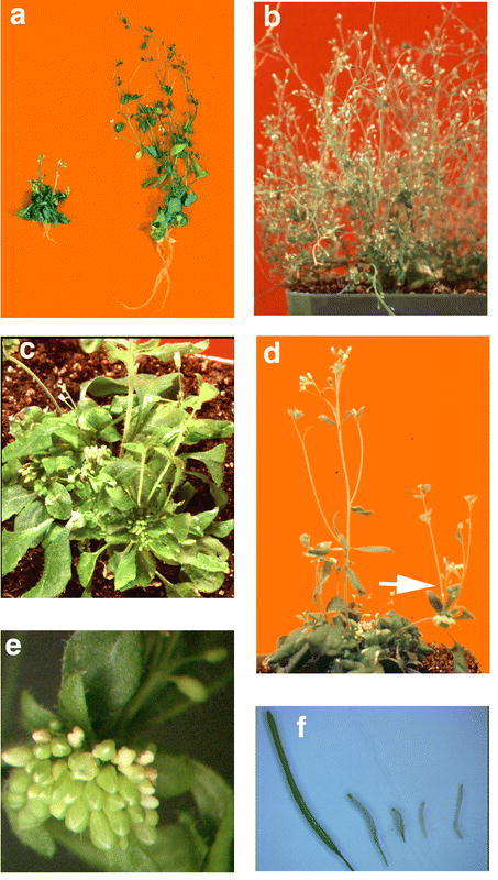
Phenotype of Arabidopsis antiTRIP-1 transgenic lines. a) Two-month-old α-TRIP-A (left) vs. wildtype (right), showing the extreme dwarfism and altered leaf morphology of the antisense line, similar to BR-deficient and – insensitive mutants. b) Six-month-old α-TRIP-D showing delayed senescence and bushy growth habit similar to the bri1 mutant. c) Two-month-old α-TRIP-C1 showing the abnormally high number of rosette leaves and the initiation of multiple floral meristems before the elongation of the inflorescence. d) The arrow shows the initiation of a new rosette of leaves and multiple inflorescences at the terminal point of a primary inflorescence in α-TRIP-C1. e) Line α-TRIP-C1 initiated an abnormally high number of floral buds from a single floral meristem. f) Siliques from antisense lines were much shorter than wildtype. Left to right, mature siliques from plants of the same age obtained from wildtype, α-TRIP-D, α-TRIP-A, α-TRIP-A, and α-TRIP-D. Adapted from Jiang and Clouse (2001).
Conclusion and Future Prospects
The use of mutant analysis in Arabidopsis coupled with GC/MS measurements of endogenous BR levels has greatly advanced our understanding of BR physiology and biosynthesis. The biosynthetic pathway to BL is nearing saturation with mutants, particularly if a knockout for the AtBR6ox gene can be recovered and a homolog of the pea C-2 hydroxylase can be identified. Measurement of endogenous BR levels under different light regimes and in specific organs may help elucidate integration of BR and light signals as would examination of BR levels in light signaling mutants. Preliminary results on the promotive effect of BRs on cell division is likely to lead to new areas of molecular research and the use of microarrays to study global aspects of BR-regulated gene expression can be expected to increase. BRI1 analysis has comprised almost the entire body of BR signal transduction knowledge, but isolation of new BR-insensitive mutants has been reported at meetings and publications should follow shortly. Further analysis of BRI1 will also be required for a complete understanding of BR signal transduction. Does BRI1 form homo- or heterodimers? Are accessory steroid-binding proteins required for BR binding? What are the in vivo autophosphorylation sites and specific phosphorylation recognition sequences in kinase domain substrates? Application of advanced techniques in mass spectrometry such as MALDI/MS and LC/MS/MS are beginning to answer some of these questions. To understand downstream events, the number and nature of cytoplasmic binding partners for the BRI-KD needs to established. Forthcoming results of yeast two-hybrid experiments should be illuminating as will further study of the TRIP-1 protein as a possible BRI1 interacting protein.
Acknowledgments
Thanks to S. Choe and K. Feldmann for providing photographs of BR mutants. Work in the author's laboratory is supported by the National Science Foundation (IBN: Integrative Plant Biology), USDA/NRI (Plant Growth and Development), and the North Carolina Agricultural Research Service.
Footnotes
Citation: Clouse S.D. (2002) Brassinosteroids. The Arabidopsis Book 1:e0009. doi:10.1199/tab.0009
elocation-id: e0009
Published on: September 30, 2002
References
- Adam G., Schneider B. Uptake, Transport and Metabolism. 1999;1(1):113–136. In Brassinosteroids: Steroidal Plant Hormones, Sakurai A, Yokota T, Clouse SD eds, (Tokyo: Springer), pp. [Google Scholar]
- Altmann T. Molecular physiology of brassinosteroids revealed by the analysis of mutants. Planta. 1999;2081(1):1–11. doi: 10.1007/s004250050528. [DOI] [PubMed] [Google Scholar]
- Anderson J. W., Beardall J. Molecular activity of plant cells. 1991. (Oxford: Blackwell Scientific)
- Asakawa S., Abe H., Nishikawa N., Natsume M., Koshioka M. Purification and identification of new acyl-conjugated teasterones in lily pollen. Biosci. Biotech. Biochem. 1996;601(1):1416–1420. [Google Scholar]
- Asami T., Min Y. K., Nagata N., Yamagishi K., Takatsuto S., Fujioka S., Murofushi N., Yamaguchi I., Yoshida S. Characterization of brassinazole, a triazole-type brassinosteroid biosynthesis inhibitor. Plant Physiol. 2000;1231(1):93–100. doi: 10.1104/pp.123.1.93. [DOI] [PMC free article] [PubMed] [Google Scholar]
- Asami T., Yoshida S. Brassinosteroid biosynthesis inhibitors. Trends Plant Sci. 1999;41(1):348–353. doi: 10.1016/s1360-1385(99)01456-9. [DOI] [PubMed] [Google Scholar]
- Azpiroz R., Wu Y., LoCascio J., Feldmann K. An Arabidopsis brassinosteroid-dependent mutant is blocked in cell elongation. Plant Cell. 1998;101(1):219–230. doi: 10.1105/tpc.10.2.219. [DOI] [PMC free article] [PubMed] [Google Scholar]
- Benveniste P. Sterol biosynthesis. Annu Rev Plant Physiol. 1986;371(1):275–308. [Google Scholar]
- Bishop G. J., Nomura T., Yokota T., Harrison K., Noguchi T., Fujioka S., Takatsuto S., Jones J. D. G., Kamiya Y. The tomato DWARF enzyme catalyses C-6 oxidation in brassinosteroid biosynthesis. Proc. Natl. Acad. Sci. USA. 1999;961(1):1761–1766. doi: 10.1073/pnas.96.4.1761. [DOI] [PMC free article] [PubMed] [Google Scholar]
- Bower M., Matias D., Fernandes-Carvalho E., Mazzurco M., Gu T., Rothstein S., Goring D. Two members of the thioredoxin-h family interact with the kinase domain of a Brassica S locus receptor kinase. Plant Cell. 1996;81(1):1641–1650. doi: 10.1105/tpc.8.9.1641. [DOI] [PMC free article] [PubMed] [Google Scholar]
- Burks E. A., Bezerra P. P., Le H., Gallie D. R., Browning K. S. Plant initiation factor 3 subunit composition resembles mammalian initiation factor 3 and has a novel subunit. J Biol Chem. 2001;2761(1):2122–2131. doi: 10.1074/jbc.M007236200. [DOI] [PubMed] [Google Scholar]
- Carpita N., Gibeaut D. Structural models of the primary cell walls in flowering plants: consistency of molecular structure with the physical properties of the walls during growth. Plant J. 1993;31(1):1–30. doi: 10.1111/j.1365-313x.1993.tb00007.x. [DOI] [PubMed] [Google Scholar]
- Catala C., Rose J. K. C., Bennett A. Auxin-regulation and spatial localization of an endo-1,4-β-D-glucanase and a xyloglucan endotransglycosylase in expanding tomato hypocotyls. Plant J. 1997;121(1):417–426. doi: 10.1046/j.1365-313x.1997.12020417.x. [DOI] [PubMed] [Google Scholar]
- Catterou M., Dubois F., Schaller H., Aubanelle L., Vilcot B., Sangwan-Norreel B. S., Sangwan R. S. Brassinosteroids, microtubules and cell elongation in Arabidopsis thaliana. II. Effects of brassinosteroids on microtubules and cell elongation in the bul1 mutant. Planta. 2001;2121(1):673–83. doi: 10.1007/s004250000467. [DOI] [PubMed] [Google Scholar]
- Chen R. H., Miettinen P. J., Maruoka E. M., Choy L., Derynck R. A WD-domain protein that is associated with and phosphorylated by the type II TGF-beta receptor. Nature. 1995;3771(1):548–52. doi: 10.1038/377548a0. [DOI] [PubMed] [Google Scholar]
- Choe S., Dilkes B. P., Fujioka S., Takatsuto S., Sakurai A., Feldmann K. A. The DWF4 gene of Arabidopsis encodes a cytochrome P450 that mediates multiple 22α-hydroxylation steps in brassinosteroid biosynthesis. Plant Cell. 1998;101(1):231–243. doi: 10.1105/tpc.10.2.231. [DOI] [PMC free article] [PubMed] [Google Scholar]
- Choe S., Dilkes B. P., Gregory B. D., Ross A. S., Yuan H., Noguchi T., Fujioka S., Takatsuto S., Tanaka A., Yoshida S., Tax F., Feldmann K. A. The Arabidopsis dwarf1 mutant is defective in the conversion of 24-methylenecholesterol to campesterol in brassinosteroid biosynthesis. Plant Physiol. 1999a;1191(1):897–907. doi: 10.1104/pp.119.3.897. [DOI] [PMC free article] [PubMed] [Google Scholar]
- Choe S., Noguchi T., Fujioka S., Takatsuto S., Tissier C. P., Gregory B. D., Ross A. S., Tanaka A., Yoshida S., Tax F. E., Feldmann K. A. The Arabidopsis dwf7/ste1 mutant is defective in the Δ7 sterol C-5 desaturation step leading to brassinosteroid biosynthesis. Plant Cell. 1999b;111(1):207–221. [PMC free article] [PubMed] [Google Scholar]
- Choe S., Tanaka A., Noguchi T., Fujioka S., Takatsuto S., Ross A. S., Tax F. E., Yoshida S., Feldmann K. A. Lesions in the sterol delta reductase gene of Arabidopsis cause dwarfism due to a block in brassinosteroid biosynthesis. Plant J. 2000;211(1):431–43. doi: 10.1046/j.1365-313x.2000.00693.x. [DOI] [PubMed] [Google Scholar]
- Choi Y-H., Fujioka S., Nomura T., Harada AY T., Takatsuto S., Sakurai A. An alternative brassinolide biosynthetic pathway via late C-6 oxidation. Phytochemistry. 1997;441(1):609–613. [Google Scholar]
- Chory J., Catterjee m, Cook R., Elich T., Fankhauser C., Li J., Nagpal P., Neff M., Pepper A., Poole D., Reed J., Vitart V. From seed germination to flowering, light controls plant development via the pigment phytochrome. Proc. Natl. Acad. Sci. USA. 1996;931(1):12066–71. doi: 10.1073/pnas.93.22.12066. [DOI] [PMC free article] [PubMed] [Google Scholar]
- Chory J., Nagpal P., Peto C. Phenotypic and genetic analysis of det2, a new mutant that affects light-regulated seedling development in Arabidopsis. Plant Cell. 1991;31(1):445–459. doi: 10.1105/tpc.3.5.445. [DOI] [PMC free article] [PubMed] [Google Scholar]
- Choy L., Derynck R. The type II transforming growth factor (TGF)-beta receptor-interacting protein TRIP-1 acts as a modulator of the TGF-beta response. J Biol Chem. 1998;2731(1):31455–62. doi: 10.1074/jbc.273.47.31455. [DOI] [PubMed] [Google Scholar]
- Clark S., Williams R., Meyerowitz E. The CLAVATA1 gene encodes a putative receptor kinase that controls shoot and floral meristem size in Arabidopsis. Cell. 1997;891(1):575–585. doi: 10.1016/s0092-8674(00)80239-1. [DOI] [PubMed] [Google Scholar]
- Clouse S. D. Molecular genetic analysis of brassinosteroid action. Physiol. Plant. 1997;1001(1):702–709. [Google Scholar]
- Clouse S. D. Plant development: A role for sterols in embryogenesis. Curr Biol. 2000;101(1):R601–4. doi: 10.1016/s0960-9822(00)00639-4. [DOI] [PubMed] [Google Scholar]
- Clouse S. D., Feldmann K. A. Molecular genetics of brassinosteroid action. 1999;1(1):163–190. In Brassinosteroids: Steroidal Plant Hormones, Sakurai A, Yokota T, Clouse SD eds, (Tokyo: Springer), pp. [Google Scholar]
- Clouse S. D., hall A. F., Langford M., McMorris T. C., Baker M. E. Physiological and molecular effects of brassinosteroids on Arabidopsis thaliana. J Plant Growth Regul. 1993;121(1):61–66. [Google Scholar]
- Clouse S. D., Langford M., McMorris T. C. A brassinosteroid-insensitive mutant in Arabidopsis thaliana exhibits multiple defects in growth and development. Plant Physiol. 1996;1111(1):671–678. doi: 10.1104/pp.111.3.671. [DOI] [PMC free article] [PubMed] [Google Scholar]
- Clouse S. D., Sasse J. M. Brassinosteroids: Essential Regulators of Plant Growth and Development. Annu. Rev. Plant Physiol. Plant Mol. Biol. 1998;491(1):427–451. doi: 10.1146/annurev.arplant.49.1.427. [DOI] [PubMed] [Google Scholar]
- Clouse S. D., Zurek D. Molecular analysis of brassinolide action in plant growth and development. 1991;1(1):122–40. In Brassinosteroids: Chemistry, Bioactivity, & Applications, Cutler HG, Yokota T, Adam G eds (Washington, D.C.: American Chemical Society), pp. [Google Scholar]
- Cosgrove D. J. Relaxation in a high-stress environment: the molecular basis of extensible cell walls and enlargement. Plant Cell. 1997;91(1):1031–1041. doi: 10.1105/tpc.9.7.1031. [DOI] [PMC free article] [PubMed] [Google Scholar]
- Cutler H. G., Yokota T., Adam G. Brassinosteroids: Chemistry, Bioactivity, & Applications. 1991. (Washington, D.C.: American Chemical Society)
- Diener A. C., Li H., Zhou W., Whoriskey W. J., Nes W. D., Fink G. R. Sterol methyltransferase 1 controls the level of cholesterol in plants. Plant Cell. 2000;121(1):853–70. doi: 10.1105/tpc.12.6.853. [DOI] [PMC free article] [PubMed] [Google Scholar]
- Ding W-M., Zhao Y-J. Effect of epi-BR on activity of peroxidase and soluble protein content of cucumber cotyledon. Acta Phytophysiologica Sinica. 1995;211(1):259–264. [Google Scholar]
- Edwards P., Ericsson J. Signaling molecules derived from the cholesterol biosynthetic pathway. Annu Rev Biochem. 1999;681(1):157–185. doi: 10.1146/annurev.biochem.68.1.157. [DOI] [PubMed] [Google Scholar]
- Ephritikhine G., Fellner M., Vannini C., Lapous D., Barbier-Brygoo H. The sax1 dwarf mutant of Arabidopsis thaliana shows altered sensitivity of growth responses to abscisic acid, auxin, gibberellins and ethylene and is partially rescued by exogenous brassinosteroid. Plant J. 1999a;181(1):303–14. doi: 10.1046/j.1365-313x.1999.00454.x. [DOI] [PubMed] [Google Scholar]
- Ephritikhine G., Pagant S., Fujioka S., Takatsuto S., Lapous D., Caboche M., Kendrick R. E., Barbier-Brygoo H. The sax1 mutation defines a new locus involved in the brassinosteroid biosynthesis pathway in Arabidopsis thaliana. Plant J. 1999b;181(1):315–20. doi: 10.1046/j.1365-313x.1999.00455.x. [DOI] [PubMed] [Google Scholar]
- Falkenstein E., Meyer C., Eisen C., Scriba P., Wehling M. Full-length cDNA sequence of a progesterone membrane-binding protein from porcine vascular smooth muscle cells. Biochem. Biophys. Res. Comm. 1996;2291(1):86–89. doi: 10.1006/bbrc.1996.1761. [DOI] [PubMed] [Google Scholar]
- Feldmann K., Marks M., Christianson M., Quatrano R. A dwarf mutant of Arabidopsis generated by T-DNA insertion mutagenesis. Science. 1989;2431(1):1351–1354. doi: 10.1126/science.243.4896.1351. [DOI] [PubMed] [Google Scholar]
- Forest M., Pugeat M. Binding Proteins of Seroid Hormones. 1986. (London: John Libbey)
- Friedrichsen D. M., Joazeiro C. A., Li J., Hunter T., Chory J. Brassinosteroid-insensitive-1 is a ubiquitously expressed leucine-rich repeat receptor serine/threonine kinase. Plant Physiol. 2000;1231(1):1247–56. doi: 10.1104/pp.123.4.1247. [DOI] [PMC free article] [PubMed] [Google Scholar]
- Fujioka S. Natural occurrence of brassinosteroids in the plant kingdom. 1999;1(1):21–45. In Brassinosteroids: Steroidal Plant Hormones, Sakurai A, Yokota T, Clouse SD eds, (Tokyo: Springer), pp. [Google Scholar]
- Fujioka S., Choi Y-H., Takatsuto S., Yokota T., Li J., Chory J., Sakurai A. Identification of castasterone, 6-deoxocastasterone, typhasterol and 6-deoxotyphasterol from the shoots of Arabidopsis thaliana. Plant Cell Physiol. 1996;371(1):1201–03. doi: 10.1093/oxfordjournals.pcp.a029074. [DOI] [PubMed] [Google Scholar]
- Fujioka S., Li J., Choi Y-H., Seto H., Takatsuto S., Noguchi T., Watanabe T., Kuriyama H., Yokota T., Chory J., Sakurai A. The Arabidopsis deetiolated2 mutant is blocked early in brassinosteroid biosynthesis. Plant Cell. 1997;91(1):1951–1962. doi: 10.1105/tpc.9.11.1951. [DOI] [PMC free article] [PubMed] [Google Scholar]
- Fujioka S., Sakurai A. Biosynthesis and metabolism of brassinosteroids. Physiol. Plant. 1997;1001(1):710–715. [Google Scholar]
- Fukuda H. Tracheary element differentiation. Plant Cell. 1997;91(1):1147–1156. doi: 10.1105/tpc.9.7.1147. [DOI] [PMC free article] [PubMed] [Google Scholar]
- Gachotte D., Meens R., Benveniste P. An Arabidopsis mutant deficient in sterol biosynthesis: heterologous complementation by ERG 3 encoding a delta 7-sterol-C-5-desaturase from yeast. Plant J. 1995;81(1):407–16. doi: 10.1046/j.1365-313x.1995.08030407.x. [DOI] [PubMed] [Google Scholar]
- Gerdes D., Wehling M., Leube B., Falkenstein E. Cloning and tissue expression of two putative steroid membrane receptors. Biol. Chem. 1998;3791(1):907–911. doi: 10.1515/bchm.1998.379.7.907. [DOI] [PubMed] [Google Scholar]
- Griffiths P. G., Sasse J. M., Yokota T., Cameron D. W. 6-Deoxotyphasterol and 3-Dehydro-6-deoxoteasterone, possible precursors to brassinosteroids in the pollen of Cupressus arizonica. Biosci. Biotech. Biochem. 1995;591(1):956–959. [Google Scholar]
- Grove M. D., Spencer G. F., Rohwedder W. K., Mandava N. B., Worley J. F., Warthen J. D., Steffens G. L., Flippen-Anderson J. L., Cook J. C. Brassinolide, a plant growth-promoting steroid isolated from Brassica napus pollen. Nature. 1979;2811(1):216–17. [Google Scholar]
- Gu T., Mazzurco M., Sulaman W., Matias D. D., Goring D. R. Binding of an arm repeat protein to the kinase domain of the S-locus receptor kinase. Proc Natl Acad Sci U S A. 1998;951(1):382–7. doi: 10.1073/pnas.95.1.382. [DOI] [PMC free article] [PubMed] [Google Scholar]
- Hai T., Schneider B., Porzel A., Adam G. Metabolism of 24-epicastasterone in cell suspension culture of Lycopersicon esculentum. Phytochem. 1996;411(1):197–201. [Google Scholar]
- Hanks S. K., Quinn A. M., Hunter T. The protein kinase family: conserved features and deduced phylogeny of the catalytic domains. Science. 1988;2411(1):42–52. doi: 10.1126/science.3291115. [DOI] [PubMed] [Google Scholar]
- Hartmann M. Plant sterols and the membrane environment. Trends Plant Sci. 1998;31(1):170–175. [Google Scholar]
- He Y-J., Xu R-J., Zhao Y-J. Enhancement of senescence by epibrassinolide in leaves of mung bean seedling. Acta Phytophysiologica Sinica. 1996;221(1):58–62. [Google Scholar]
- He Z., Wang Z. Y., Li J., Zhu Q., Lamb C., Ronald P., Chory J. Perception of brassinosteroids by the extracellular domain of the receptor kinase BRI1. Science. 2000;2881(1):2360–3. doi: 10.1126/science.288.5475.2360. [DOI] [PubMed] [Google Scholar]
- Heldin C. Dimerization of cell surface receptors in signal transduction. Cell. 1995;801(1):213–224. doi: 10.1016/0092-8674(95)90404-2. [DOI] [PubMed] [Google Scholar]
- Hewitt F. R., Hough T., O'Neill P., Sasse J. M., Williams E. G., Rowan K. S. Effect of brassinolide and other growth regulators on the germination and growth of pollen tubes of Prunus avium using a multiple hanging drop assay. Aust. J. Plant Physiol. 1985;11(1):201–11. [Google Scholar]
- Hu Y., Bao F., Li J. Promotive effect of brassinosteroids on cell division involves a distinct CycD3-induction pathway in Arabidopsis. Plant J. 2000;241(1):693–701. doi: 10.1046/j.1365-313x.2000.00915.x. [DOI] [PubMed] [Google Scholar]
- Hunter T., Sefton B. M. Protein Phosphorylation: Part B. 1991. (San Diego: Academic Press) [PubMed]
- Husselstein T., Schaller H., Gachotte D., Benveniste P. Delta7-sterol-C5-desaturase: molecular characterization and functional expression of wild-type and mutant alleles. Plant Mol Biol. 1999;391(1):891–906. doi: 10.1023/a:1006113919172. [DOI] [PubMed] [Google Scholar]
- Iwasaki T., Shibaoka H. Brassinosteroids act as regulators of tracheary-element differentiation in isolated Zinnia mesophyll cells. Plant Cell Physiol. 1991;321(1):1007–1014. [Google Scholar]
- Jang J. C., Fujioka S., Tasaka M., Seto H., Takatsuto S., Ishii A., Aida M., Yoshida S., Sheen J. A critical role of sterols in embryonic patterning and meristem programming revealed by the fackel mutants of Arabidopsis thaliana. Genes Dev. 2000;141(1):1485–97. [PMC free article] [PubMed] [Google Scholar]
- Jiang J., Clouse S. D. Expression of a plant gene with sequence similarity to animal TGF-β Receptor Interacting Protein is regulated by brassinosteroids and required for normal plant development. Plant J. 2001;261(1):35–45. doi: 10.1046/j.1365-313x.2001.01007.x. [DOI] [PubMed] [Google Scholar]
- Kang J. G., Yun J., Kim D. H., Chung K. S., Fujioka S., Kim J. I., Dae H. W., Yoshida S., Takatsuto S., Song P. S., Park C. M. Light and brassinosteroid signals are integrated via a dark-induced small G protein in etiolated seedling growth. Cell. 2001;1051(1):625–36. doi: 10.1016/s0092-8674(01)00370-1. [DOI] [PubMed] [Google Scholar]
- Kauschmann A., Jessop A., Koncz C., Szekeres m, Willmitzer L., Altmann T. Genetic evidence for an essential role of brassinosteroids in plant development. Plant J. 1996;91(1):701–713. [Google Scholar]
- Khripach V. A., Zhabinskii V. N., de Groot A. E. Brassinosteroids: A New Class of Plant Hormones. 1999. (San Diego: Academic Press)
- Kim S-K., Abe H., Little C. H. A., Pharis R. P. Identification of two brassinosteroids from the cambial region of Scots pine (Pinus silvestris) by gas chromatography-mass spectrometry, after detection using a dwarf rice lamina inclination bioassay. Plant Physiol. 1990;941(1):1709–1713. doi: 10.1104/pp.94.4.1709. [DOI] [PMC free article] [PubMed] [Google Scholar]
- Kitani Y. Induction of parthenogenetic haploid plants with brassinolide. Jpn. J. Genet. 1994;691(1):35–9. [Google Scholar]
- Klahre U., Noguchi T., Fujioka S., Takatsuto S., Yokota T., Nomura T., Yoshid S., Chua N-H. The Arabidopsis DIMINUTO/DWARF1 gene encodes a protein involved in steroid synthesis. Plant Cell. 1998;101(1):1677–1690. doi: 10.1105/tpc.10.10.1677. [DOI] [PMC free article] [PubMed] [Google Scholar]
- Kobe B., Deisenhofer J. The leucine-rich repeat: a versatile binding motif. Trends Biochem Sci. 1994;191(1):415–21. doi: 10.1016/0968-0004(94)90090-6. [DOI] [PubMed] [Google Scholar]
- Koka C. V., Cerny R. E., Gardner R. G., Noguchi T., Fujioka S., Takatsuto S., Yoshida S., Clouse S. D. A putative role for the tomato genes DUMPY and CURL-3 in brassinosteroid biosynthesis and response. Plant Physiol. 2000;1221(1):85–98. doi: 10.1104/pp.122.1.85. [DOI] [PMC free article] [PubMed] [Google Scholar]
- Kuriyan J., Cowburn D. Molecular peptide recognition domains in eukaryotic signaling. Annu Rev Biophys Biomol Struct. 1997;261(1):259–288. doi: 10.1146/annurev.biophys.26.1.259. [DOI] [PubMed] [Google Scholar]
- Lease K., Ingham E., Walker J. C. Challenges in understanding RLK function. Curr Opin Plant Biol. 1998;11(1):388–92. doi: 10.1016/s1369-5266(98)80261-6. [DOI] [PubMed] [Google Scholar]
- Li J., Biswas M. G., Chao A., Russell D. W., Chory J. Conservation of function between mammalian and plant steroid 5alpha-reductases. Proc Natl Acad Sci U S A. 1997;941(1):3554–9. doi: 10.1073/pnas.94.8.3554. [DOI] [PMC free article] [PubMed] [Google Scholar]
- Li J., Chory J. A putative leucine-rich repeat receptor kinase involved in brassinsteroid signal transduction. Cell. 1997;901(1):929–938. doi: 10.1016/s0092-8674(00)80357-8. [DOI] [PubMed] [Google Scholar]
- Li J., Lease K. A., Tax F. E., Walker J. C. BRS1, a serine carboxypeptidase, regulates BRI1 signaling in Arabidopsis thaliana. Proc Natl Acad Sci U S A. 2001;981(1):5916–21. doi: 10.1073/pnas.091065998. [DOI] [PMC free article] [PubMed] [Google Scholar]
- Li J., Nagpal P., Vitart V., McMorris T. C., Chory J. A role for brassinosteroids in light-dependent development of Arabidopsis. Science. 1996;2721(1):398–401. doi: 10.1126/science.272.5260.398. [DOI] [PubMed] [Google Scholar]
- Liao P. C., Leykam J., Andrews P. C., Gage D. A., Allison J. An approach to locate phosphorylation sites in a phosphoprotein: mass mapping by combining specific enzymatic degradation with matrix-assisted laser desorption/ionization mass spectrometry. Anal Biochem. 1994;2191(1):9–20. doi: 10.1006/abio.1994.1224. [DOI] [PubMed] [Google Scholar]
- Mandava N. B. Plant growth-promoting brassinosteroids. Annu. Rev. Plant Physiol. Plant Molec. Biol. 1988;391(1):23–52. [Google Scholar]
- Mathur J., Molnar G., Fujioka S., Takatsuto S., Sakurai A., Yokota T., Adam G., Voigt B., Nagy F., Maas C., Schell J., Koncz C., Szekeres M. Transcription of the Arabidopsis CPD gene, encoding a steroidogenic cytochrome P450, is negatively controlled by brassinosteroids. Plant J. 1998;141(1):593–602. doi: 10.1046/j.1365-313x.1998.00158.x. [DOI] [PubMed] [Google Scholar]
- Mayer U., Torres-Ruiz R., Berleth T., Misera S., Jurgens G. Mutations affecting the body organization in the Arabidopsis embryo. Nature. 1991;1(1):402–407. [Google Scholar]
- Mayumi K., Shibaoka H. A possible double role for brassinolide in the re-orientation of cortical microtubules in the epidermal cells of Azuki bean epicotyls. Plant Cell Physiol. 1995;361(1):173–81. [Google Scholar]
- McMichael R. J., Klein R., Salvucci M., Huber S. Identification of the major regulatory phosphorylation site in sucrose-phosphate synthase. Arch. Biochem Biophys. 1993;3071(1):248–252. doi: 10.1006/abbi.1993.1586. [DOI] [PubMed] [Google Scholar]
- Mitchell J. W., Mandava N. B., Worley J. F., Plimmer J. R., Smith M. V. Brassins: a new family of plant hormones from rape pollen. Nature (London) 1970;2251(1):1065–66. doi: 10.1038/2251065a0. [DOI] [PubMed] [Google Scholar]
- Morillon R., Catterou M., Sangwan R. S., Sangwan B. S., Lassalles J. P. Brassinolide may control aquaporin activities in Arabidopsis thaliana. Planta. 2001;2121(1):199–204. doi: 10.1007/s004250000379. [DOI] [PubMed] [Google Scholar]
- Nagata N., Min Y. K., Nakano T., Asami T., Yoshida S. Treatment of dark-grown Arabidopsis thaliana with a brassinosteroid-biosynthesis inhibitor, brassinazole, induces some characteristics of light-grown plants. Planta. 2000;2111(1):781–90. doi: 10.1007/s004250000351. [DOI] [PubMed] [Google Scholar]
- Nakajima N., Shida A., Toyama S. Effects of brassinosteroid on cell division and colony formation of Chinese cabbage mesophyll protoplasts. Japanese J. Crop Science. 1996;651(1):114–118. [Google Scholar]
- Neff M. M., Nguyen S. M., Malancharuvil E. J., Fujioka S., Noguchi T., Seto H., Tsubuki M., Honda T., Takatsuto S., Yoshida S., Chory J. BAS1: A gene regulating brassinosteroid levels and light responsiveness in Arabidopsis. Proc Natl Acad Sci U S A. 1999;961(1):15316–23. doi: 10.1073/pnas.96.26.15316. [DOI] [PMC free article] [PubMed] [Google Scholar]
- Noguchi T., Fujioka S., Choe S., Takatsuto S., Tax F. E., Yoshida S., Feldmann K. A. Biosynthetic pathways of brassinolide in Arabidopsis. Plant Physiol. 2000;1241(1):201–9. doi: 10.1104/pp.124.1.201. [DOI] [PMC free article] [PubMed] [Google Scholar]
- Noguchi T., Fujioka S., Choe S., Takatsuto S., Yoshida S., Yuan H., Feldmann K. A., Tax F. E. Brassinosteroid-insensitive dwarf mutants of Arabidopsis accumulate brassinosteroids. Plant Physiol. 1999a;1211(1):743–52. doi: 10.1104/pp.121.3.743. [DOI] [PMC free article] [PubMed] [Google Scholar]
- Noguchi T., Fujioka S., Takatsuto S., Sakurai A., Yoshida S., Li J., Chory J. Arabidopsis det2 is defective in the conversion of (24R)-24-Methylcholest-4-En-3-One to (24R)-24-Methyl-5α-Cholestan-3-One in brassinosteroid biosynthesis. Plant Physiol. 1999b;1201(1):833–839. doi: 10.1104/pp.120.3.833. [DOI] [PMC free article] [PubMed] [Google Scholar]
- Nomura T., Sato T., Bishop G. J., Kamiya Y., Takatsuto S., Yokota T. Accumulation of 6-deoxocathasterone and 6-deoxocastasterone in Arabidopsis, pea and tomato is suggestive of common rate-limiting steps in brassinosteroid biosynthesis. Phytochemistry. 2001;571(1):171–8. doi: 10.1016/s0031-9422(00)00440-4. [DOI] [PubMed] [Google Scholar]
- Oh M-H., Clouse S. D. Brassinolide affects the rate of cell division in isolated leaf protoplasts of Petunia hybrida. Plant Cell Reports. 1998;171(1):921–924. doi: 10.1007/s002990050510. [DOI] [PubMed] [Google Scholar]
- Oh M. H., Ray W. K., Huber S. C., Asara J. M., Gage D. A., Clouse S. D. Recombinant Brassinosteroid Insensitive 1 Receptor-Like Kinase Autophosphorylates on Serine and Threonine Residues and Phosphorylates a Conserved Peptide Motif in Vitro. Plant Physiol. 2000;1241(1):751–766. doi: 10.1104/pp.124.2.751. [DOI] [PMC free article] [PubMed] [Google Scholar]
- Oh M. H., Romanow W. G., Smith R. C., Zamski E., Sasse J., Clouse S. D. Soybean BRU1 encodes a functional xyloglucan endotransglycosylase that is highly expressed in inner epicotyl tissues during brassinosteroid-promoted elongation. Plant Cell Physiol. 1998;391(1):124–130. [Google Scholar]
- Park O., Schaefer T., Nathans D. In vitro activation of Stat3 by epidermal growth factor receptor kinase. Proc. Natl. Acad. Sci. USA. 1996;931(1):13704–13708. doi: 10.1073/pnas.93.24.13704. [DOI] [PMC free article] [PubMed] [Google Scholar]
- Sakurai A. Biosynthesis. 1999;1(1):91–112. In Brassinosteroids: Steroidal Plant Hormones, Sakurai A, Yokota T, Clouse SD eds, (Tokyo: Springer), pp. [Google Scholar]
- Sakurai A., Yokota T., Clouse S. D. Brassinosteroids: Steroidal Plant Hormones. 1999. (Tokyo: Springer)
- Sasse J. Physiological Actions of Brassinosteroids. 1999;1(1):137–161. In Brassinosteroids: Steroidal Plant Hormones, Sakurai A, Yokota T, Clouse SD eds, (Tokyo: Springer), pp. [Google Scholar]
- Sasse J. M. The case for brassinosteroids as endogenous plant hormones. 1991;1(1):158–166. In Brassinosteroids: Chemistry, Bioactivity, & Applications, Cutler HG, Yokota T, Adam G eds (Washington, D.C.: American Chemical Society), pp. [Google Scholar]
- Sasse J. M. Recent progress in brassinosteroid research. Physiol. Plant. 1997;1001(1):696–701. [Google Scholar]
- Schneider B., Kolbe A., Porzel A., Adam G. A metabolite of 24-epibrassinolide in cell suspension cultures of Lycopersicon esculentum. Phytochem. 1994;361(1):319–321. [Google Scholar]
- Schrick K., Mayer U., Horrichs A., Kuhnt C., Bellini C., Dangl J., Schmidt J., Jurgens G. FACKEL is a sterol C-14 reductase required for organized cell division and expansion in Arabidopsis embryogenesis. Genes Dev. 2000;141(1):1471–84. [PMC free article] [PubMed] [Google Scholar]
- Schumacher K., Chory J. Brassinosteroid signal transduction: still casting the actors. Curr Opin Plant Biol. 2000;31(1):79–84. doi: 10.1016/s1369-5266(99)00038-2. [DOI] [PubMed] [Google Scholar]
- Schumacher K., Vafeados D., McCarthy M., Sze H., Wilkins T., Chory J. The Arabidopsis det3 mutant reveals a central role for the vacuolar H(+)-ATPase in plant growth and development. Genes Dev. 1999;131(1):3259–70. doi: 10.1101/gad.13.24.3259. [DOI] [PMC free article] [PubMed] [Google Scholar]
- Sekimata K., Kimura T., Kaneko I., Nakano T., Yoneyama K., Takeuchi Y., Yoshida S., Asami T. A specific brassinosteroid biosynthesis inhibitor, Brz2001: evaluation of its effects on Arabidopsis, cress, tobacco, and rice. Planta. 2001. In Press. [DOI] [PubMed]
- Shimada Y., Fujioka S., Miyauchi N., Kushiro M., Takatsuto S., Nomura T., Yokota T., Kamiya Y., Bishop G. J., Yoshida S. Brassinosteroid-6-oxidases from arabidopsis and tomato catalyze multiple c-6 oxidations in brassinosteroid biosynthesis. Plant Physiol. 2001;1261(1):770–9. doi: 10.1104/pp.126.2.770. [DOI] [PMC free article] [PubMed] [Google Scholar]
- Steber C. M., McCourt P. A role for brassinosteroids in germination in arabidopsis. Plant Physiol. 2001;1251(1):763–9. doi: 10.1104/pp.125.2.763. [DOI] [PMC free article] [PubMed] [Google Scholar]
- Steffens G. L. U.S. Department of Agriculture Brassins Project: 1970–1980. In Brassinosteroids: Chemistry, Bioactivity, & Applications. 1991;1(1):2–17. Cutler HG, Yokota T, Adam G eds (Washington, D.C.: American Chemical Society), pp. [Google Scholar]
- Stone J., Collinge M., Smith R., Horn M., Walker J. Interaction of a protein phosphatase with an Arabidopsis serine-threonine receptor kinase. Science. 1994;2661(1):793–795. doi: 10.1126/science.7973632. [DOI] [PubMed] [Google Scholar]
- Szekeres M., Nemeth K., Koncz-kalman Z., Mathur J., Kauschmann A., Altmann T., Redei G. P., Nagy F., Schell J., Koncz C. Brassinosteroids rescue the deficiency of CYP90, a cytochrome P450, controlling cell elongation and de-etiolation in Arabidopsis. Cell. 1996;851(1):171–182. doi: 10.1016/s0092-8674(00)81094-6. [DOI] [PubMed] [Google Scholar]
- Takahashi T., Gasch A., Nishizawa N., Chua N. The DIMINUTO gene of Arabidopsis is involved in regulating cell elongation. Genes Devel. 1995;91(1):97–107. doi: 10.1101/gad.9.1.97. [DOI] [PubMed] [Google Scholar]
- Tominaga R., Sakurai N., Kuraishi S. Brassinolide-induced elongation of inner tissues of segments of squash (Cucurbita maxima Duch.) hypocotyls. Plant Cell Physiol. 1994;351(1):1103–06. [Google Scholar]
- Torii K., Mitsukawa N., Oosumi T., Matsuura Y., Yokoyama R., Whittier R., Komeda Y. The Arabidopsis ERECTA gene encodes a putative receptor protein kinase with extracellular leucine-rich repeats. Plant Cell. 1996;81(1):735–746. doi: 10.1105/tpc.8.4.735. [DOI] [PMC free article] [PubMed] [Google Scholar]
- Toroser D., Huber S. 3-Hydroxy-3-methylglutaryl-coenzyme A reductase kinase and sucrose-phosphate synthase kinase activities in cauliflower florets: Ca2+ dependence and substrate specificities. Arch. Biochem Biophys. 1998;3551(1):291–300. doi: 10.1006/abbi.1998.0740. [DOI] [PubMed] [Google Scholar]
- Trotochaud A., Hao T., Wu G., Yang Z., Clark S. The CLAVATA1 receptor-like kinase requires CLAVATA3 for its assembly into a signalling complex that includes KAPP and a Rho-related protein. Plant Cell. 1999;111(1):393–405. doi: 10.1105/tpc.11.3.393. [DOI] [PMC free article] [PubMed] [Google Scholar]
- Wang T-W., Cosgrove D. J., Arteca R. N. Brassinosteroid stimulation of hypocotyl elongation and wall relaxation in pakchoi (Brassica chinensis cv Lei-choi). Plant Physiol. 1993;1011(1):965–968. doi: 10.1104/pp.101.3.965. [DOI] [PMC free article] [PubMed] [Google Scholar]
- Wang Z. Y., Seto H., Fujioka S., Yoshida S., Chory J. BRI1 is a critical component of a plasma-membrane receptor for plant steroids. Nature. 2001;4101(1):380–3. doi: 10.1038/35066597. [DOI] [PubMed] [Google Scholar]
- Winter J., Schneider B., Strack D., Adam G. Role of a cytochrome P450-dependent monooxygenase in the hydroxylation of 24-epibrassinolide. Phytochemistry. 1997;451(1):233–237. [Google Scholar]
- Xu W., Campbell P., Vargheese A. K., Braam J. The Arabidopsis XET-related gene family: environmental and hormonal regulation of expression. Plant J. 1996;91(1):879–889. doi: 10.1046/j.1365-313x.1996.9060879.x. [DOI] [PubMed] [Google Scholar]
- Xu W., Purugganan M. M., Polisenksy D. H., Antosiewicz D. M., Fry S. C., Braam J. Arabidopsis TCH4, regulated by hormones and the environment, encodes a xyloglucan endotransglycosylase. Plant Cell. 1995;71(1):1555–1567. doi: 10.1105/tpc.7.10.1555. [DOI] [PMC free article] [PubMed] [Google Scholar]
- Yokota T. The structure, biosynthesis and function of brassinosteroids. Trends Plant Science. 1997;21(1):137–143. [Google Scholar]
- Yokota T. The history of brassinosteroids: Discovery to isolation of biosynthesis and signal transduction mutants. 1999;1(1):1–20. In Brassinosteroids: Steroidal Plant Hormones, Sakurai A, Yokota T, Clouse SD eds, (Tokyo: Springer), pp. [Google Scholar]
- Yokota T., Ogino Y., Suzuki H., Takahashi N., Saimoto H., Fujioka S., Sakurai A. Metabolism and biosynthesis of brassinosteroids. 1991;1(1):86–96. In Brassinosteroids: Chemistry, Bioactivity, & Applications, Cutler HG, Yokota T, Adam G eds (Washington, D.C.: American Chemical Society), pp. [Google Scholar]
- Zhao Y-J., Xu R-J., Luo W-H. Inhibitory effects of abscisic acid on epibrassinolide-induced senescence of detached cotyledons in cucumber seedlings. Chin. Sci. Bull. 1990;351(1):928–931. [Google Scholar]
- Zurek D. M., Clouse S. D. Molecular cloning and characterization of a brassinosteroid-regulated gene from elongating soybean (Glycine max L.) epicotyls. Plant Physiol. 1994;1041(1):161–170. doi: 10.1104/pp.104.1.161. [DOI] [PMC free article] [PubMed] [Google Scholar]
- Zurek D. M., Rayle D. L., McMorris T. C., Clouse S. D. Investigation of gene expression, growth kinetics, and wall extensibility during brassinosteroid-regulated stem elongation. Plant Physiol. 1994;1041(1):505–513. doi: 10.1104/pp.104.2.505. [DOI] [PMC free article] [PubMed] [Google Scholar]


