Introduction
Purine and pyrimidine nucleotides are major energy carriers, subunits of nucleic acids and precursors for the synthesis of nucleotide cofactors such as NAD and SAM. Despite the obvious importance of these molecules, we still have much to learn about how these nucleotides are synthesized and metabolized by plants. Moreover, of the research that has been done in this area relatively little has used genetic analysis to evaluate the function(s) of specific enzymes.
The pathways for the synthesis of nucleotides in plant cells are similar to those found in animals and microorganisms. This conclusion is based primarily on the results of studies using in vivo radiotracers, specific inhibitors of nucleotide synthesis and on analyses of the kinetic parameters of purified enzymes involved in nucleotide synthesis that are unlikely to have similar demands for purine and pyrimidine nucleotides have been used in this research. A more comprehensive understanding of the role(s) of specific nucleotide biosynthetic enzymes throughout plant development and factors that regulate their activity/expression is still lacking. Ultimately this information will explain how the requirements of different plants are met, such as those of ureide-producing legumes (Schubert and Boland, 1990) or those synthesizing caffeine (Suzuki et al., 1992; Ashihara and Crozier, 1999).
There are two principal routes for the synthesis of nucleotides: the de novo and the salvage pathways (Figures 1 and 2, Figures 3 and 4, respectively). Using 5-phosphoribosyl-1-pyrophosphate (PRPP), the de novo pathway enzymes build purine and pyrimidine nucleotides from “scratch” using simple molecules such as CO2, amino acids and tetrahydrofolate. This route of nucleotide synthesis has a high requirement for energy as compared that of the salvage pathway. For example, five of the 12 steps of de novo purine synthesis require hydrolysis of ATP or GTP but only one salvage cycle reaction uses ATP. The enzymes of both of these biosynthetic routes are classified as “housekeeping” enzymes because they perform basic, cellular activities and are assumed to be present in low, constitutive levels in all cells. Whereas the de novo pathway is thought to reside in plastids, salvage cycle enzymes may be localized in more than one compartment.
Figure 1.
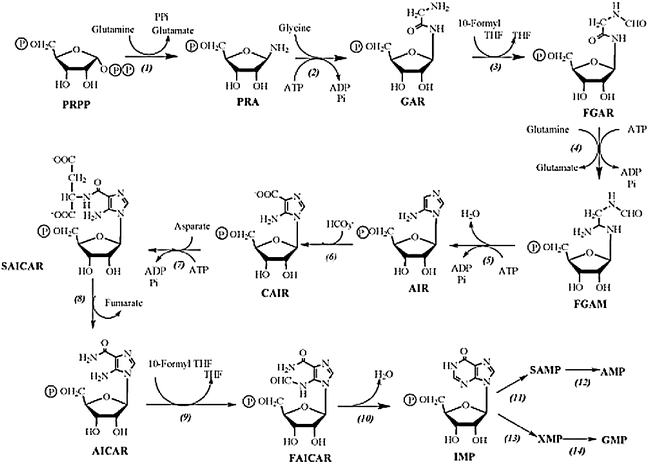
De novo biosynthetic pathway of purine nucleotides in plants. Enzymes shown are: amido phosphoribosyltransferase, (2) GAR synthetase, (3) GAR formyl transferase, (4) FGAM synthetase, (5) AIR synthetase, (6) AIR carboxylase, (7) SAICAR synthetase, (8) adenylosuccinate lyase, (9) AICAR formyl transferase, (10) IMP cyclohydrolase, (11) SAMP synthetase, (12) adenylosuccinase, (13) IMP dehydrogenase, (14) GMP synthetase.
Figure 2.
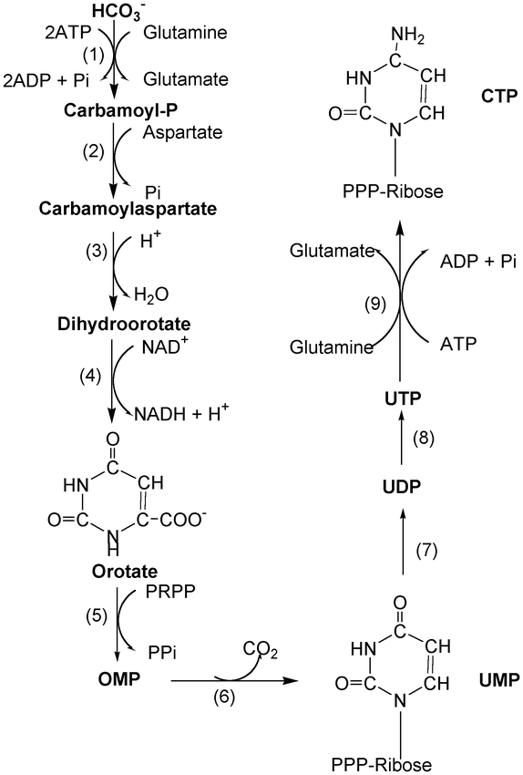
De novo biosynthetic pathway of pyrimidine nucleotides in plants. Enzymes shown are: (1) Carbamoyl phosphate synthetase, (2) aspartate transcarbamoylase, (3) dihydroorotase, (4) dihydroorotate dehydrogenase, (5)-(6) UMP synthase (orotate phosphoribosyltransferase plus orotidine-5′-phosphate decarboxylase), (7) UMP kinase, (8) nucleoside diphosphate kinase, (9) CTP synthetase.
Figure 3.
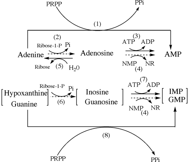
Salvage reactions of purine bases and nucleosides in plants. Enzymes shown are: (1) adenine phosphoribosyltransferase, (2) adenosine phosphorylase, (3) adenosine kinase, (4) adenosine phosphorylase, (5) nucleoside nucleosidase, (6) inosine-guanosine phosphorylase, (7) inosine-guanosine kinase, (8) hypoxanthine-guanine phoshoribosyltransferase. Solid arrows: major reactions; dashed arrows: minor reactions.
Figure 4.
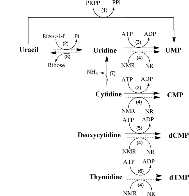
Pyrimidine salvage and related pathways in plants. Enzymes shown are: (1) Uracil phosphoribosyltransferase, (2) uridine phosphorylase, (3) uridine kinase, (4) nucleoside phosphotransferase, (5) deoxycytidine kinase, (6) thymidine kinase, (7) cytidine deaminase, (8) uridine nucleosidase. Solid arrows: major reactions; dashed arrows: minor reactions.
The following will summarize what is currently known about nucleotide biosynthesis and metabolism in Arabidopsis. Detailed descriptions of the early research in this area as well as additional references to studies of these pathways in plants other than Arabidopsis can be found in Takahashi and Suzuki (1977), Wasternack (1982), Rose and Last (1994) and Crozier and Ashihara (1999).
De novo purine nucleotide metabolism
The de novo pathway leading to the synthesis of AMP and GMP begins with the transfer of an amido group from glutamine to PRPP (Figure 1). Since PRPP is used for the both de novo and salvage synthesis of purine and pyrimidine nucleotides as well as for the synthesis of NAD, histidine and tryptophan, any stress that alters PRPP availability affects multiple pathways. Given the essential nature of PRPP, all free-living organisms contain at least one gene encoding PRPP synthetase (PRS; EC 2.7.6.1) (Krath and Hove-Jensen, 1999). Recently four PRS cDNAs were isolated from Arabidopsis by functional complementation of an Escherichia coli mutant lacking PRS activity (Genbank accessions X83764 (PRS1), X92974 (PRS2), AJ012406 (PRS3), AJ012407 (PRS4)) (Krath et al., 1999). A fifth PRS sequence that predicts a protein with high amino acid sequence homology to the other Arabidopsis PRSs, particularly with PRS1 and 2 was located on chromosome II and designated PRS5 (Krath et al., 1999). Kinetic characterization of the gene products of PRS1-4 indicated that PRS3 and 4 represent a novel class of PRSs since their activities are independent of inorganic phosphate (Pi). A phylogenetic comparison of 45 putative PRS amino acid sequences from 31 organisms also suggested that PRS3 and 4 are a divergent family, distinct from any of the other PRS sequences (Krath et al., 1999). The subcellular localization of the Arabidopsis PRSs has not yet been determined but may resemble the compartmentation of spinach PRSs that have been localized to the cytosol, chloroplast and mitochondria (Krath and Hove-Jensen, 1999).
In animal cells amido phosphoribosyltransferase or PRPP amidotransferase (ATase or PRAT; EC 2.4.2.14) catalyzes the first step in de novo purine synthesis and is sensitive to feedback regulation by purine ribonucleotides produced by the salvage cycle. Inhibition of ATase in cultured fibroblasts regulates not only purine de novo synthesis but also the rates of DNA and protein synthesis and cell growth (Yamaoka et al., 2001). While it is known that plant ATase is sensitive to feedback regulation (Ito et al., 1994) the impact of this inhibition on plant cellular metabolism has not been examined thoroughly. Isolation of cDNAs derived from two Arabidopsis ATase genes (Genbank accessions D28868 and D28869) of similar sequence by Ito and colleagues (1994) should be helpful in clarifying the activity and regulation of this enzyme. The two ATase sequences, designated AtATase1 and 2, were recovered in a screen for sequences preferentially transcribed in young floral buds. Whereas AtATase1 transcript levels are highest in flowers and roots and absent in leaves, AtATase2 transcripts are most abundant in leaves, only moderately expressed in flowers and very weakly accumulated in roots. The predicted amino acid sequences of these genes are most similar to the [4Fe-4S] cluster-dependent group of ATases that are activated by cleavage of a propeptide (Ito et al., 1994 and references therein). Since the Arabidopsis sequences contain the conserved residues for the propeptide cleavage site it is likely, but unproven, that the plant enzyme is activated similarly. The putative propeptides of the Arabidopsis sequences are particularly long and so may also contain additional functions such as a signal for targeting to the chloroplast (Ito et al., 1994). Senecoff et al. (1996) reported cloning an Arabidopsis ATase gene that they designated PUR1 since its product catalyzes the first step in de novo purine synthesis. However, the relationship of PUR1 to the genes described by Ito et al. (1994) has not been described. PUR1 transcripts are found in all organs with the highest levels accumulating in flowers, leaves and stems (Senecoff et al., 1996).
Using the same functional complementation strategy Schnorr et al (1994) cloned two additional cDNAs encoding purine pathway enzymes: glycineamide ribonucelotide (GAR) synthetase (Genbank accession: X74766; PUR2; EC 6.3.4.13) and GAR formyl transferase (Genbank accession X74767; PUR3; EC 2.1.2.2), respectively. Each cDNA is reported to code for a product with a single enzymatic domain, a structure similar to prokaryote purine enzymes whereas eukaryote genes typically encode bifunctional (PUR2, PUR5) (Henikoff, 1987) or trifunctional (PUR2, PUR5, PUR3) (Aimi et al, 1990; Henikoff, 1987; Henikoff and Furlong, 1983) enzymes. The prokaryotic, monofunctional enzyme structure predicted by the Arabidopsis PUR2 and PUR3 genes is consistent with the outcome of a phylogenetic comparison of their predicted amino acid sequences with those from other organisms, which shows these to be most similar to the corresponding sequences from bacteria (Schnorr et al., 1994).
PUR5 which encodes 5-aminoimidazole ribonucleotide (AIR) synthetase (Genbank accession: L12457; EC 6.3.3.1) was actually the first de novo plant purine gene to be characterized (Senecoff and Meagher, 1993). Based on Southern analysis, AIR synthetase is a single copy gene in the Arabidopsis genome. The region upstream of the first exon of PUR5 lacks a TATA box but does contain two initiator (INR) sequences (CTCANTCT) just downstream of the transcription start site that are thought to contribute to transcription initiation of PUR5. This region directs the synthesis of a 1.5 kb transcript that encodes a monofunctional protein with a basic, hydrophobic transit sequence consistent with the transport of PUR5 into chloroplasts. Phylogenetic analysis of eight other AIR synthetase amino acid sequences from animals, fungi and bacteria indicated that the Arabidopsis sequence has the highest homology with those from bacteria.
Senecoff et al. (1993) isolated cDNA sequences that suppress E. coli auxotrophs in purB, C and H (corresponding to PUR8/12, PUR7, and PUR9/10, respectively) but only purC complementing clones have described in detail (Senecoff et al., 1996). These clones encode 5-aminoimidazole-4-N-succinocarboxyamide ribonucleotide (SAICAR) synthetase (Genbank accession: U05599; EC 6.3.2.6), a single copy gene in the Arabidopsis genome. The virtual translation of the 1472 bp PUR7 cDNA is a 411 amino acid sequence that contains an N-terminal chloroplast transit sequence of 80–90 amino acids. Only weak conservation was found among SAICAR synthetase amino acid sequences from fungal, animal and bacterial sources with stronger similarity with the only other plant sequence from Vigna aconitifolia (Senecoff et al., 1996). Transcripts hybridizing to the PUR7 cDNA were found in all organs with the levels in flowers higher than in leaves and stems which are higher than in roots, siliques and pollen. The upstream region of the PUR7 gene lacks obvious signals for transcription initiation (no TATA box in −30 to −35 bp region and no INR sequences near the transcription start site), although it is clear that promoter elements are present since transgenic plants containing a PUR7::GUS translational fusion show high levels of GUS expression in actively-dividing regions of the root, shoot and flowers. Mitotically active cells nearest the stem also express the PUR7::GUS reporter (Senecoff et al., 1996). When these plants were treated with auxin, strong GUS expression was observed in the adventitious lateral roots that developed in response to auxin exposure. Growth of young seedlings in the presence of inhibitors of de novo purine biosynthesis (azaserine and diazooxynorleucine) was slower and was associated with increased GUS expression directed from the PUR7 upstream region. The authors suggested that the developmental delay associated with the application of de novo purine inhibitors may be due to a decrease in purine nucleotide levels and this may lead to increased PUR7 expression (Senecoff et al., 1996) although this remains to be investigated.
PUR11, which encodes adenylosuccinate synthetase (Genbank accession U49389; AdSS or PUR11; EC 6.3.4.4), was cloned by Fonne-Pfister et al. in1996. This enzyme catalyzes the first step of the two-step conversion of IMP to AMP and is of particular interest as it is inhibited by the antibiotic hadacidin (Stayton et al., 1983) and the microbial phytotoxin hydantocidin (Siehl et al., 1996). The structure of Arabidopsis AdSS was recently determined and found to be essentially identical to that of Triticum aestivum AdSS except for minor differences in regions away from the active site (Prade et al., 2000). Interestingly, although the structure of the Arabidopsis enzyme differs only slightly from that of the E. coli AdSS, these differences impart distinct kinetic properties to these enzymes, particularly with respect to binding of the co-factor, GTP (Prade et al., 2000).
The conversion of IMP to XMP is the rate-limiting step in the de novo synthesis of guanine nucleotides and is catalyzed by IMP dehydrogenase (IMPDH or PUR13; EC 1.1.1.205). The Arabidopsis gene encoding this enzyme was recently cloned and found to be 69% similar to human Type II IMPDH after allowing for conservative substitutions Collart et al. (1996). This enzyme is of particular interest due to its role in the production of ureides in legumes (see Purine nucleotide catabolism section).
Thus there are reports of Arabidopsis cDNA or genomic sequences corresponding to 12 of the 14 steps involved in de novo purine nucleotide synthesis depicted in Figure 1; genes coding for formyl glycine amidine ribonucleotide (FGAM) synthetase; (EC 6.3.5.3; PUR4) and GMP synthetase (EC 6.3.4.1; PUR14) have yet to be isolated. Of the genes that have been isolated, relatively few have been characterized in detail. The identification of mutants of these genes will provide insight into their roles in plant growth and the relationship of purine metabolism and other biochemical pathways (Thorneycroft et al., 2001).
Salvage synthesis of purine nucleotides
The salvage cycle interconverts purine bases, nucleosides and nucleotides released as by-products of cellular metabolism or from the catabolism of nucleic acids or nucleotide cofactors. This strategy for purine nucleotide synthesis is energetically favorable for a cell since only one salvage reaction requires ATP (phosphorylation of nucleosides to nucleotides). For example, bases and nucleosides released from storage organs during germination or by senscencing leaves are recycled by this pathway (for review see Ashihara and Crozier, 1999). Operation of the salvage pathway also reduces the levels of purine bases and nucleosides that may otherwise be inhibitory to other metabolic reactions.
Guanine/Guanosine recycling
The pathway that is believed to function in the salvage of guanine and guanosine is shown schematically in Figure 3. Unfortunately, there has been limited research on guanine/guanosine salvage cycles in plants and none of these studies used Arabidopsis. A recent, comprehensive study of guanosine metabolism in Catharanthus roseus cell cultures showed that guanosine is either recycled into guanine nucleotides or catabolized by conventional pathways to xanthine and allantoin, as occurs in animals and microorganisms (Ashihara et al., 1997). Descriptions of several of these activities including guanine phosphoribosyltransferase, guanosine nucleosidase and gunanosine deaminase from various plant sources have been reported (reviewed in Ashihara et al., 1997).
Adenine recycling
The enzymes that salvage adenine (Ade) and adenosine (Ado) have been studied to a greater extent than other purine enzymes, in part because the salvage cycle is thought to contribute also to the metabolism of cytokinins (CKs; Mok and Mok, 2001). Since CK bases/nucleosides are proposed to be the active form of this growth regulator, metabolism by the salvage cycle enzymes could affect the level of active hormone in a plant cell. Although progress has been made on characterizing adenine salvage metabolism, it remains unclear as to whether plant cells utilize these enzymes for CK interconversion in vivo. Further genetic analysis coupled with more sensitive measurements of the CK constituents in relevant mutants will be necessary to resolve this issue.
The principal route for adenine recycling is mediated by adenine phosophoribosyl-transferase (APT; EC 2.4.2.7) which converts adenine and PRPP to AMP and PPi. There are five sequences annotated as coding for APT or APT-like enzymes in the Arabidopsis genome. These have been designated APT1-5 (Genbank accessions L19637, X96867, AL033545, AL049730, AL360314, respectively). At present there is evidence for the expression of only APT1, 2 and 3. No ESTs specific for APT4 or 5 have been identified and those that are listed in their Genbank records are more similar to the other APT sequences. Also, transcripts for APT4 are not detectable by RT-PCR analysis of leaf or flower RNA (Moffatt, unpublished data). It should be noted that APT5 is less than 12% identical to APT1 at the amino acid level, raising the possibility that APT5 may utilize a substrate other than adenine, in vivo. APT1 is by far the most abundant isoform since an APT1 mutant (apt1–3) lacks 99% of the APT activity detected in wild-type leaves (Moffatt and Somerville, 1988).
The cDNAs encoding APT1, 2 and 3 were overexpressed in E. coli to compare the kinetic properties of each isoform on adenine and three CK substrates (zeatin, isopentenyladenine, benzyladenine) (M Allen, W Qin, F Moreau, BA Moffatt, submitted). All three isoforms bind adenine very efficiently based on their KM values (0.8–2.6 µM) although the Vmax estimates indicate that APT1 is approximately 30–50 times more efficient than either APT2 or 3 in converting adenine to AMP. The APT1KMs for these CKs are extremely high, around 1–2 mM, while the KMs of APT2 and 3 range from 15–370 mM depending upon the CK substrate, suggesting that APT1 is less likely to contribute to CK interconversion. However, while APT2 and 3 have much higher affinities for CKs than does APT1, APT1 has almost a 10-fold higher Vmax for each CK as compared with the other isoforms. Thus the parameter Vmax/KM, which is an estimate of catalytic potential of an enzyme, predicts that the three APT isoforms are very similar in their utilization of these CK substrates in vitro (M Allen, W Qin, F Moreau, BA Moffatt, submitted). Isolation of mutants deficient in APT2 or APT3 activities and direct measurement of the absolute levels of CKs and their route of synthesis in wild type and the apt1–3 mutant will be required to address whether any of these APTs actually contribute to CK interconversion in vivo.
None of the predicted amino acid sequences for the Arabidopsis APTs appear to contain transit signaling peptides although APTs have been localized to plastids and mitochondria in other plants (Hirose and Ashihara, 1982; Le Floc'h and Lafleuriel, 1983; Wasternack et al., 1985; Koshiishi et al., 2001). To investigate the subcellular localization of APT1, 2 and 3, peptide antibodies specific for each isoform were prepared and used to probe immunoblots of subcellular fractions of Arabidopsis leaves. Based on these analyses, APT1 and 3 are cytosolic while the data for APT2 were inconclusive (M Allen, W Qin, F Moreau, BA Moffatt, submitted).
Arabidopsis APT-deficient mutants were isolated using a direct selection scheme first developed for animal systems. For its application to plants, mutagenized seed were germinated in the presence the adenine analogue 2,6-diaminopurine (diap), a compound that is converted to a toxic nucleotide only by APT. Mutants lacking APT activity develop long roots and true leaves in the presence of diap. Using this selection several nuclear recessive mutant alleles of APT1 were recovered, the most deficient of which, apt1–3, is completely male sterile (Moffatt and Somerville, 1988). The APT1 cDNA (Moffatt et al., 1992) and gene (Moffatt et al., 1994) were subsequently isolated. The gene lacks traditional promoter elements and the predicted protein has no obvious chloroplast transit sequence. The apt1–3 mutation eliminates the splice acceptor site of the third intron of APT1 creating a BstNI RFLP (Gaillard et al., 1998).
The ability of apt1–3 seedlings to metabolize adenine and the CK benzyladenine (BA) to their corresponding nucleotides was evaluated by in vivo feeding studies (Moffatt et al., 1991). BA was rapidly taken up by wild-type seedlings and converted to BAMP and to BA 7- and 9-glucosides (which are considered to be storage forms of CKs). While mutant seedlings took up the BA effectively, its conversion to BAMP was reduced 8-fold and there was a higher accumulation of the glucoside forms relative to the wild type. Conversion of the riboside benzyladenosine (BAR) to BAMP was not altered in the mutant. Assuming that exogenously fed CKs are metabolized in a mechanism similar to the endogenous hormone, these results suggest that APT1 can contribute to the interconversion of CK bases to CK nucleotides in vivo. Kinetic characterization of homogeneous APT1 activity in vitro also suggested that it is capable of utilizing CK substrates, although its affinity for BA is quite low, based on a KM of 730 mM for this substrate (Lee and Moffatt, 1993). As mentioned above, further characterization of the profile of CKs in this mutant will be necessary to elucidate whether it APT1 contributes to their interconversion.
Since the apt1–3 mutant germinates more slowly than wild type and induces callus less efficiently, the involvement of APT in these processes was also investigated (Lee and Moffatt, 1994). APT activity increases early during germination and callus induction and then declines to basal levels within about 10 days. In mature plants, APT activity, expressed as a specific activity or on a dry weight basis is highest in organs with meristems (roots and flowers). A similar expression pattern is observed in plants transformed with a construct expressing b-glucuronidase (GUS) from the APT1 promoter region. GUS activity in the APT1p::GUS lines is particularly high during cambial development in roots and stems (SR Regan, A Smith, L Pereira L, BA Moffatt, unpublished). Increased APT activity in meristematic regions or rapidly dividing cells is consistent with the recovery of multiple APT sequences from EST libraries prepared from cambial tissue of poplar (Sterky et al., 1999) and the increases in APT transcripts detected in microarray analysis of RNA from leaves versus cell cultures (http://genome-www4.stanford.edu/cgi-bin/MDEV/mdev.pl; experiments # 6922, 6923, 6925, 6927, 9722) and in leaves versus flowers (experiment #2371). At this point it is not clear whether these increases in APT activity occur to meet metabolic demand for adenine recycling into adenylates, interconversion of CKs, or both.
The basis of the male sterile phenotype of APT1-deficient mutants has been investigated by light microscopy (Regan and Moffatt, 1990) and more recently by electron microscopy (Zhang et al., 2001). Histochemical staining indicates that the first defects in pollen development in apt1–3 mutants occurs when the microspores are released from the tetrad, just after meiosis. At this stage mutant microspores have abnormal cell walls that do not stain normally with histochemical stains for intine development. Subsequent vacuole formation is delayed and the microspores begin to visibly deteriorate; very few undergo the mitotic divisions. Vital stains indicate that mutant microspores are less metabolically active following meiosis. Subsequent ultrastructural analysis by electron microscopy revealed changes in both the tapetum and the microspores prior to the onset of meiosis. The microspores of the mutant appear to initiate meiosis earlier than in the wild type. Based on studies in other plants, CKs may contribute to the signal for the onset of meiosis suggesting that the apt1–3 mutants may experience a transient increase in CKs due to reduced metabolism by APT1. The later defects in pollen development are most easily explained by a deficiency in adenylates. Thus the male sterililty in these mutants may be a result of defects in both CK and Ade metabolism.
In situ hybridization has been used to investigate why APT2 and 3 do not compensate for APT1 deficiency in floral organs even though transcripts for each enzyme are abundant in flowers (C Zhang, L Pereira, BA Moffatt, unpublished data). The results clearly indicate that each isoform is predominantly expressed in different tissues: APT1 transcripts are found in all cells but to a much higher extent in microspores just prior to meiosis, APT2 transcripts are present at very low levels in the receptacle while APT3 transcripts are present predominantly in the ovule. The transcript levels of each APT gene increase in the apt1–3 mutant although the physiological basis for this increase has not yet been identified.
Adenosine recycling
The reutilization of Ado into AMP by the salvage pathway augments intracellular adenylate pools while simultaneously reducing the level of free Ado. Ado arises not only from the catabolism of nucleic acids and nucleotide cofactors, but also as a by-product of methylation reactions that use S-adenosylmethionine (SAM) as the methyl donor. For example, pectin, lignin, phosphatidylcholine and nucleic acids are all methylated in reactions that are catalyzed by specific methyltransferases dependent on SAM. One molecule of S-adenosylhomocysteine (SAH) is produced for each methyl group that is transferred from SAM. Since SAH is a competitive inhibitor of SAM-dependent methyltransferases, it must be continuously metabolized by SAH hydrolase to maintain transmethylation activities. SAH hydrolase converts SAH to Ado and homocysteine; the homocysteine is recycled to methionine and SAM while the Ado is used for nucleotide synthesis. The reaction catalyzed by SAH hydrolase is reversible and its equilibrium lies in the direction of SAH synthesis; it is only drawn in the direction of SAH hydrolysis by the removal of the products Ado and homocysteine. Moreover, since both homocysteine and Ado are product inhibitors of SAH hydrolase thus they must also be steadily removed in order to regenerate SAM and the adenylate pools. Should Ado levels increase, this would inhibit SAH hydrolase and lead to an increase in SAH that would inhibit transmethylases. Thus Ado metabolism is critical for the maintainence of methyl utilization and recycling (for review see Moffatt and Weretilnyk, 2001).
Ado is recycled by Ado kinase (ADK; EC 2.7.1.20), nucleoside phosphotransferase (EC 2.7.1.77) or Ado nucleosidase (EC 3.2.2.7) (Figure 3). Alternatively it can be converted to inosine by Ado deaminase (Figure 5; ADA; EC 3.5.4.4). Although animal cells rely principally on ADA for Ado recycling, plants primarily use ADK. This conclusion is based on several observations. Both ADA (Yabuki and Ashihara 1991; BA Moffatt, unpublished) and nucleoside phosphotransferase (Hirose and Ashihara, 1984) activities are very low (or below the limit of detection) relative to ADK (Moffatt et al., 2000). There is only one Arabidopsis EST with sequence similarity to ADAs of other organisms whereas 29 ADK ESTs are present currently in Genbank; a severe phenotype is associated with ADK deficiency (BA Moffatt, Y Stevens, M Allen, J Snider, PS Summers, EA Weretilnyk, L Martin-McCaffrey, LA Pereira, M. Todorova, C Wagner, unpublished) and presumably these plants would be less abnormal if there were other enzymes capable of metabolizing Ado efficiently. Auer (1999) reported the in vitro characterization of a nucleosidase activity in Arabidopsis that prefers Ado but also accepts several CK nucleosides as substrates. The cym mutant that is deficient in this nucleosidase activity develops normally but young seedlings are impaired in their metabolism of exogenously-fed BAR (Auer, 1999). Further characterization of this mutant will reveal the in vivo role of this nucleosidase in Ado and CK metabolism.
Figure 5.
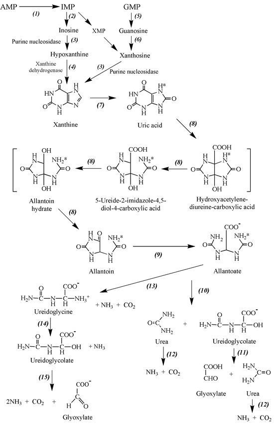
Catabolism of purine nucleotides in plants. Enzymes shown are: (1) AMP deaminase, (2) IMP dehydrogenase, (3) 5′-nucleotidase, (4) inosine-guanosine nucleosidase, (5) guanosine deaminase, (6) guanine deaminase, (7) xanthine dehydrogenase, (8) uricase, (9) allantoinase, (10) allantoicase, (11) ureidoglycolate lyase, (12) urease, (13) allantoin deaminase, (14) ureidoglycine amidohydrolase, (15) ureidoglycolate hydrolase.
ADK-deficient lines created by sense and antisense silencing have wavy leaves and short internodes and stamen filaments (BA Moffatt, Y Stevens, M Allen, J Snider, PS Summers, EA Weretilnyk, L Martin-McCaffrey, LA Pereira, M Todorova, C Wagner, unpublished). This phenotype is similar to that of tobacco lines deficient in SAH hydrolase activity (Tanaka et al., 1997). In both cases, there is reduction in SAM-dependent methylation: ADK-deficient plants have less methylated pectin in their seed mucilage and SAH hydrolase-deficient lines have less methylated DNA. The current hypothesis to explain the phenotype of the ADK lines is that reduced Ado recycling leads to Ado inhibition of SAH hydrolase activity and SAH accumulation, ultimately causing inhibition of SAM-dependent transmethylation activities.
ADK and SAH hydrolase transcript levels and activities increase when methylation demand increases, such as in plants that accumulate methylated osmolytes in response to salinity stress or in lignifying cells (Weretlinyk et al., 2001). This suggests that although ADK is a housekeeping enzyme, its level of expression is dynamic so as to meet changes in methylation activity and maintain methyl recycling. The direct correlation between the level of pectin methylation and ADK activity in the ADK-deficient lines indicates that ADK has become limiting for methyl recycling activity in these plants.
Further characterization of ADK- and SAH hydrolase-deficient lines will reveal which aspects of plant metabolism rely on Ado recycling and how ADK activity changes in response to methylation demand.
The Arabidopsis genome encodes two ADK proteins that are 92% identical at the amino acid level (Genbank accessions, ADK1: AF180896, ADK2: AF180897) (Moffatt et al., 2000). These genes are transcribed in all organs tested, with the levels of ADK1 transcripts being slightly higher in most cases. ADK enzyme activity when expressed per mg protein is highest in stems and maturing flowers. Each ADK cDNA was over-expressed in E. coli to evaluate the catalytic activity of its protein product towards the substrates Ado and the CK isopentyladenosine ([9R]iP). The two isoforms have almost indistinguishable activity on Ado: KM and KM /Vmax for Ado of 0.3–0.5 mM and 5.4–22 L min-1mg-1 protein, respectively. Both enzymes prefer Ado as a substrate over [9R]iP as evidenced by their 10-fold lower KMs towards Ado. The only notable differences between the two isoforms are that ADK2 has a 10-fold higher Vmax on [9R]iP and it is sensitive to substrate inhibition by Ado concentrations above 2 mM whereas ADK1 is not sensitive at 5 mM. Since the endogenous CK levels in Arabidopsis are about 103-fold lower than are the apparent KMs of these ADKs for [9R]iP (Åstot et al., 1999), it is likely that their primary substrate in vivo is Ado.
There is at least one other ribokinase encoded in the Arabidopsis genome that is capable of metabolizing both CK nucleosides and Ado in vitro. If CK nucleosides are the active form of the hormone this ribokinase would act to reduce CK levels. Interestingly plants lacking this enzyme activity exhibit a high CK phenotype (Parsons, et al., 1999). Conclusive proof that this ribokinase is the principal enzyme that converts CK ribosides to nucleotides awaits its direct biochemical analysis.
Catabolism of purine nucleotides
The end products of purine catabolism are different in different species. For example, uric acid is the end product of higher primates including man, however, allantoin is formed in other mammals (Henderson and Paterson, 1973). In most plants, purine nucleotides are degraded via ureides, allantoin and allantoate to NH3 and CO2 by the conventional purine catabolic pathway (Figure 5). In specific organs (e.g., roots) of ureide-accumulating plants, allantoin and/or allantoate are the end products of this pathway and they are translocated to other parts of the plant, such as shoots and leaves, where they are degraded completely. So, in contrast to biosynthesis of purine nucleotides, catabolism of purines is diversified in different species and organs. Furthermore, various enzymes that participate in each step of catabolism exist in nature, and some enzymes commonly found in animals are missing in plants. A typical example is ADA, which is widely distributed in animals, but this enzyme is not present in most plants. In some bacteria, Ade deaminase can participate in the deamination of Ade ring (see Ashihara and Crozier, 1999). There is no research on the purine catabolic pathway in A. thaliana and only a few putative genes encoding the enzymes of purine catabolism have been characterized.
The initial step of Ade nucleotide catabolism is deamination of AMP to IMP, catalyzed by AMP deaminase; the product is dephosphorylated to inosine, which, in turn, is hydrolysed to hypoxanthine (Figure 5). These conversions are catalyzed by 5′-nucleotidase (and/or phosphatase) and inosine/guanosine nucleosidase (Atkins et al., 1989). ATP is essential for higher plant AMP deaminase activity, and thus, catabolism of adenylates is dependent on the cellular ATP level (Yabuki and Ashihara, 1991; 1992). Glycosidic bond cleavage of purine nucleosides is accomplished either hydrolytically or phosphorolytically in nature. In plants hydrolytic cleavage is the most common. An alternative route of xanthine formation has been proposed to occur in soybean nodules. In nodule cells IMP is initially converted to XMP by IMP dehydrogenase and then dephosphorylated (Schubert and Boland, 1990). Therefore, two possible routes of xanthine formation from IMP may be operative in plants (i) IMP Æ inosine Æ hypoxanthine Æ xanthine and (ii) IMP Æ XMP Æ xanthosine Æ xanthine (Figure 5).
The major pathway for the catabolism of guanine nucleotides begins with a dephosphorylation reaction that yields guanosine. Guanosine deaminase then catalyzes the conversion of guanosine to xanthosine, which is further catabolized to xanthine by purine nucleosidase (Figure 5). Hypoxanthine and/or xanthine are converted to uricate by xanthine oxidoreductase. There are two forms of xanthine oxidoreductase. One form has a requirement for molecular oxygen and is called xanthine oxidase (EC 1.2.3.2). The other form, xanthine dehydrogenase (EC 1.2.1.37), is an NAD-dependent enzyme. In higher plants, oxidation of hypoxanthine and xanthine seems to be catalyzed by xanthine dehydrogenase, although the two forms of xanthine oxidoreductase may be interconverted (see Nguyen, 1986). Allopurinol (4-hydroxypyrazolo(3,4-d)pyrimidine), a specific inhibitor of xanthine oxidoreductase (Weir and Fischer, 1970) is often used as an inhibitor of purine catabolism in studies with higher plants (Fujiwara and Yamaguchi, 1978; Ashihara, 1983; Hammer et al., 1985).
Several intermediates in the degradation of uric acid are either known or their existence in plants has been postulated (Figure 5). Allantoin amidohydrolyase (allantoinase) catalyzes the hydrolysis of the internal amide bond of allantoin resulting in its conversion to allantoate. This enzyme is found in many plant species (Schubert and Boland, 1990). The classic pathway is the “allantoate amidohydrolase (allantoicase)” pathway in which allantoate is hydrolysed to urea and ureidoglycolate. Ureidoglycolate is further degraded to glyoxylate and urea. Urea formed via this pathway may be hydrolysed to ammonia and CO2 by urease (Figure 5, steps 10–12). However, recent studies suggest the existence of an alternative route, the “allantoate deiminase (allantoate amidohydrolase)” pathway (Winkler et al., 1988; Schubert and Boland, 1990). In this pathway allantoate is hydrolysed to CO2, NH3 and ureidoglycine. Ureidoglycine is unstable and can be deaminated, either spontaneously or enzymatically, to ureidoglycolate which is cleaved by ureidoglycolate amidohydrolase to produce NH3, CO2 and glyoxylate (steps 13–15). In the earlier studies, urea was often found as a degradation product of allantoin, however, Winkler et al. (1988) pointed out non-enzymatic degradation of allantoate to urea can occur.
Two distinct purine nucleosidase enzymes, Ado nucleosidase and inosine-guanosine nucleosidase (EC 3.2.2.2), appear to be participating in ribose cleavage of purine nucleosides in plant cells. The former is specific for Ado, while the latter enzyme hydrolyses guanosine, inosine and xanthosine (Le Floc'h and Lafleuriel, 1981; Guranowski, 1982). Ado nucleosidase seems to be growth specific enzyme, because its activity is absent in seeds and embryos, but appears during germination of seeds and somatic embryos (Guranowski and Barankiewicz, 1979; Stasolla et al. 2001). Ade is not degraded further, because Ade deaminase is not present in plants. On the other hand, guanine seems to be catabolized to xanthine and enters the purine catabolic pathway, as guanine deaminase is present in plant cells (Ashihara and Crozier, 1999). The Arabidopsis genes encoding these enzymes have not yet been isolated or characterized.
De novo synthesis of pyrimidine nucleotides
De novo pyrimidine nucleotide biosynthesis which is also referred to as the “orotate pathway” is usually defined as the formation of UMP from carbamoyl phosphate (CP). Although the sequence of events for biosynthesis of pyrimidine nucleotides in plants is essentially the same as that in animals and microorganisms (Wagner and Baker, 1992), the organization, the control mechanism, and subcellular localization of the enzymes of this pathway in plants are different from those in other organisms. The orotate pathway consists of the six reactions as shown in Figure 2. The initial reaction catalyzed by CP synthetase (CPS) is the formation of CP by combination of carbonate, ATP and an amino group from glutamine. Three additional reactions are necessary to form the pyrimidine ring from CP. The phosphoribosyl group of PRPP is added to the pyrimidine base, orotate, forming orotidine 5′-monophosphate (OMP) which is decarboxylated to make UMP, the first pyrimidine nucleotide. UMP is subsequently phosphorylated to UDP and UTP. The transfer of an amino group from glutamine to UTP by CTP synthetase leads to the synthesis of CTP.
Here we review the biochemical properties and genes of each enzyme involved in the pyrimidine nucleotide biosynthesis de novo.
Carbamoyl phosphate synthetase (CPS, EC 6.3.5.5)
CP is the substrate for the biosynthesis of both pyrimidine nucleotides and arginine and thus this compound is not a unique starting material for pyrimidine base formation. Nevertheless, the formation of CP is the most important regulatory step in pyrimidine biosynthesis. Most eukaryotes except plants have two types of CPS. Ammonia-specific CPS (CPS I) contributes to arginine biosynthesis and its activity is stimulated by N-acetyl-L-glutamate that is produced as an intermediate of ornithine biosynthesis. In mammals, this enzyme is located in mitochondria of ureotelic liver and is aimed primarily at the formation of urea. A second CPS (CPS II), which preferentially utilizes glutamine as a substrate and does not require N-acetyl-L-glutamate as a co-factor, contributes to the synthesis of pyrimidines. In contrast to CSP I, CSP II is present in the cytosol of all dividing cells of mammals (Tatibana, 1978).
Biochemical studies indicate that higher plants have only a single form of CPS which is proposed to provide CP for the biosynthesis of both pyrimidine nucleotides and arginine (Wasternack, 1982, Sasamoto and Ashihara, 1988). Thus, plant CPS is subject to multiple types of regulation. Its activity is inhibited by UMP and activated by ornithine and PRPP (Ong and Jackson, 1972; O'Neal and Naylor, 1976; Kanamori et al., 1980). These compounds provide feed-back (UMP) and feed-forward (PRPP) control of pyrimidine biosynthesis. UMP inhibition of CPS may be overcome by ornithine leading to the utilization of CP for arginine biosynthesis (O'Neal and Naylor, 1976).
The properties of plant CPS are very similar to those of E. coli CPS, where a single CPS supplies CP to both the pyrimidine and arginine pathways and its activity is regulated by UMP and ornithine (Nygaard and Saxild, 2000). These effectors bind to the large subunit of E. coli CPS, which is encoded by the carB gene. The small subunit of this bacterial enzyme, encoded by the carA gene, exhibits a glutamine amidotransferase (GAT) activity that hydrolyzes glutamine to glutamate, providing ammonia to the large subunit. Molecular cloning of CPS from plants indicates a similar gene structure in these organisms. CPS large (carA) and small subunits (carB) are encoded by individual genes in the Arabidopsis nuclear genome (Williamson et al., 1996, Brandenberg et al., 1998). Alignment of the deduced sequence for the Arabidopsis small subunit with sequences for small subunit or GAT domains of other CPS sequences in Genbank suggests that the plant protein is synthesized as a precursor containing a chloroplast transit sequence (Newman et al., 1994). This is consistent with the biochemical evidence for the chloroplast localization of CPS activity (Shibata et al., 1986). The putative mature Arabidopsis small subunit protein sequence is 69% identical to an Alnus root nodule carA protein (Lundquist et al. 1996) and is highly homologous to the E. coli CPS small subunit protein (46% amino acid identity). The deduced amino acid sequence of Arabidopsis carB is also highly homologous to the E. coli carB product (56% identity). The calculated molecular masses of carA and carB proteins are 40 kDa and 120 kDa, respectively; which are similar to those that have been reported for other CPS small and large subunits. There has been one report suggesting the possible existence of both CPS I- and CPS II-type CPS in alfalfa (Maley et al., 1992), however, this has not yet been confirmed.
In most eukaryotes including mammals, CPS II resides within two superdomains of a multifunctional protein that contains CPS II, aspartate transcarbamoylase (ATC) and dihydroorotase (DHO) activities (Christopherson and Szabados, 1997). The protein is abbreviated CAD to reflect the first letter of its constituent enzymes. However there are no reports of a similar multifunctional protein being present in plants or E. coli.
Aspartate transcarbamoylases (ATC, EC 2.1.3.2)
Conversion of CP to carbamoylaspartate, the first committed step of de novo pyrimidine biosynthesis, is catalyzed by aspartate transcarbamoylase (ATC). E. coli ATC is subjected to allosteric regulation and is the rate-limiting step for UMP synthesis (see Henderson and Patterson, 1973). However, this step is not likely to the major rate-limiting site for pyrimidine biosynthesis in animals and plants because the activity of this enzyme is in great excess over that of the other enzymes of the pathway (Henderson and Patterson, 1973, Kanamori et al., 1980). However, high concentrations of UMP inhibit the activity of plant ATC in vitro, by binding directly to the catalytic subunits of plant ATC homotrimer (Cole and Yon, 1984). In this situation cells need not to produce UMP, and CP, a substrate of ATC, is then used for the arginine biosynthesis. Thus, inhibition of ATC by UMP seems to be important in partitioning CP between pyrimidine and arginine synthesis.
Williamson and Slocum (1994) cloned cDNAs from pea that encode two different isoforms of ATC (pyrB1 and pyrB2), by complementation of an E. coli delta pyrB mutant. The two cDNAs encode polypeptides of 386 and 385 amino acid residues, respectively that contain typical chloroplast transit peptide sequences. Northern analyses indicates that the pyrB1 and pyrB2 transcripts are 1.6 kb in size and are differentially expressed in pea tissues. The small transcript sizes and data from biochemical studies indicate that plant ATCs are simple homotrimers of 36kDa catalytic subunits (Bartlett et al., 1994; Khan et al., 1999). Williamson et al. (1996) cloned a cDNA encoding a third pea ATC (pyrB3), whose deduced amino acid sequence is very close to that of pyr B2 (96% identity). cDNAs encoding ATCs have been cloned also from wheat (Bartlett et al., 1994) and Arabidopsis (Nasr et al., 1994) although the latter have not been characterized.
Dihydroorotase (DHO, EC 3.5.2.3)
Dihydroorotase (DHO) is the third enzyme in the highly conserved de novo pyrimidine biosynthetic pathway. This enzyme catalyzes the reversible conversion of carbamoyl aspartate into dihydroorotic acid. Sequences encoding DHO have been isolated from various organisms including A. thaliana (Nasr et al., 1994). As outlined above, the dihydroorotase of mammals (Simmer et al., 1990), Drosophila melanogaster (Freund and Jarry, 1987) and Dictyostelium discoideum (Faure et al., 1989), is part of a multifunctional protein called CAD. On the other hand, the DHOs of plants, S. cerevisiae (Guyonvarch et al., 1988), Chlorella (Dunn et al., 1977) and some fungi (Spanos et al., 1992), are monomeric proteins that are not physically linked with other enzymes.
The full-length Arabidopsis cDNA encoding DHO is 1,362 nucleotides long and predicts a protein of 377 amino acids (Genbank accession AF000146) which shares high identity with prokaryotic dihydroorotase enzymes and moderate identity with the eukaryotic enzymes (Zhou et al., 1997). The three conserved domains found in other DHOs (Guyonvarch et al., 1988) are also found in the protein encoded by this Arabidopsis cDNA: Domain A (DWHLRDGDL), Domain B (AIVMPNLKPPVTS) and Domain C (FLGTDSAPHERSRK). The histidines of Domain A are thought to coordinate the catalytic zinc that is associated with the enzyme (Brown and Collins, 1991). This suggests that the plant DHO has a catalytic mechanism similar to that of the E. coli and S. cerevisiae enzymes (Zhou et al., 1997).
Dihydroorotate dehydrogenase (DODH; EC 1.3.99.11)
The fourth reaction of de novo pyrimidine biosynthesis is the conversion of DHO to orotate. DODH is thought to catalyze this reaction (Jones, 1980). DODH was found to be located on the outer surface of the inner membrane of mitochondria in mammals (Jones, 1980). Although there are no detailed studies of a similar enzyme in plants, Miersch et al. (1986) suggested that tomato DODH is also located in mitochondria.
Uridine 5′-monophosphate synthase (UMPS, EC 2.4.2.10 plus 4.1.1.23)
Orotate is converted to UMP in two successive reactions catalyzed by orotate phosphoribosyl transferase (OPRT) and orotidine 5′-monophosphate decarboxylase (ODCase). As the intermediate of these steps, orotidine-5′-monophosphate, was not detected in plant tissue, and these two activities co-purified through several purification steps, it was suggested that these two enzymes form a complex in vivo (Ashihara, 1978; Walther et al., 1984). In fact, recent results demonstrate that OPRT and ODCase reside in a single polypeptide that is termed UMP synthase. This bifunctional enzyme catalyzes the last two steps of the de novo pyrimidine pathway in plants as well as mammals (Santoso and Thornburg, 1992). This structure improves the efficiency of these reactions by channeling the product of the first reaction to the second enzyme without dissociation from the complex. In most organisms, except some parasitic protozoans, the N-terminal portion of this bifunctional enzyme has sequence identity with OPRT while the C-terminal region has identity with ODCase (Suttle et al., 1988, Schoeber et al., 1993, Nasr et al., 1994, Maier et al., 1995). In some parasitic protozoans the order of the activities within the enzyme is reversed (Gao et al., 1999) suggesting that a bifunctional UMPS has arisen more than once during the course of evolution.
Santos and Thornburg (1992) isolated a Nicotiana tabacum UMPS cDNA with an open reading frame of 461 amino acids. Southern blot analysis indicates that there is only one UMPS sequence in the N. plumbaginifolia genome and two in that of N. tabacum. More recently rice UMPS cDNAs have been recovered from the collection of MAFF (Ministry of Agriculture, Forestry and Fisheries) DNA database (Japanese Rice Genome Project) based on their sequence homology with the N. tabacum cDNA. Two different rice UMPS cDNAs, Os-umps1a (AF210322) and Os-ump2 (AF210325) were characterized. While Os-umps1a appears to encode both enzyme activities, the Os-ump2 gene product is predicted to have only ODC activity due to an internal deletion within the OPRT region of the enzyme (Maier et al., 1995; Park and Thornburg, 2000). A phylogenetic analysis of 11 different UMPS amino acid sequences revealed three clades: one containing the mammalian sequences, one formed of plant sequences (with branches for monocots and dicots) and a third consisting of the slime mold and Drosophila sequences (Park and Thornburg, 2000). The evolutionary implications of the mosaic pyrimidine-biosynthetic pathway in eukaryotes have been described by Nara et al. (2000). During evolution of eukaryotes, plants and fungi in particular may have secondarily acquired the characteristic enzymes of this pathway. This conclusion is based in part on the finding that phylogenetic classification of plant pyrimidine biosynthetic enzymes is highly chimaeric. For example, although the two CPS subunits cluster with a clade including sequences from cyanobacteria and red algal chloroplasts, ACT sequences do not fall in the clade with cyanobacteria and DHO sequences group within a clade containing proteobacterial sequences. In fungi, DHO and OPRT cluster with their corresponding proteobacterial counterparts.
Formation of uridine-5′-triphosphate (UTP) and cytosine nucleotides from UMP
UMP, the product of the de novo pyrimidine nucleotide biosynthetic pathway, is further phosphorylated by kinases to form UTP. Cytidine-5′-triphosphate (CTP) is formed by an amination of UTP. The activities of enzymes that participate the conversion of UMP to UTP are very high in plant cells (Hirose and Ashihara, 1984) and as a result, the level of uracil nucleotides is equilibrated in cells and tissues. UMP kinase and a non-specific nucleoside diphosphate kinase have been characterized in plants as follows.
UMP kinase (UMPK, EC 2.7.4.4)
UMPK catalyzes a phosphoryl group transfer from ATP to either UMP or CMP to produce UDP and CDP, respectively. This enzyme has been studied from a variety of bacterial sources (Valentin-Hansen, 1978, Yamanaka et al., 1992, Serina et al., 1995, Serina et al., 1996). The bacterial enzyme is allosterically regulated by both GTP and UTP (Serina et al., 1995). Recently, an A. thaliana cDNA encoding UMPK was isolated, expressed in E. coli and the recombinant UMPK was characterized (Zhou et al., 1998, Zhou and Thornburg, 1998). The plant UMPK is insensitive to GTP and UTP. Eukaryotic UMPKs all share a conserved glycine-rich sequence in their N-terminal regions which is referred to as the phosphate-binding loop and may play a role in ATP binding and/or enzyme catalysis (Muller-Dieckmann and Schulz, 1994, Muller-Dieckmann and Schulz, 1995, Scheffzek et al., 1996). Site-specific mutations within this glycine-rich conserved region of the Arabidopsis enzyme resulted in significant changes in its catalytic activity. Mutations that showed reduced ATP binding showed increased UMP binding (Zhou and Thornburg, 1998). The cDNAs encoding UMPK of rice and Arabidopsis share roughly equivalent identity to the enzymes from yeast (45.5% and 49.7%), Dictyostelium (48.9% and 50.5%), and mammals (55.6% and 53.0%, respectively) (Park et al., 1999).
Nucleoside diphosphate kinase (NDPK, EC 2.7.4.6)
Conversion of UDP to UTP is performed by NDPK. Substrate specificity of NDPK is low, and the g-phosphate group of ATP (the most abundant nucleotide in cells) is rapidly redistributed to other nucleotides to form various nucleoside triphosphates. Thus the reaction of nucleoside diphosphate kinases can be generalized as follows: NDP + ATP ´ NTP + ADP. The activity of NDPK is very high in most organisms including plants (Hirose and Ashihara, 1984), and the equilibrium constant is almost unity. NDPK activity is not known to be regulated by any allosteric effectors In addition to NDPK's primary role as a catalyst, many other biological phenomena appear to rely on NDPK activities. NDPK has a protein kinase activity, which can phosphorylate both serine/threonine and histidine/aspartate residues (Engel et al., 1995; Wagner and Vu, 1995; Freije et al., 1997; Wagner et al., 1997). Other functions, such as activating G-proteins, have also been suggested (Bominaar et al., 1993). In humans, NDPK was identified as the tumor suppressor, nm23 (DeLaRosa et al., 1995). Yi et al. (1998) cloned and sequenced a nucleoside diphosphate kinase 2 (ndpk2) from A. thaliana which encodes a protein of 231 amino acids. This amino acid sequence shows high similarity to two other plant NDPK2 genes (73% spinach NDPK2 and 70% pea NDPK2). A BLAST search of the Arabidopsis genome shows that the genomic DNA sequence of an Arabidopsis MDC12 clone (Accession No AB008265) contains a partial sequence of the ndpk2 gene and indicates that the ndpk2 gene is located on chromosome 5. Reports suggest that gene might encode a phytochrome-interacting protein and its further characterization will shed light on the role of this multifunctional protein in plant development (Yi et al., 1998).
CTP synthetase (CTPS, EC 6.3.4.2)
CTP formation from UTP is catalyzed by CTPS in the following reaction: UTP + ATP + glutamine à CTP + ADP + glutamate + Pi. This reaction requires ATP and glutamine in addition to GTP which is a strong activator of this enzyme (Weinfeld et al., 1978). A gene encoding a CTP synthetase-like protein has been annotated on chromosome 4 of A. thaliana (Atg02120) but has not yet been characterized.
Pyrimidine salvage
As the de novo pyrimidine biosynthetic pathway is energy consuming, plant cells reutilize pyrimidine bases and nucleosides derived from the preformed nucleotides (Figure 4). Of the bases, only uracil is directly reused via a specific phosphoribosyltransferase whereas the pyrimidine nucleosides, uridine, cytidine and deoxycytidine are exclusively salvaged to their respective nucleotides, UMP, CMP and dCMP. High activity of uridine/cytidine kinase and nucleoside phosphotransferase in plants may contribute the salvage of these nucleosides (Kanamori-Fukuda et al., 1981).
Uracil Phosphoribosyltransferase (UPRT, EC 2.4.2.9)
Uracil is converted directly into UMP by the action of UPRT which transfers the phosphoribosyl moiety from PRPP to uracil to form UMP (Bressan et al., 1978).
Zhou et al. (1998) isolated an A. thaliana cDNA encoding a UPRT of 198 amino acids which is structurally and functionally similar to other phosphoribosyltransferases and is predicted to be localized in the cytoplasm. This subcellular localization is consistent with that of the enzyme catalyzing the next step in pyrimidine metabolism, UMP kinase. Comparison of the Arabidopsis UPRT sequence with those identified from sources including four species of bacteria and three species of eukaryotes revealed that sequences clustered into two distinct gene subfamilies. A. thaliana UPRT1 is most closely related to the Toxoplasma gondii and S. cerevisiae enzymes.
Uridine kinase (UK, EC 2.7.1.48)
Uridine and cytidine are converted to UMP and CMP by UK. The activity of UK is usually higher than that of UPRT and thus uridine is more efficiently salvaged to UMP than is uracil (e.g. Kanamori-Fukuda et al. 1981; Ashihara et al., 2000). UK has been partially purified from higher plants and its enzymatic properties have been well characterized (Deng and Ives, 1972 and 1975). However, there have been no reports of the isolation of UK coding sequences or genes. Deoxycytidine kinase (EC 2.7.1.74) and thymidine kinase (EC 2.7.1.21) are also present in plants, but details of their activities have yet to be described.
Thymidine kinase (TK, EC 2.7.1.22)
Genes encoding TK have been identified in prokaryote and mammalian genomes, but have not been found in the genome of Saccharomyces cerevisiae. Ullah et al. (1999) isolated a rice gene encoding TK based on its sequence similarity with the genes from prokaryote and mammalian genomes. Although the expression of animal TK genes is correlated with cell cycle activity (Dou and Pardee, 1996) this does not appear to be the case for the rice gene since TK mRNA levels are higher in mature tissues than in meristematic tissues. It was speculated that TK in plants might function in DNA repair in cells exposed to UV light (Ullah et al., 1999).
Nucleoside phosphotransferase (NPT, EC 2.7.1.77)
Non-specific nucleoside phosphotransferase activity also participates in the salvage of pyrimidine nucleosides and is widely distributed in plants (Deng and Ives 1972). Thymidine kinase activity measured in crude plant extracts is due, at least in part, to the activity of NPT and phosphatases (Arima et al., 1971; Mullin & Fites, 1978).
Biosynthesis of thymidine nucleotides
Thymidine nucleotide is synthesized from dUMP by the following reaction catalyzed by thymidylate synthase (TS, EC 1.5.1.3) : dUMP + N5, N10-methyltetrahydrofolate à dTMP + dihydrofolate. In this reaction, N5, N10-methyltetrahydrofolate which is produced by dihydrofolate reductase (DHFR, EC 1.5.1.3) acts both as donor of the methyl group and reducing agent. Although in most organisms including fungi and mammals, TS and DHFR are distinct monofunctional proteins, a bifunctional TS-DHFR has been discovered in protozoa (Ferone and Roland, 1980; Meek et al., 1985). In plants, the occurrence of both monofunctional and bifunctional polypeptides has been described (Cella and Parisi, 1993). However, sequences encoding only a bifunctional TS-DHFR polypeptide have been recovered from the genome of A. thaliana (Lazar et al., 1993), Daucus carota (Luo et al., 1993) and Glycine max (Wang et al., 1995).
Catabolism of pyrimidine nucleotides
Pyrimidine nucleotides seem to be catabolised to pyrimidine bases via their nucleosides. Although both reductive and oxidative degradation pathways of pyrimidine bases have been demonstrated in nature, pyrimidine bases, uracil and thymine, are mainly catabolised by the former pathway in plants (Wasternack, 1978). As shown in Figure 6, uracil and thymine are catabolized by the three sequential reactions catalyzed by dihydrouracil dehydrogenase, dihydropyrimidinase and ß-ureidopropionase. The endproducts of this pathway are either ß-alanine or ß-aminoisobutyrate depending upon whether uracil or thymine are catabolised by this pathway. In both cases, NH3 and CO2 are released as byproducts. Uracil catabolism, may be an important source of ß-alanine, a precursor for the pantothenate moiety of coenzyme A (Wasternack, 1978; Walsh et al., 2001). Since cytosine is not the substrate of this pyrimidine reductive pathway and plants lack cytosine deaminase, catabolism of cytosine nucleotides may proceed via uridine. Cytidine formed from CMP can be converted to uridine via cytidine deaminase. An Arabidopsis cDNA encoding cytidine deaminase (AT-CDA1) was recently cloned (Vincenzetti et al., 1999) and found to encode polypeptide of 301 amino acids which had an estimated molecular mass of 32.5 kDa. In contrast, the molecular mass of recombinant AT-CDA1 estimated by gel filtration was 63 kDa, indicating that the Arabidopsis enzyme is a dimer of two identical subunits (Vincenzetti et al., 1999). The predicted protein lacks transit sequences for either chloroplasts or mitochondria, suggesting it is localized in the cytosol. Interestingly uridine has been reported to be capable of promoting cell division in pea roots (Smit et al., 1995), raising the possibility that CDA may play a role in nodulation of legume plants (Vincenzetti et al., 1999). The absence of cytosine deaminase activity in plants has led to its development as a negative selectable marker in several plants including Arabidopsis (Perera et al. 1993).
Figure 6.
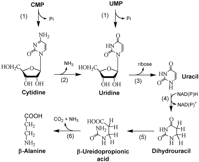
Catabolism of pyrimidine nucleotides in plants. Enzymes shown are: (1) 5′-nucleotidase, (2) cytidine deaminase, (3) uridine nucleosidase, (4) dihydrouracil dehydrogenase, (5) dihydropyriminase, (6) b-ureidopropionase.
Little is known about the first two enzymes of pyrimidine catabolism in plants (Wasternack, 1978). The third enzyme, ß-ureidopropionase (ß-UP, EC 3.5.1.6) was recently characterized by Walsh et al. (2001). Native ß-UP partially purified from the etiolated maize shoots had Kms of 11 and 6 mM for ß-ureidopropionate and ß-ureidoisobutylate, respectively. The pH optimum for the enzyme was broad (6.0–7.2) and its native molecular mass estimated by size exclusion chromatography was approximately 440 kD. An Arabidopsis gene encoding a protein of 405 amino acids that has 55% homology with ß-UP from rat liver has been described (Kvalnes-Krick and Traut, 1993, GenBank accession no. Q03248).
Summary and perspectives
These investigations of nucleotide biosynthesis and metabolism in plants have provided a framework for more detailed analyses of specific enzymes and the isolation of the corresponding genes. The availability of the complete genome sequence of Arabidopsis will aid the search for coding sequences of purine and pyrimidine biosynthesis and catabolism enzymes based on their sequence similarity to counterparts identified in other organisms. Collections of Arabidopsis mutants created by T-DNA insertions and the development of tilling, a PCR-based screening procedure to recover chemically-induced mutants (McCallum et al., 2001), are valuable resources for the genetic analysis of these genes.
Housekeeping enzymes, such as those involved in nucleotide metabolism, are often thought to be expressed at a consistent level in all cells. However, it is now evident that activities of constitutively expressed genes are very dynamic so as to meet the metabolic demands of plant cells. Since these changes can have a profound effect on plant development there is a new appreciation of the importance of housekeeping activities in plant survival such as in response to stress. Identification of the mechanisms by which plant cells monitor and respond to their basic metabolic requirements will rely on the integration of transcript, protein and metabolite profiles of different cell types (Fiehn et al., 2001). Such studies must also consider the effects of changes in the cell cycle and circadian rhythms. For the enzymes that are encoded by multiple genes, the expression, subcellular localization and activity of each member of the family will need to be evaluated since they are often expressed in a tissue specific manner. Unfortunately the modest expression levels of housekeeping genes complicate their analysis since small differences in activity (2–3 fold) that may have large metabolic impacts are difficult to reproducibly detect. Also techniques such as in situ hybridization for monitoring transcript abundance in different tissues are not easily applied to constitutively expressed genes since cell density and volume changes affect the intensity of the signal. However, as more sensitive techniques for measuring the levels of proteins and transcript are applied to the study of purine and pyrimidine nucleotide metabolism, the functional significance of these housekeeping genes in plant development will become more evident.
Acknowledgments
The authors are grateful to Dr. Alan Crozier for preparing the figures. This research was supported by a Natural Sciences and Engineering Research Council research grant (to B.M.).
Footnotes
Citation: Moffatt B.A., and Ashihara H. (2002) Purine and Pyrimidine Nucleotide Synthesis and Metabolism. The Arabidopsis Book 1:e0018. doi:10.1199/tab.0018
elocation-id: e0018
Published on: April 4, 2002
References
- Ashihara H. Changes in the activities of the de novo and salvage pathways of pyrimidine nucleotide biosynthesis during germination of black gram (Phaseolus mungo) seeds. Z. Pflanzenphysiol. 1977;811(1):199–211. [Google Scholar]
- Ashihara H. Orotate phosphoribosyltransferase and orotidine-5′-monophosphate decarboxylase of black gram (Phaseolus mungo) seedlings. Z. Pflanzenphysiol. 1978;871(1):225–241. [Google Scholar]
- Ashihara H. Changes in activities of purine salvage and ureide synthesis during germination of black gram (Phaseolus mungo) seeds. Z. Pflanzenphysiol. 1983;1131(1):47–60. [Google Scholar]
- Ashihara H., Crozier A. Biosynthesis and metabolism of caffeine and related purine alkaloids in plants. Adv. Bot. Res. 1999;301(1):117–205. [Google Scholar]
- Ashihara H., Takasawa Y., Suzuki T. Metabolic fate of guanosine in higher plants. Physiol. Plant. 1997;1001(1):90–916. [Google Scholar]
- Ashihara H., Stasolla C., Loukanina N., Thorpe T. A. Purine and pyrimidine metabolism in cultured white spruce (Picea glauca) cells: Metabolic fate of 14C-labeled precursors and activity of key enzymes. Physiol. Plant. 2000;1081(1):25–33. [Google Scholar]
- Åstot C., Dolczal K., Moritz T., Sandberg G. Precolumn derivatization and capillary liquid chromatography/frit-fast atom bombardment mass spectrometric analysis of cytokinins in Arabidopsis thaliana. J. Mass Spectrom. 1998;331(1):892–902. doi: 10.1002/(SICI)1096-9888(199809)33:9<892::AID-JMS701>3.0.CO;2-N. [DOI] [PubMed] [Google Scholar]
- Atkins C. A., Storer P. J., Shelp B. J. Purification and properties of purine nucleosidase from N2-fixing nodules of cowpea (Vigna unguiculata). J. Plant Physiol. 1989;1341(1):447–452. [Google Scholar]
- Auer C. The Arabidopsis mutation cym changes cytokinin metabolism, adenosine nucleosidase activity and plant phenotype. Biologia Plant. 1999;421(1):S2. (Suppl) [Google Scholar]
- Bäckström D., Sjöberg R-M., Lundberg L. G. Nucleotide sequence of the structural gene for dihydroorotase of Escherichia coli K-12. Eur. J. Biochem. 1986;1601(1):77–82. doi: 10.1111/j.1432-1033.1986.tb09942.x. [DOI] [PubMed] [Google Scholar]
- Bartlett T. J., Aibangbee A., Bruce I. J., Donovan P. J., Yon R. J. Endogenous polypeptide-chain length and partial sequence of aspartate transcarbamoylase from wheat, characterized by immunochemical and cDNA methods. Biochim. Biophys. Acta. 1994;12071(1):187–193. doi: 10.1016/0167-4838(94)00068-9. [DOI] [PubMed] [Google Scholar]
- Bominaar A., Molijn A. C., Pestel M., Veron M., Van H. P. Activation of G-proteins by receptor-stimulated nucleoside diphosphate kinase in Dictyostelium. EMBO J. 1993;121(1):2275–2279. doi: 10.1002/j.1460-2075.1993.tb05881.x. [DOI] [PMC free article] [PubMed] [Google Scholar]
- Brandenburg S. A., Williamson C. L., Slocum R. D. Characterization of a cDNA encoding the small subunit of Arabidopsis carbamoyl phosphate synthetase (Accession No. U73175). (PGR98-087). Plant Physiol. 1998;1171(1):717. [Google Scholar]
- Bressan R. A., Murray M. G., Gale J. M., Ross C. W. Properties of pea seedling uracil phosphoribosyltransferase and its distribution in other plants [pinto beans, soybeans, cereals]. Plant Physiol. 1978;611(1):442–446. doi: 10.1104/pp.61.3.442. [DOI] [PMC free article] [PubMed] [Google Scholar]
- Brown D. C., Collins K. D. Dihydroorotase from Escherichia coli. J. Biol. Chem. 1991;2661(1):1597–1604. [PubMed] [Google Scholar]
- Christopherson R. I., Jones M. E. The effects of pH and inhibitors upon the catalytic activity of the dihydroorotase of multienzymatic protein pyr1-3 from mouse ehrlich ascites carcinoma. J. Biol. Chem. 1980;2551(1):3358–3370. [PubMed] [Google Scholar]
- Cole S. C. J., Yon R. J. Ligand-mediated conformational changes in wheat germ aspartate transcarbamoylase EC 2.1.3.2 indicated by proteolytic susceptibility. Biochem. J. 1984;2211(1):289–296. doi: 10.1042/bj2210289. [DOI] [PMC free article] [PubMed] [Google Scholar]
- Collart F. R., Osipiuk J., Trent J., Olsen G. J., Huberman E. Cloning and characterization of the gene encoding IMP dehydrogenase from Arabidopsis thaliana. Gene. 1996;1741(1):217–220. doi: 10.1016/0378-1119(96)00045-5. [DOI] [PubMed] [Google Scholar]
- De La Rosa A., Williams R. L., Steeg P. S. Nm23/nucleoside diphosphate kinase: toward a structural and biochemical understanding of its biological functions. Bioessays. 1995;171(1):53–62. doi: 10.1002/bies.950170111. [DOI] [PubMed] [Google Scholar]
- Dunn J. H., Jervis H. H., Wilkins J. H., Meredith M. J., Smith K. T., Flora J. B., Schmidt R. R. Coordinate and non-coordinate accumulation of aspartate tanscarbamylase and dihydroorotase in synchronous Chlorella cells growing on different nitrogen sources. Biochim. Biophys. Acta. 1977;4851(1):301–313. doi: 10.1016/0005-2744(77)90166-8. [DOI] [PubMed] [Google Scholar]
- Engel M., Veron M., Theisinger B., Lacombe M., Seib T., Dooley S., Welter C. A novel serine/threonine-specific protein phosphotransferase activity of Nm23/nucleoside diphosphate kinase. Eur. J. Biochem. 1995;2341(1):200–207. doi: 10.1111/j.1432-1033.1995.200_c.x. [DOI] [PubMed] [Google Scholar]
- Faure M., Camonis J. H., Jacquet M. Molecular characterization of a Dictyostelium discoideum gene encoding a multifunctional enzyme of the pyrimidine pathway. Eur. J. Biochem. 1989;1791(1):345–358. doi: 10.1111/j.1432-1033.1989.tb14560.x. [DOI] [PubMed] [Google Scholar]
- Ferone R., Roland S. Dihydrofolate reductase:thymidine synthase, a bifunctional polypeopide from Chrithidia fasciculate. Proc. Natl. Acad. Sci., USA. 1980;841(1):8434–8438. doi: 10.1073/pnas.77.10.5802. [DOI] [PMC free article] [PubMed] [Google Scholar]
- Fiehn O., Kloska S., Altmann T. Integrated studies on plant biology using multiparallel techniques. Current Opinion in Biotechnology. 2001;121(1):82–86. doi: 10.1016/s0958-1669(00)00165-8. [DOI] [PubMed] [Google Scholar]
- Fonne-Pfister R., Chemla P., Ward E., Girardet M., Kreuz K. E., Honzatko R. B., Fromm H. J., Schär H-P., Grütter M. G., Cowan-Jacob S. W. The mode of action and the structure of a herbicide in complex with its target: binding of activated hydantocidin to the feedback regulation site of adenylosuccinate synthetase. Proc. Natl. Acad. Sci. USA. 1996;931(1):9431–9436. doi: 10.1073/pnas.93.18.9431. [DOI] [PMC free article] [PubMed] [Google Scholar]
- Freije J. M. P., Blay P., MacDonald N. J., Manrow R. E., Steeg P. S. Site-directed mutation of Nm23-H1 mutations lacking motility suppressive capacity upon transfection are deficient in histidine-dependent protein phosphotransferase pathways in vitro. J. Biol. Chem. 1997;2721(1):5525–5532. doi: 10.1074/jbc.272.9.5525. [DOI] [PubMed] [Google Scholar]
- Freund J. N., Jarry B. P. The rudimentary gene of Drosophila melanogaster encodes four enzymic functions. J. Mol. Biol. 1987;1931(1):1–13. doi: 10.1016/0022-2836(87)90621-8. [DOI] [PubMed] [Google Scholar]
- Fujiwara S., Yamaguchi M. Effect of allopurinol [4-hydroxypyrazolo(3,4-d) pyrimidine] on the metabolism of allantoin in soybean plants. Plant Physiol. 1978;621(1):134–138. doi: 10.1104/pp.62.1.134. [DOI] [PMC free article] [PubMed] [Google Scholar]
- Gaillard C., Moffatt B. A., Blacker M., Laloue M. Male sterility associated with APRT deficiency in Arabidopsis thaliana results from a mutation in the gene APT1. Mol. Gen. Genet. 1998;2571(1):348–353. doi: 10.1007/s004380050656. [DOI] [PubMed] [Google Scholar]
- Gao G., Nara T., Nakajima-Shimada J., Aoki T. Novel organization and sequences of five genes encoding all six enzymes for de novo pyrimidine biosynthesis in Trypanosoma cruzi. J. Mol. Biol. 1999;2851(1):149–161. doi: 10.1006/jmbi.1998.2293. [DOI] [PubMed] [Google Scholar]
- Guranowski A. Purine catabolism in plants. Purification and some properties of inosine nucleosidase from yellow lupin (Lupinus leteus L.) seeds. Plant Physiol. 1982;701(1):344–349. doi: 10.1104/pp.70.2.344. [DOI] [PMC free article] [PubMed] [Google Scholar]
- Guranowski A., Barankiewicz J. Purine salvage in cotyledons of germinating lupin seeds. FEBS Lett. 1979;1041(1):95–98. doi: 10.1016/0014-5793(79)81091-1. [DOI] [PubMed] [Google Scholar]
- Guyonvarch A., Nguyen-Juilleret M., Hubert J. C., Lacroute F. Structure of the Saccharomyces cerevisiae URA4 gene encoding dihydroorotase. Mol. Gen. Genet. 1988;2121(1):134–141. doi: 10.1007/BF00322456. [DOI] [PubMed] [Google Scholar]
- Hammer B., Lui J., Widholm J. M. Effect of allopurinol on the utilization of purine degradation pathway intermediates by tobacco cell cultures. Plant Cell Reports. 1985;41(1):304–306. doi: 10.1007/BF00269884. [DOI] [PubMed] [Google Scholar]
- Henderson J. F., Paterson A. R. P. Nucleotide Metabolism: An Introduction (New York, Academic Press). 1973;1(1):1–304. [Google Scholar]
- Hirose F., Ashihara H. Adenine phosphoribosyltransferase activity in mitochondria of Catharanthus roseus cells. Z. Naturforsch. 1982;371(1):1288–1289. [Google Scholar]
- Hirose F., Ashihara H. Changes in activity of enzymes involved in purine “salvage” and nucleic acid degradation during growth of Catharanthus roseus cells in suspension culture. Physiol. Plant. 1984;601(1):532–538. [Google Scholar]
- Ito T., Shiraishi H., Okada K., Shimura Y. Two amidophosphoribosyltransferase genes of Arabidopsis thaliana expressed in different organs. Plant Mol. Biol. 1994;261(1):529–533. doi: 10.1007/BF00039565. [DOI] [PubMed] [Google Scholar]
- Jones M. E. Pyrimidine nucleotide biosynthesis in animals: Gene, enzyme and regulation of UMP biosynthesis. Annu. Rev. Biochem. 1980;491(1):253–279. doi: 10.1146/annurev.bi.49.070180.001345. [DOI] [PubMed] [Google Scholar]
- Kanamori I., Ashihara H., Komamine A. Subcellular distribution and activity of enzymes involved in uridine-5′-monophosphate synthesis in Vinca rosea cells in suspension cultures. Z. Pflanzenphysiol. 1980;931(1):437–448. [Google Scholar]
- Kanamori-Fukuda I., Ashihara H., Komamine A. Pyrimidine nucleotide biosynthesis in Vinca rosea cells: Changes in the activity of de novo and salvage pathways during growth in a suspension culture. J. Exp. Bot. 1981;321(1):69–78. [Google Scholar]
- Khan A. I., Chowdhry B. Z., Yon R. J. Wheat-germ aspartate transcarbamoylase: revised purification, stability and re-evaluation of regulatory kinetics in terms of the Monod-Wyman-Changeux model. Eur. J. Biochem. 1999;2591(1):71–78. doi: 10.1046/j.1432-1327.1999.00005.x. [DOI] [PubMed] [Google Scholar]
- Koshiishi C., Kato A., Yama S., Crozier A., Ashihara H. A new caffeine biosynthetic pathway in tea leaves: utilization of adenosine released from S-adenosyl-L-methionine cycle. FEBS Lett. 2001;4991(1):50–54. doi: 10.1016/s0014-5793(01)02512-1. [DOI] [PubMed] [Google Scholar]
- Krath B. N., Hove-Jensen B. Organellar and cytosolic localization of four phosphoribosyl diphosphate isozymes in spinach. Plant Physiol. 1999;1191(1):497–505. doi: 10.1104/pp.119.2.497. [DOI] [PMC free article] [PubMed] [Google Scholar]
- Krath B. N., Eriksen T. A., Poulsen T. S., Hove-Jensen B. Cloning and sequencing of cDNAs specifying a novel class of phosphoribosyl diphosphate synthase in Arabidopsis thaliana. Biochim. Biophy. Acta. 1999;14301(1):403–408. doi: 10.1016/s0167-4838(99)00022-9. [DOI] [PubMed] [Google Scholar]
- Kvalnes-Krick K. L., Traut T. W. Cloning, sequencing, and expression of a cDNA encoding b-alanine synthase from rat liver. J. Biol. Chem. 1993;2681(1):5686–5693. [PubMed] [Google Scholar]
- Le Floc'h F., Lafleuriel J. The role of mitochondria in the recycling of adenine into purine nucleotides in the Jerusalem artichoke (Helianthus tuberosus). Z. Pflanzenphysiol. 1983;1131(1):61–71. [Google Scholar]
- Le Floc'h F., Lafleuriel J. The purine nucleosidases of Jerusalem artichoke shoots. Phytochemistry. 1981;201(1):2127–2129. [Google Scholar]
- Lee D., Moffatt B. A. Purification and charcterization of adenine phosphoribosyltransferase from Arabidopsis thaliana. Physiol. Plant. 1993;871(1):483–492. [Google Scholar]
- Lee D., Moffatt B. A. Adenine salvage activity during callus induction and plant growth. Physiol. Plant. 1994;901(1):739–747. [Google Scholar]
- Lundquist P-O., Pawlowski K., Vance C. P. Carbamoylphosphate synthase: Sequence analysis and nodule enhanced expression in nitrogen-fixing Alnus incana root nodules. Plant Physiol. 1996;1111(1):326. (Suppl.) [Google Scholar]
- Maier T., Zhou L., Thornburg R. Nucleotide sequence of a cDNA encoding UMP synthase from Nicotiana tabacum (GenBank U22260). Plant Physiol. 1995;1081(1):1747. [Google Scholar]
- Maley J. A., Hyman B. C., Lovatt C. J. Evidence for two carbamylphosphate synthetases in plants. Plant Physiol. 1992;991(1):90. (Suppl) [Google Scholar]
- McCallum C. M., Comai L., Greene E. A., Henikoff S. Targeting induced local lesions in genomes (TILLING) for plant functional genomics. Plant Physiol. 2001;1231(1):439–442. doi: 10.1104/pp.123.2.439. [DOI] [PMC free article] [PubMed] [Google Scholar]
- Meek T. D., Garvey E. P., Santi D. V. Purification and characterization of a bifunctional thymidylate synthase-dihydrofolic reductase from methotrexate resistant Leishmania tropica. Biochemistry. 1985;241(1):678–686. doi: 10.1021/bi00324a021. [DOI] [PubMed] [Google Scholar]
- Miersch J., Krauss G-J., Metzger U. Properties and subcellular localization of dihydroorotate dehydrogenase in cells of tomato suspension culture. J. Plant Physiol. 1986;1221(1):55–66. [Google Scholar]
- Moffatt B. A., Somerville C. R. Positive selection for male-sterile mutants of Arabidopsis lacking adenine phosphoribosyltransferase activity. Plant Physiol. 1988;861(1):1150–1154. doi: 10.1104/pp.86.4.1150. [DOI] [PMC free article] [PubMed] [Google Scholar]
- Moffatt B. A., Weretilnyk E. A. Sustaining S-adenosylmethionine-dependent methyltransferase activity in plant cells. Physiol. Plant. 2001. in press.
- Moffatt B. A., McWhinnie E. A., Agarwal S. K., Schaff D. A. The adenine phosphoribosyltranferase-encoding gene of Arabidopsis thaliana. Gene. 1994;1431(1):211–216. doi: 10.1016/0378-1119(94)90098-1. [DOI] [PubMed] [Google Scholar]
- Moffatt B. A., McWhinnie E. A., Burkhard W. E., Pasternak J. J., Rothstein S. J. A complete cDNA for adenine phosphoribosyltransferase from Arabidopsis thaliana. Plant Mol. Biol. 1992;181(1):653–662. doi: 10.1007/BF00020008. [DOI] [PubMed] [Google Scholar]
- Moffatt B. A., Pethe C., Laloue M. Metabolism of benzyladenine is impaired in a mutant of Arabidopsis thaliana lacking adenine phosphoribosyltransferase activity. Plant Physiol. 1991;951(1):900–908. doi: 10.1104/pp.95.3.900. [DOI] [PMC free article] [PubMed] [Google Scholar]
- Moffatt B. A., Wang L., Allen M. S., Stevens Y. Y., Qin W., Snider J., von Schwartzenberg K. Adenosine kinase of Arabidopsis thaliana: kinetic properties and gene expression. Plant Physiol. 2000;1241(1):1775–1785. doi: 10.1104/pp.124.4.1775. [DOI] [PMC free article] [PubMed] [Google Scholar]
- Mok D. W. S., Mok M. C. Cytokinin metabolism and action. Annu. Rev. Plant Physiol. Plant Mol. Biol. 2001;521(1):89–118. doi: 10.1146/annurev.arplant.52.1.89. [DOI] [PubMed] [Google Scholar]
- Muller-Dieckmann H. J., Schulz G. E. The structure of uridylate kinase with its substrates, showing the transition state geometry. J. Mol. Biol. 1994;2361(1):361–367. doi: 10.1006/jmbi.1994.1140. [DOI] [PubMed] [Google Scholar]
- Muller-Dieckmann H. J., Schulz G. E. Substrate specificity and assembly of the catalytic center derived from two structures of ligated uridylate kinase. J. Mol. Biol. 1995;2461(1):522–530. doi: 10.1006/jmbi.1994.0104. [DOI] [PubMed] [Google Scholar]
- Nara T., Hshimoto T., Aoki T. Evolutionary implications of the mosaic pyrimidine-biosynthetic pathway in eukaryotes. Gene. 2000;2571(1):209–222. doi: 10.1016/s0378-1119(00)00411-x. [DOI] [PubMed] [Google Scholar]
- Nasr F., Bertauche N., Dufour M-E., Minet M., Lacroute F. Heterospecific cloning of Arabidopsis thaliana cDNAs by direct complementation of pyrimidine auxotrophic mutants of Saccharomyces cerevisiae I. Cloning and sequence analysis of two cDNAs catalyzing the second, fifth and sixth steps of the de novo pyrimidine biosynthesis pathway. Mol. Gen. Genet. 1994;2441(1):23–32. doi: 10.1007/BF00280183. [DOI] [PubMed] [Google Scholar]
- Newman T., deBruijn F. J., Green P., Keegstra K., Kende H., McIntosh L., Ohlrogge J., Raikhel N., Somerville S., Thomashow M., Retzel E., Somerville C. Genes galore: A summary of methods for accessing results from large-scale partial sequencing of anonymous Arabidopsis cDNA clones. Plant Physiol. 1994;1061(1):1241–1255. doi: 10.1104/pp.106.4.1241. [DOI] [PMC free article] [PubMed] [Google Scholar]
- Nguyen J. Plant xanthine dehydrogenase: its distribution, properties and function. Physiol. Vég. 1986;241(1):263–281. [Google Scholar]
- Nygaard P., Saxild H. H. Nucleotide metabolism. 2000;1(1):418–430. In Encyclopedia of Microbiology, 2nd Ed., (San Diego, Academic Press), pp. [Google Scholar]
- O'Neal T. D., Naylor A. W. Some properties of pea leaf carbamoyl phosphate synthetase of Alaska pea (Pisum sativum L. cv. Alaska). Biochem. J. 1976;1131(1):271–279. doi: 10.1042/bj1130271. [DOI] [PMC free article] [PubMed] [Google Scholar]
- Ong B. L., Jackson J. F. Pyrimidine nucleotide biosynthesis in Phaseolus aureus. Enzymatic aspects of the control of carbamoyl phosphate synthesis and utilization. Biochem. J. 1972;1291(1):583–593. doi: 10.1042/bj1290583. [DOI] [PMC free article] [PubMed] [Google Scholar]
- Park S., Thornburg R. W. Characterization of UMP kinase cDNAs from Oryza sativa (AF187062, AF187063). (PGR99-174). Plant Physiol. 1999;1211(1):1383. [Google Scholar]
- Park S., Thornburg R. W. Characterization of a cDNA encoding UMP synthase from Oryza sativa (AF210322, AF210323, AF210324, and AF210325) (PGR00-043). Plant Physiol. 2000;1221(1):1457. [Google Scholar]
- Parsons R. L., Behringer F. J., Medford J. I. The SCHIZOID gene regulates differentiation and cell division in Arabidopsis thaliana shoots. Planta. 2000;2111(1):34–42. doi: 10.1007/s004250000255. [DOI] [PubMed] [Google Scholar]
- Perera R. J., Linard C. G., Signer E. R. Cytosine deaminase as a negative selectable marker for Arabidopsis. Plant Mol. Biol. 1993;231(1):793–799. doi: 10.1007/BF00021534. [DOI] [PubMed] [Google Scholar]
- Prade L., Cowan-Jacob S. W., Chemla P., Potter S., Ward E., Fonne-Pfister R. Structures of adenylosuccinate synthetase from Triticum aestivum and Arabidopsis thaliana. J. Mol. Biol. 2000;2961(1):569–577. doi: 10.1006/jmbi.1999.3473. [DOI] [PubMed] [Google Scholar]
- Regan S. M., Moffatt B. A. Cytochemical analysis of pollen development in wild-type Arabidopsis and a male-sterile mutant. Plant Cell. 1990;21(1):877–889. doi: 10.1105/tpc.2.9.877. [DOI] [PMC free article] [PubMed] [Google Scholar]
- Rose A. B., Last R. L. Molecular genetics of amino acid, nucleotide, and vitamin biosynthesis. 1994;1(1):835–880. In Arabidopsis, E.M. Meyerowitz and C.R. Somerville, eds. (Plainview: Cold Spring Harbor Laboratory Press), pp. [Google Scholar]
- Santoso D., Thornburg R. Isolation and characterization of UMP synthase mutants from haploid cell suspension of Nicotiana tabacum. Plant Physiol. 1992;991(1):1216–1225. doi: 10.1104/pp.99.3.1216. [DOI] [PMC free article] [PubMed] [Google Scholar]
- Sasamoto H., Ashihara H. Biosynthesis of pyrimidine nucleotides and arginine in a suspension culture of Catharanthus roseus. Int. J. Biochem. 1988;201(1):87–92. [Google Scholar]
- Schachman H. K., Pauza C. D., Navre M., Karels M. J., Wu L., Yang Y. R. Location of amino acid alterations in mutants of aspartate transcarbamoylase: structural aspects of interallelic complementation. Proc. Natl. Acad. Sci. USA. 1984;811(1):115–119. doi: 10.1073/pnas.81.1.115. [DOI] [PMC free article] [PubMed] [Google Scholar]
- Scheffzek K., Kliche W., Weismüller L., Reinstein J. Crystal structure of the complex of UMP/CMP kinase from Dictyostelium discoideum and the bisubstrate inhibitor P1-(5′-adenosyl) P5-(5′-uridyl) pentaphosphate (UP5A) and Mg2+ at 2.2 A: implications for water-mediated specificity. Biochemistry. 1996;351(1):9716–9727. doi: 10.1021/bi960642s. [DOI] [PubMed] [Google Scholar]
- Schnorr K. M., Nygaard P., Laloue M. Molecular characterization of Arabidopsis thaliana cDNAs encoding three purine biosynthetic enzymes. Plant J. 1994;61(1):113–121. doi: 10.1046/j.1365-313x.1994.6010113.x. [DOI] [PubMed] [Google Scholar]
- Schoeber S., Simon D., Schwenger B. Sequence of the cDNA encoding bovine uridine monophosphate synthase. Gene. 1993;1241(1):307–308. doi: 10.1016/0378-1119(93)90412-v. [DOI] [PubMed] [Google Scholar]
- Schubert K. R., Boland M. J. The ureides. 1990;1(1):197–282. In The Biochemistry of Plants, Vol. 16, B.J. Miflin and P.J Lea, eds. (San Diego:Academic Press), pp. [Google Scholar]
- Senecoff J. F., McKinney E. C., Meagher R. B. De novo purine synthesis in Arabidopsis thaliana. II. The PUR7 gene encoding 5¢-phosphoribosyl-4-(N-succinocarboxamide)-5-aminoimidazole synthetase is expressed in rapidly dividing tissues. Plant Physiol. 1996;1121(1):905–917. doi: 10.1104/pp.112.3.905. [DOI] [PMC free article] [PubMed] [Google Scholar]
- Senecoff J. F., Meagher R. B. Isolating the Arabidopsis thaliana genes for de novo purine synthesis by suppression of Escherichia coli mutants. I. 5¢-phosphoribosyl-5-aminoimidazole synthetase. Plant Physiol. 1993;1021(1):387–399. doi: 10.1104/pp.102.2.387. [DOI] [PMC free article] [PubMed] [Google Scholar]
- Serina L., Blondin C., Krin E., Sismeiro O., Danchin A., Sakamoto H., Giles A-M., Bârzu O. Escherichia coli UMP-kinase, a member of the aspartokinase family is a hexamer regulated by guanine nucleotides and UTP. Biochemistry. 1995;341(1):5066–5074. doi: 10.1021/bi00015a018. [DOI] [PubMed] [Google Scholar]
- Serina L., Bucurenci N., Giles A-M., Surewicz W. K., Favian H., Mantsch H. H., Takahashi M., Petrescu I., Batelier G., Bârzu O. Structural properties of UMP-kinase from Escherichia coli: Modulation of protein solubility by pH and UTP. FEBS Lett. 1996;2261(1):7003–7011. doi: 10.1021/bi960062v. [DOI] [PubMed] [Google Scholar]
- Shibata H. H., Ochiai H., Sawa Y., Miyoshi S. Localization of carbamoyl phosphate synthetase and aspartate carbamoyltransferase in chloroplasts. Plant Physiol. 1986;801(1):126–129. doi: 10.1104/pp.80.1.126. [DOI] [PMC free article] [PubMed] [Google Scholar]
- Siehl D. L., Subramanian M. V., Walters E. W., Lee S-F., Anderson R. J., Toschi A. G. Adenoylsuccinate synthetase: site of action of Hydantocidin, a microbial phytotoxin. Plant Physiol. 1996;1101(1):753–758. doi: 10.1104/pp.110.3.753. [DOI] [PMC free article] [PubMed] [Google Scholar]
- Simmer J. P., Kelly R. E., Rinkler A. G., Jr, Zimmermann B. H., Scully J. L., Kim H., Evans D. R. Mammalian dihydroorotase: nucleotide sequence, peptide sequences and evolution of the dihydroorotase domain of the multifunctional protein CAD. Proc. Natl. Acad. Sci. USA. 1990;871(1):174–178. doi: 10.1073/pnas.87.1.174. [DOI] [PMC free article] [PubMed] [Google Scholar]
- Smit G., de Koser C. C., Schripsema J., Spaink H. P., van Brussel A. A., Kijne J. W. Uridine, a cell division factor in pea roots. Plant Mol. Biol. 1995;291(1):869–873. doi: 10.1007/BF00041176. [DOI] [PubMed] [Google Scholar]
- Spanos A., Kanuga N., Holden D. W., Banks G. R. The Ustilago maydis pyr3 gene: sequence and transcriptional analysis. Gene. 1992;1171(1):73–79. doi: 10.1016/0378-1119(92)90492-8. [DOI] [PubMed] [Google Scholar]
- Stasolla C., Loukanina N., Ashihara H., Yeung E. C., Thorpe T. A. Ascorbic acid changes the pattern of purine metabolism during germination of white spruce somatic embryos. Tree Physiol. 2001;211(1):359–367. doi: 10.1093/treephys/21.6.359. [DOI] [PubMed] [Google Scholar]
- Stayton M. M., Rudolph R. F., Fromm H. J. Regulation, genetics and properties of adenylosuccinate synthetase: a review. Curr. Top. Cell. Regul. 1983;221(1):103–141. doi: 10.1016/b978-0-12-152822-5.50008-7. [DOI] [PubMed] [Google Scholar]
- Sterky F., Regan S., Karlsson J., Hertzberg M., Rohde A., Holmberg A., Amini B., Bhalerao R., Larsson M., Villarroel R., Van Montagu M., Sandberg G., Olsson O., Teeri T. T., Boerjan W., Gustafsson P., Uhlén M., Sundberg B., Lundeberg J. Gene discovery in the wood-forming tissues of poplar: Analysis of 5,692 expressed sequence tags. Proc. Natl. Acad. Sci. USA. 1999;951(1):13330–13335. doi: 10.1073/pnas.95.22.13330. [DOI] [PMC free article] [PubMed] [Google Scholar]
- Stougaard J. Substrate-dependent negative selection in plants using a bacterial cytosine deaminase gene. Plant J. 1993;31(1):755–761. [Google Scholar]
- Suttle D. P., Bugg B. Y., Winkler J. K., Kanalas J. J. Molecular cloning and nucleotide sequence for the complete coding region of human UMP synthase. Proc. Natl. Acad. Sci. USA. 1988;851(1):1754–1758. doi: 10.1073/pnas.85.6.1754. [DOI] [PMC free article] [PubMed] [Google Scholar]
- Suzuki T., Takahashi E. Biosynthesis of purine nucleotides and methylated purines in higher plants. Drug Met. Rev. 1977;61(1):213–242. doi: 10.3109/03602537708997480. [DOI] [PubMed] [Google Scholar]
- Suzuki T., Ashihara H., Waller G. R. Purine and purine alkaloid metabolism in Camellia and Coffea plants. Phytochemistry. 1992;311(1):2575–2584. [Google Scholar]
- Tanaka H., Masuta C., Uehara K., Kataoka J., Koiwai A., Noma M. Morphological changes and hypomethylation of DNA in transgenic tobacco expressing antisense RNA of the S-adenosyl-L-homocysteine hydrolase gene. Plant Mol. Biol. 1997;351(1):981–986. doi: 10.1023/a:1005896711321. [DOI] [PubMed] [Google Scholar]
- Tatibana M. Interrelationship of purine and pyrimidine metabolism. 1978;1(1):125–154. In Uric Acid, W.N. Kelley and I.M. Weiner, eds (Berlin, Germany: Springer-Verlag), pp. [Google Scholar]
- Valentin-Hansen P. Uridine-cytidine kinase from Escherichia coli. Methods Enzymol. 1978;511(1):308–314. doi: 10.1016/s0076-6879(78)51041-0. [DOI] [PubMed] [Google Scholar]
- Vincenzetti S., Cambi A., Neuhard J., Schnorr K., Grelloni M., Vita A. Cloning, expression, and purification of cytidine deaminase from Arabidopsis thaliana. Protein Express. Purif. 1999;151(1):8–15. doi: 10.1006/prep.1998.0959. [DOI] [PubMed] [Google Scholar]
- Wagner K. G., Backer A. I. Dynamics of nucleotides in plants studied on a cellular basis. Intl. Rev. Cytol. 1992;1341(1):1–84. [Google Scholar]
- Wagner P. D., Vu N-D. Phosphorylation of ATP-citrate lyase by nucleoside diphosphate kinase. J. Biol. Chem. 1995;2701(1):21758–21764. doi: 10.1074/jbc.270.37.21758. [DOI] [PubMed] [Google Scholar]
- Wagner P. D., Steeg P. S., Vu N-D. Two-component kinase-like activity of nm23 correlates with its motility-suppressive activity. J. Biol. Chem. 1997;941(1):9000–9005. doi: 10.1073/pnas.94.17.9000. [DOI] [PMC free article] [PubMed] [Google Scholar]
- Walther R., Wald K., Glund K., Tewes A. Evidence that a single polypeptide catalyzes the two step conversion of orotate to UMP in cells from tomato suspension culture. J. Plant Physiol. 1984;1161(1):301–311. doi: 10.1016/S0176-1617(84)80109-1. [DOI] [PubMed] [Google Scholar]
- Wasternack C. Metabolism of pyrimidines and purines. 1982;1(1):261–301. In Encyclopedia of Plant Physiology, 14B. Parthier and D. Boulter, eds (Berlin, Germany: Springer-Verlag), pp. [Google Scholar]
- Wasternack C., Guranowski A., Glund K., Tewes A., Walther R. Subcellular localization of some purine-metabolizing enzymes in tomato (Lycopersicon esculentum) cells grown in suspension. J. Plant Physiol. 1985;1201(1):19–28. [Google Scholar]
- Weers B. D., Thornburg R. W. Characterization of the cDNA and gene for an Arabidopsis thaliana uracil phosphoribosyltransferase (Accession No. AF116860). (PGR99-044). Plant Physiol. 1999;1191(1):1567. [Google Scholar]
- Weinfeld H., Savage C. R., Jr, McPartland R. P. CTP synthase of bovine calf liver. Meth. Enzymol. 1978;511(1):84–90. doi: 10.1016/s0076-6879(78)51015-x. [DOI] [PubMed] [Google Scholar]
- Weir E., Fischer J. R. The effect of allopurinol on the excretion of oxypurines by the chick. Biochim. Biophys. Acta. 1970;2221(1):556–557. doi: 10.1016/0304-4165(70)90155-8. [DOI] [PubMed] [Google Scholar]
- Weretilnyk E. A., Alexander K. J., Drebenstedt M., Snider J. D., Summers P. S., Moffatt B. A. Maintaining methylation activities during salt stress. The involvement of adenosine kinase. Plant Physiol. 2001;1251(1):856–865. doi: 10.1104/pp.125.2.856. [DOI] [PMC free article] [PubMed] [Google Scholar]
- Williamson C. L., Slocum R. D. Molecular cloning and characterization of the pyrB1 and pyrB2 genes encoding aspartate transcarbamoylase in pea (Pisum sativum L.). Plant Physiol. 1994;1051(1):377–384. doi: 10.1104/pp.105.1.377. [DOI] [PMC free article] [PubMed] [Google Scholar]
- Williamson C. L., Lake M. R., Slocum R. D. A cDNA encoding carbamoyl phosphate synthetase large subunit (carB) from Arabidopsis (Accession No. U40341) (PGR96-055). Plant Physiol. 1996;1111(1):1354. [Google Scholar]
- Williamson C. L., To L-L., Slocum R. D. Characterization of a cDNA (Accession No. U05293) encoding a third aspartate transcarbamoylase (pyrB3) from pea (PGR96-063). Plant Physiol. 1996;1121(1):446. [Google Scholar]
- Winkler R. G., Blevins D. G., Polacco J. C., Randall D. D. Ureide catabolism in nitrogen-fixing legumes. Trends Biochem. Sci. 1988;131(1):97–100. doi: 10.1016/0968-0004(88)90049-7. [DOI] [PubMed] [Google Scholar]
- Yabuki N., Ashihara H. Catabolism of adenine nucleosides in suspension-cultured plant cells. Biochim. Biophys. Acta. 1991;10731(1):474–480. doi: 10.1016/0304-4165(91)90218-6. [DOI] [PubMed] [Google Scholar]
- Yabuki N., Ashihara H. AMP deaminase and the control of adenylate catabolism in suspension-cultured Catharanthus roseus cells. Phytochemistry. 1992;311(1):1905–1909. [Google Scholar]
- Yamanaka K., Ogura T., Niki H., Hiraga S. Identification and characterization of the smbA gene, a suppressor of the mukB null mutant of Escherichia coli. J. Bacteriol. 1992;1741(1):7517–7526. doi: 10.1128/jb.174.23.7517-7526.1992. [DOI] [PMC free article] [PubMed] [Google Scholar]
- Yamaoka T., Yano M., Kondo M., Sasaki H., Hino S., Katashima R., Moritani M., Itakura M. Feedback inhibition of amidophosphoribosyltransferase regulates the rate of cell growth via purine nucleotide, DNA and protein synthesis. J. Biol Chem. 2001;2761(1):21285–21291. doi: 10.1074/jbc.M011103200. [DOI] [PubMed] [Google Scholar]
- Yi H., Song P-S., Choi G. Cloning and characterization of nucleoside diphosphate kinase 2 from Arabidopsis thaliana (Accession No. AF017640) (PGR98-056). Plant Physiol. 1998;1161(1):1604. [Google Scholar]
- Zhang C., Guinel F., Moffatt B. A. A comparative ultrastructural study of pollen development in Arabidopsis thaliana, ecotype Columbia and the male-sterile mutant, apt 1-3. Protoplasma. 2001. in press. [DOI] [PubMed]
- Zhou L., Thornburg R. W. Site specific mutagenesis of conserved residues in the phosphate binding loop of the Arabidopsis UMP/CMP kinase alter ATP and UMP binding. Arch. Biochem. Biophys. 1998;3581(1):297–302. doi: 10.1006/abbi.1998.0874. [DOI] [PubMed] [Google Scholar]
- Zhou L., Lacroute F., Thornburg R. Characterization of the Arabidopsis thaliana cDNA encoding dihydroorotase (Accession No. AF000146) (PGR97-115). Plant Physiol. 1997;1141(1):1569. [Google Scholar]
- Zhou L., Lacroute F., Thornburg R. Cloning, expression in Escherichia coli and characterization of Arabidopsis thaliana uridine 5′-monophosphate/cytidine 5′-monophosphate kinase. Plant Physiol. 1998;1171(1):245–253. doi: 10.1104/pp.117.1.245. [DOI] [PMC free article] [PubMed] [Google Scholar]


