Abstract
Background
B-cell chronic lymphocytic leukemia is a clinically heterogeneous disease; some patients rapidly progress and die within a few years of diagnosis, whereas others have a long life expectancy with minimal or no treatment. Telomere length and telomerase levels have been proposed as prognostic factors; however, very few cases have been characterized for both parameters and no study has analyzed the prognostic value of the telomere/telomerase profile.
Design and Methods
One hundred and seventy-three cases of chronic lymphocytic leukemia were characterized for telomere lengths and telomerase levels by real-time polymerase chain reaction. Data were correlated with established prognostic markers, IGVH mutational status and chromosomal aberrations, and clinical outcome.
Results
Telomere lengths were inversely correlated with telomerase levels (rs= −0.213; P=0.012), and most of the cases of chronic lymphocytic leukemia with high levels (above median) of telomerase had short (below median) telomeres (P=0.0001). Telomerase levels were higher and telomeres were shorter in unmutated IGVH cases than in mutated IGVH ones (P<0.0001). Chronic lymphocytic leukemias with 11q, 17p deletion or 12 trisomy had significantly higher levels of telomerase and shorter telomeres than those with no chromosomal aberration or the sole 13q deletion (P<0.001). Telomere length/telomerase level profiles identified subgroups of patients with different clinical outcomes (P<0.0001), even within the subsets of chronic lymphocytic leukemia defined by IGVH mutational status or chromosomal aberrations. Short telomere/high telomerase profile was independently associated with more rapid disease progression.
Conclusions
Comprehensive analyses of telomeres, telomerase, chromosomal aberrations, and IGVH mutational status delineate groups of chronic lymphocytic leukemias with distinct biological characteristics and clinical outcomes. The telomere/telomerase profile may be particularly useful in refining the prognosis of chronic lymphocytic leukemia patients with mutated IGVH and no high-risk chromosomal aberrations.
Keywords: B-CLL, telomere, telomerase, chromosomal aberrations
Introduction
B-cell chronic lymphocytic leukemia (B-CLL), one of the most common adult leukemias, is characterized by a highly heterogeneous clinical course. Some patients rapidly progress and die within a few months after diagnosis, whereas others live for several years with minimal or no treatment.1 A comprehensive prognostic characterization of patients with B-CLL and identification of reliable prognostic markers is essential for tailoring therapeutic strategies.2
Specific molecular alterations in gene expression and protein activity are thought to underlie the variability in disease outcome.3–5 Among molecular indicators, the presence or absence of somatic mutations in the immunoglobulin heavy-chain variable gene (IGVH) appears to be the best prognostic discriminator, with the unmutated IGVH profile (<2% difference from germ-line) being associated with an aggressive clinical course.6,7 B-CLL is also characterized by a genomic instability that gives rise to several chromosomal aberrations, 11q, 13q, 17p deletions and 12 trisomy being the most relevant. While 11q and 17p deletions have been associated with rapid disease progression, the absence of chromosomal abnormalities and the presence of 13q deletion as the sole abnormality are associated with a better prognosis.8–10 Despite the established prognostic value of these bio-markers, stratification based on these parameters fails to cover the complex heterogeneity of B-CLL.
Telomeres, which are specialized protective structures at the end of eukaryotic chromosomes, are progressively shortened during each round of cell replication because of end-replication problems of DNA polymerase.11 The progressive shortening of telomeres is a key mechanism in controlling cellular replicative potential; when telomere erosion reaches a critical point, cells cease to proliferate and undergo senescence or apoptosis.12 While maintenance of telomere length by telomerase is critical for preserving the replicative potential of cancer cells, further erosion of telomeres may impair their function in protecting chromosome ends, resulting in genetic instability, a key event in the initiation of carcinogenesis.13–15 Recently, short telomeres have been associated with genetic complexity and short survival in B-CLL patients.16–18
Telomerase, a ribonucleoprotein complex containing an internal RNA template (hTR) and a catalytic protein with telomere-specific reverse transcriptase activity (hTERT), maintains telomere length by adding hexameric TTAGGG repeats to the chromosomal ends, thus compensating for the continued replicative loss of telomeres.19 hTERT is the rate-limiting component of the telomerase complex, and its expression correlates with telomerase activity.20,21 Telomerase activity, usually absent from normal somatic cells, is essential for unlimited cell growth and plays a critical role in tumorigenesis.22 Several studies found a relationship between levels of hTERT expression, telomerase activity and clinical aggressiveness of a variety of malignancies, including leukemia and lymphoma.23–25 It has also been demonstrated that telomerase activity, or hTERT expression, may be a prognostic indicator of overall survival in B-CLL.26–28 To date, only one study has analyzed both telomeres and telomerase in the same B-CLL patients and showed that patients with unmutated IGVH had shorter telomeres and higher telomerase activity than mutated IGVH cases.29 No study has yet analyzed both hTERT levels and telomere length in a large cohort of patients with B-CLL, nor the interplay of hTERT expression/telomere length in relation to other known prognostic factors and disease outcome.
The aim of this study was to evaluate the interplay between hTERT expression and telomere length, their relationship with other biological variables and their impact on clinical outcome.
Design and Methods
Patients
Peripheral blood cells were collected from 173 B-CLL patients (116 males and 57 females) who attended two institutions (Department of Clinical and Experimental Medicine, Hematology Section, Padova, and Department of Hematology, Vicenza). The median age was 62 (range, 38 to 81) years. All samples were collected at the time of diagnosis, and all patients were untreated at the time of sampling. Flow cytometry and fluorescence in situ hybridization (FISH) studies were performed on freshly collected peripheral blood samples, while IGVH mutational status, telomere length and hTERT levels were determined on paired frozen samples collected at the same time point. The median (interquartile) follow-up time from blood sampling was 45 (31–73) months. The decision to give primary treatment was taken following general practice assessments.30 Time from diagnosis to first treatment (TTFT) was considered as a marker for time to disease progression. Informed consent was obtained according to the Helsinki declaration and the study was approved by the local Ethics Committees.
IGVH mutation analysis and CD38 and ZAP-70 expression
IGVH gene status was assessed as previously described.28 The cut-off of 2% mutations was employed to define unmutated (< 2%) and mutated (>2%) IGVH cases. Expression of CD38 and ZAP-70 proteins was performed by flow cytometry, as previously described.28
Fluorescence in situ hybridization
Fluorescence in situ hybridization (FISH) was performed on standard cytogenetic preparations from peripheral blood. The slides were hybridized with the multicolour probe sets LSI p53/LSI ATM and LSI D13S319/LSI 13q34/ CEP12 (Vysis-Abbott, Des Plaines, IL, USA) according to the manufacturer’s protocol. Three hundred interphase nuclei were analyzed for each probe. The cut-off for positive values (mean of normal control ±3 standard deviation) determined from ten cytogenetically normal samples was 4% for centromere 12 trisomy (+12), and 10% for del 11q22.3(11q-), del13q14.3 (13q-) and del 17p13.1 (17p-). The B-CLL cases with 11q- or 17p- and 13q- (n=12) were included in the group of 11q-,17p-.
Determination of telomere length and quantification of hTERT transcripts
Telomere length was determined by real-time polymerase chain reaction (PCR), exactly as previously described,31 and values were expressed as telomere/single copy-gene (T/S) ratio. T/S values were converted to kilobases (Kb) using the adjusted formula y=1.53x+3.62.31 All hTERT transcripts in B-CLL samples were quantified by real-time PCR using the AT1 and AT2 primer pair, exactly as previously described,28 and hTERT values were normalised for 106 copies of the housekeeping gene GAPDH.28
Quantification of telomerase activity by real-time polymerase chain reaction
Two million cells were lysed in 50–60 μL of CHAPS buffer (0.5% CHAPS, 10 mM Tris HCl, pH 7.5, 1 mM MgCl2, 1 mM EGTA, 0.1 mM phenylmethyl-sulfonyl fluoride, 5 mM β-mercaptoethanol, and 10% glycerol) and incubated at 4 °C for 30 min. The lysate was then centrifuged at 12000 g for 30 min at 4 °C, and the supernatant was collected as previously reported.26 The protein concentration was measured using NanoDrop spectrophotometry (ND-1000; Celbio). Telomerase activity was evaluated by a real-time PCR method,32 using 250 ng of cellular protein extract for each sample. Threshold cycle values (Ct) of the samples were plotted against a standard curve generated from serial five-fold dilutions starting from 2 μg protein extract from telomerase-positive BL41 cells. Each sample was analyzed in duplicate and values are expressed as relative units.
Statistical analyses
The distribution of continuous variables, such as hTERT levels, telomere length and age, were compared by the Kruskal-Wallis test and the associations among nominal variables were analyzed by the χ2 test. For each variable, TTFT analysis was performed using the Kaplan-Meier method and compared by the log-rank test. hTERT levels and telomere length were analyzed as dichotomous variables (cut-off: ≤median or >median). Hazard ratios for each category were estimated using univariate Cox proportional hazards models with low risk as the reference class. TTFT analysis was also performed to explore the interplay of hTERT and telomere length, with inclusion of the interaction term. The independent role of hTERT/telomere interplay in predicting TTFT was tested using a Cox proportional hazards model adjusting separately for cytogenetic categories and IGVH mutational status. This choice was due to the dataset reduction driven by the fact that not all subjects had both FISH and IGVH mutational data. The proportionality assumption was tested by including time-dependent covariates in each model. All tests were two-sided, and a P value less than 0.05 was considered statistically significant. Statistical analyses were performed using SAS version 9.1 (SAS Institute, Cary, NC, USA).
Results
hTERT expression and telomere length in cases of B-cell chronic lymphocytic leukemia
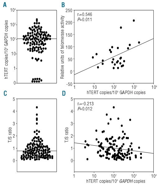
The median (interquartile, IQR) hTERT level, determined in 151 B-CLL samples, was 106 (40–250) copies/106 GAPDH (Figure 1A). In agreement with a previous observation,28 levels of hTERT mRNA significantly correlated with telomerase activity (rs=0.546, P=0.011) (Figure 1B). The median (IQR) telomere length, determined in 164 B-CLL samples, was 0.81 (0.41–1.30) corresponding to 4.85 (4.24–5.61) Kb (Figure 1C). These values were similar to those reported by other authors.16,17,33 Of interest, hTERT levels were inversely correlated with telomere length (rs =−0.213; P=0.012) (Figure 1D).
Figure 1.
Levels of hTERT transcripts and telomere lengths in B-CLL samples. (A) hTERT levels (hTERTcopies/106 GAPDH copies) in B-CLL samples. The line indicates the median value. (B) Relationship between hTERT levels and relative units of telomerase activity. (C) Telomere length (T/S ratio) in B-CLL samples. The line indicates the median value. (D) Inverse relationship between hTERT levels and telomere length.
hTERT expression and telomere length in relation to IGVH mutational status and CD38 and ZAP-70 expression
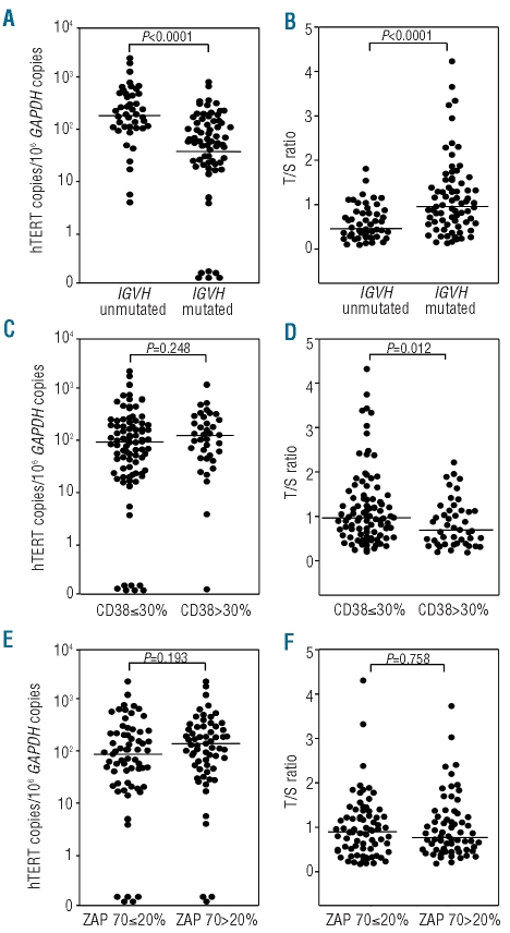
hTERT transcript levels and T/S values were compared to the IGVH mutational profile, which was available in 138 cases, 60% with mutated IGVH and 40% with unmutated IGVH. Levels of hTERT were significantly higher in IGVH unmutated B-CLL than in mutated B-CLL [median (IQR) 205 (107–489) versus 56 (22–153) copies; P<0.0001] (Figure 2A). In contrast, T/S lengths were significantly lower in unmutated IGVH than mutated IGVH cases [median (IQR) 0.44 (0.28–0.81) versus 1.00 (0.58–1.50); P<0.0001] (Figure 2B). hTERT transcript levels and T/S values were also compared with CD38 and ZAP-70 expression, available for 154 and 158 B-CLL cases, respectively. CD38 expression was low (≤30%) or high (>30%) in 69% and 31% of the cases, respectively. In agreement with previous observations,28 hTERT values were higher in CD38 high-positive rather than in CD38 low-positive cases [median (IQR) 132 (46–296) versus 98 (27–206)] (Figure 2C), but the difference was not statistically significant. In contrast, T/S values were significantly lower in CD38 high-positive than in CD38 low-positive cases [median (IQR) 0.58 (0.28–1.05) versus 0.88 (0.48–1.40); P=0.012] (Figure 2D). Moreover, 52% and 48% of the cases had low (≤20%) or high (>20%) ZAP-70 expression. hTERT levels were higher in ZAP-70 high-positive samples than in low-positive samples [median (IQR) 128 (45–296) versus 96 (23–222)] (Figure 2E), but these differences were not statistically significant. T/S values did not differ significantly between ZAP-70 low- or high-positive samples [median (IQR) 0.81 (0.38–1.19) versus 0.75(0.41–1.17)] (Figure 2F).
Figure 2.
Levels of hTERT transcripts (hTERTcopies/106 GAPDH copies) and telomere lengths (T/S ratio) in B-CLL samples, according to (A, B) IGVH mutational status, (C, D) CD38 expression and (E, F) ZAP-70 expression.
hTERT expression and telomere length in relation to genomic aberrations
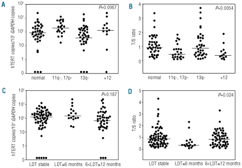
FISH analysis was performed in 125 cases; 19% had 11q- or 17p-, 34% had the 13q- as the sole chromosomal aberration, 10% were +12, and 37% of the cases had no chromosomal abnormalities and were considered as normal. hTERT levels were significantly higher in B-CLL with 11q- or 17p- and lower in B-CLL with 13q- [median (IQR) 94 (40–250), 235 (107–618), 159 (87–359), and 56 (20–166) in normal, 11q-/ 17p-, +12, and 13q- B-CLL, respectively; P=0.0067] (Figure 3A). Within groups, hTERT levels did not differ significantly between cases with no chromosomal abnormalities and 13q deletion, and between cases with the known high-risk 11q- or 17p- and +12 (Online Supplementary Table S1). T/S values were lower in 11q-, 17p-, or +12 B-CLL and higher in 13q- or normal B-CLL [median (IQR) 0.44 (0.23–0.81), 0.39 (0.24–0.71), 0.92 (0.42–1.61), and 0.94 (0.61–1.46) in 11q-/ 17p-, +12, 13q- and normal B-CLL, respectively; P=0.0054] (Figure 3B, and Online Supplementary Table S1).
Figure 3.
Levels of hTERT transcripts (hTERTcopies/106 GAPDH copies) and telomere lengths (T/S ratio) in B-CLL samples, according to (A, B) the chromosomal categories (normal, 11q- 17p-, 13q- as the sole abnormality, and +12), and (C, D) lymphocyte doubling time (LDT). The P value refers to the overall trend of all groups in each graph. Pairwise comparisons between hTERT, T/S and chromosomal categories or LDT are shown in Online Supplementary Tables S1 and S2.
Distribution of hTERT level and telomere length in relation to lymphocyte doubling time
Lymphocyte doubling time (LDT) was available for 142 cases. The majority of cases (53%) had a stable LDT, 36% had a LDT between 6 and 12 months, while 11% had a higher proliferation rate (LDT≤6 months). hTERT expression was higher in patients with an LDT of 6 months or less than in patients with a stable LDT or LDT between 6 and 12 months, but the differences were not statistically significant [median (IQR) 150 (101–438), 128 (45–237), and 87 (25–250) respectively; P=0.187] (Figure 3C and Online Supplementary Table S2). Of interest, telomere lengths were significantly lower in cases with a LDT of 6 months or less than in cases of B-CLL with a stable LDT or LDT between 6 and 12 months [median (IQR) 0.36 (0.27–0.67), 0.88 (0.49–1.32), and 0.81 (0.39–1.31), respectively; P= 0.024] (Figure 3D and Online Supplementary Table S2).
Distribution of hTERT level and telomere length in subsets of B-cell chronic lymphocytic leukemia defined by IGVH mutational status and chromosomal aberrations
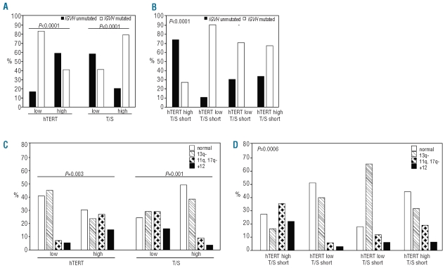
hTERT was defined as high or low, and telomeres as long or short according to their values above and below the median. High hTERT levels and short telomeres were more frequent in unmutated IGVH B-CLL cases than mutated ones (Figure 4A). Chromosomal aberrations 11q-, 17p- or +12 were more frequent in B-CLL cases with high hTERT than low hTERT and in those with short telomeres rather than long ones (Figure 4C).
Figure 4.
hTERT level, telomere length, and hTERT level/telomere length profile according to (A, B) IGVH mutational status (mutated or unmutated), and (C, D) chromosomal categories (normal, 11q-or 17p-, 13q- as the sole chromosomal abnormality, and +12). hTERT low: ≤median value; hTERT high: >median value; T/S short ≤median value; T/S long>median value.
Most of the B-CLL with low hTERT levels had long telomeres (66%), while the majority of the B-CLL with high hTERT levels had short telomeres (67%) (P=0.0001). Thus, two prevalent groups of B-CLL were identified: one with high hTERT level/short telomeres (33%) and one with low hTERT level/long telomeres (34%). Fewer cases had high hTERT level/long telomeres (17%) or low hTERT level/short telomeres (16%). The B-CLL cases with high hTERT/short telomeres were characterized by unmutated IGVH status (73%) (Figure 4B), and chromosomal abnormalities (74%) (Figure 4D). Notably, the most frequent genomic aberrations in B-CLL with high hTERT/short telomeres were 11q-, 17p-, or +12 (57%), while 40% of B-CLL cases with low hTERT/long telomeres and 65% of those with low hTERT/short telomeres had the 13q- (Figure 4D).
High hTERT and short telomeres as prognostic markers of poor clinical outcome
Disease progression, estimated as TTFT, was more rapid in B-CLL with high hTERT levels than in those with low hTERT levels [median (95% CI) 31 (19;50) versus 104 (66;-) months; P<0.0001] (Figure 5A). Cases with short telomeres had a worse clinical outcome than those with long telomeres [median (95% CI) 35 (15;54) versus 104 (63;-) months; P<0.0001] (Figure 5B). When B-CLL patients were stratified according to both hTERT and telomere values, four different groups were identified: B-CLL with high hTERT/short telomeres had the worst clinical outcome with a median TTFT of 15 (4;40) months, while cases with low hTERT/long telomeres had a slower disease progression with a median TTFT of 104 (66;-) months. B-CLL patients with low hTERT/short telomeres or high hTERT/long telomeres had intermediate clinical outcomes (Figure 5C, and Online Supplementary Table S3); disease progression between these two groups was not statistically different (P=0.588) and we, therefore, combined the two groups in the subsequent analyses.
Figure 5.
Curves of treatment-free survival [time from diagnosis to first treatment (TTFT)], according to (A) hTERT level, (B) telomere length, and (C) hTERT level/telomere length profile. hTERT low: ≤median value; hTERT high: >median value; T/S short: ≤median value; T/S long: >median value. The median (95% CI) of TTFT and hazard ratios (95%) are provided in Online Supplementary Table S3.
Prognostic value of hTERT level and telomere length in relation to IGVH mutational status and chromosomal abnormalities
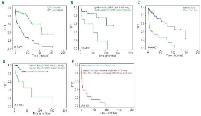
Disease progression was faster in unmutated than mutated IGVH cases [median (95% CI) 19 (8;33) months versus 107 (63;-) months (P<0.0001)] (Figure 6A, and Online Supplementary Table S3). B-CLL patients with low hTERT levels or long telomeres and mutated IGVH had a significantly better prognosis than patients with high hTERT levels or short telomeres and unmutated IGVH (Online Supplementary Figure S1A,B and Online Supplementary Table S4). Multivariate analyses confirmed the independent value of the hTERT/telomere interplay in relation to IGVH mutational status. High hTERT/short telomere and low hTERT/long telomere profiles discriminate two subgroups of B-CLL with different clinical outcomes within both the IGVH mutated and unmutated cases. This is of particular relevance in the mutated IGVH cases; patients with high hTERT/short telomere B-CLL had a poor prognosis with a median TTFT of 49 (4;108) months, which is significantly shorter than that of B-CLL cases with a low hTERT/long telomere profile (Figure 6B, and Online Supplementary Table S4).
Figure 6.
Curves of treatment-free survival [time from diagnosis to first treatment (TTFT)], according to (A) IGVH mutational status, (B) IGVH mutational status and hTERT level/telomere length profile, (C) high-risk (11q- or 17p-, +12) and low-risk (normal, 13q-) chromosomal categories, (D) chromosomal categories and hTERT level/telomere length profile, (E) IGVH mutational status, chromosomal categories, and hTERT level/telomere length profile. hTERT low: ≤median value; hTERT high: >median value; T/S short ≤median value; T/S long: >median value. The median (95% CI) of TTFT and hazard ratios are provided in Online Supplementary Table S3 (panel A, C), S4 (panel B) and S5 (panel E).
As far as concerns chromosomal categories, 11q- or 17p-B-CLL had the worst clinical outcome, while 13q- B-CLL had the best prognosis [median (95% CI) 3 (2;13) months versus (77;-) months; P<0.0001] (Online Supplementary Table S3). The median TTFT of 13q- cases did not differ significantly from that of B-CLL with normal cytogenetics; thus, B-CLL with these two chromosomal profiles had a significantly longer disease-free interval than those with 11q-, 17p- or +12 abnormalities (Figure 6C and Online Supplementary Table S3). Stratification of patients according to hTERT level or telomere length and chromosomal profile revealed that B-CLL cases with high hTERT levels or short telomeres and high-risk abnormalities (i.e. 11q-, 17p-, +12) had a poorer prognosis than cases with low hTERT or long telomeres and a low-risk genomic profile (i.e. normal or 13q-) (Online Supplementary Figure S1C,D and Online Supplementary Table S5). Multivariate analyses confirmed the independent value of the hTERT/telomere interplay in relation to chromosomal status. A high hTERT/short telomere profile identified subgroups of patients with the poorest clinical outcome; this is of particular relevance in the B-CLL cases with no or low-risk abnormalities. Within this group, disease progression was significantly more rapid in B-CLL with high hTERT/short telomeres than in those with low hTERT/long telomere (Figure 6D, and Online Supplementary Table S5).
Given the sample number, the independent role of hTERT/telomere interplay in predicting TTFT was tested separately for IGVH mutational status and chromosomal categories. However, stratification of cases analyzed for tested risk factors showed that B-CLL patients with unmutated IGVH status, 11q-,17p-, or +12 chromosomal abnormalities and high hTERT/short telomeres developed the disease (17/18) within a median (95% CI) time of 2 (1;16) months; in contrast, none of the 22 patients with any of the above risk factors progressed clinically within a median follow-up of 42 months (Figure 6E).
Discussion
This study was the first analysis of both hTERT levels and telomere length and their relationship with IGVH mutational status and chromosomal aberrations in a large cohort of B-CLL patients. Although the main function of hTERT is to stabilize telomere length,12,20,21,34 an inverse relationship between hTERT levels and telomere lengths has been found in B-CLL cases. Notably, B-CLL cases with high telomerase levels and short telomeres were frequently characterized by an unmutated IGVH status and high-risk chromosomal aberrations. Conversely, B-CLL cases with low telomerase levels and long telomeres were associated with mutated IGVH and low-risk abnormalities.
All together these findings may have important implications for the pathogenesis of B-CLL. During the T-cell-mediated germinal center (GC) experience, B cells activate telomerase and exhibit telomere lengthening.35,36 Somatic hypermutation of IG genes is a physiological process occurring in the GC; thus, B-CLL cases with unmutated IGVH genes have likely originated from pre-GC B lymphocytes and those with mutated IGVH genes from GC-experienced B lymphocytes. Our data show that mutated IGVH GC-experienced B-CLL had longer telomeres than the unmutated IGVH B-CLL, in agreement with previous results.16,29,33,37 Of interest, the unmutated IGVH B-CLL cases with short telomeres had higher levels of hTERT than the mutated IGVH cases with long telomeres. This finding highlights the concept that telomere length in tumors, rather than being associated with hTERT levels, reflects the initial kinetics of telomere erosion by cell proliferation.13,38,39 Our finding that short telomeres are associated with chromosomal abnormalities supports the concept that erosion of telomeres may impair their function in protecting chromosome ends, resulting in genetic instability13,15 and reinforces the concept that activation of hTERT is subsequent to telomere shortening, particularly in a subgroup of B-CLL cases.40 This finding is consistent with recent evidence of shortest telomeres in a subset of patients with early-stage disease.18 Our results also demonstrated that B-CLL with the highest proliferative index (i.e. LDT <6 months) had the shorter telomere length, a finding that supports the notion of telomere shortening at each cell division cycle. Thus, in B-CLL with short telomeres, high hTERT levels are essential to maintain the replicative potential of tumor cells. Conversely, the pre-existing activation of telomerase in GC-experienced B lymphocytes may explain the long telomeres in mutated IGVH B-CLL cases, despite the low levels of hTERT.
While high-risk chromosomal aberrations were more frequent in B-CLL with short telomeres and high hTERT levels, the distribution of the 13q deletion was intriguing. It was also detected in B-CLL with long telomeres, and was predominant in B-CLL cases with low hTERT levels. Most of the 13q- B-CLL cases had a stable LDT; this slow kinetics of cellular division may require low levels of hTERT to preserve the replicative potential. MicroRNA 15 and 16 are localized in 13q14,41 but molecular aberrations underlying the 13q deletion have not been fully characterized.42 How the 13q deletion affects the cell cycle and levels of hTERT remains a very interesting issue to be addressed.
In agreement with results of previous studies, we found that unmutated IGVH status,6,7 11q- or 17p- and +12 aberrations,8–10 high levels of hTERT,26–28 and short telomere length16,17,29,33 were all associated with a poor clinical outcome. The finding that the 13q-, characterized by low levels of hTERT, was associated with an even better prognosis than that of the group with normal cytogenetics supports the notion that hTERT may contribute to lymphomagenesis beyond just preservation of telomere length.43–45 In addition, the study of all these biomarkers in the same B-CLL cohort allowed us to identify new potential risk profiles. The poorest prognosis was found in the group with high hTERT levels and short telomeres, whereas B-CLL with low hTERT levels and long telomeres had a more indolent clinical behavior. It should be stressed that even within the group of B-CLL patients with unmutated IGVH and/or high risk chromosomal aberrations who had the poorest clinical outcome,46 those with low levels of hTERT and long telomeres had a better outcome. Most importantly, the evaluation of hTERT and telomere length might help clinicians in the management of B-CLL patients with mutated IGVH and/or no high-risk chromosomal aberrations since cases with high hTERT/short telomere B-CLL will progress more rapidly and might require therapy earlier than those with low hTERT/long telomeres.
In conclusion, our data demonstrate for the first time that the combined parameter telomere/telomerase is a strong predictor of progression in B-CLL patients. This is important for deepening the knowledge of the pathogenesis of B-CLL as well as for the management of the disease and the development of new therapeutic strategies.
Acknowledgments
This research was supported by the Regione Veneto-Ricerca Finalizzata, Fondazione Berlucchi per la Ricerca sul Cancro, AIRC-Associazione Italiana Ricerca sul Cancro, and Fondazione Cariverona. RE is a recipient of grants from AIRC and Fondazione Cariverona. We are grateful to Gianni Pizzolo for critical discussion. We thank Barbara Filippi for expert technical assistance with the FISH study, Lisa Smith for editorial assistance and Pierantonio Gallo for the artwork.
Footnotes
Authorship and Disclosures
The information provided by the authors about contributions from persons listed as authors and in acknowledgments is available with the full text of this paper at www.haematologica.org.
Financial and other disclosures provided by the authors using the ICMJE (www.icmje.org) Uniform Format for Disclosure of Competing Interests are also available at www.haematologica.org.
The online version of this article has a Supplementary Appendix.
References
- 1.Kipps TJ. Chronic lymphocytic leukemia. Curr Opin Hematol. 2000;7(4):223–4. doi: 10.1097/00062752-200007000-00005. [DOI] [PubMed] [Google Scholar]
- 2.Friese CR, Earle C, Magazu LS, Brown JR, Neville BA, Hevelone ND, et al. Timeliness and quality of diagnostic care for Medicare recipients with chronic lymphocytic leukemia. Cancer. 2011;117(7):1470–7. doi: 10.1002/cncr.25655. [DOI] [PMC free article] [PubMed] [Google Scholar]
- 3.Plass C, Byrd JC, Raval A, Tanner SM, de la Chapelle A. Molecular profiling of chronic lymphocytic leukemia: genetics meets epigenetics to identify predisposing genes. Br J Haematol. 2007;139(5):744–52. doi: 10.1111/j.1365-2141.2007.06875.x. [DOI] [PubMed] [Google Scholar]
- 4.Montserrat E, Moreno C. Genetic lesions in chronic lymphocytic leukemia: clinical implications. Curr Opin Oncol. 2009;21(6):609–14. doi: 10.1097/CCO.0b013e328331b702. [DOI] [PubMed] [Google Scholar]
- 5.Kienle D, Benner A, Laufle C, Winkler D, Scneider C, Buhler A, et al. Gene expression factors as predictors of genetic risk and survival in chronic lymphocytic leukemia. Haematologica. 2010;95(1):102–9. doi: 10.3324/haematol.2009.010298. [DOI] [PMC free article] [PubMed] [Google Scholar]
- 6.Damle RN, Wasil T, Fais F, Ghiotto F, Valletto A, Allen SL, et al. IgV gene mutation status and CD38 expression as novel prognostic indicators in chronic lymphocytic leukemia. Blood. 1999;94(6):1840–7. [PubMed] [Google Scholar]
- 7.Hamblin TJ, Davis Z, Gardiner A, Oscier DG, Stevenson FK. Unmutated IgVH genes are associated with a more aggressive form of chronic lymphocytic leukemia. Blood. 1999;94(6):1848–54. [PubMed] [Google Scholar]
- 8.Döhner H, Stilgenbauer S, Benner A, Leupolt E, Kröber A, Bullinger L, et al. Genomic aberrations and survival in chronic lymphocytic leukemia. N Engl J Med. 2000;343(26):1910–6. doi: 10.1056/NEJM200012283432602. [DOI] [PubMed] [Google Scholar]
- 9.Rossi D, Cerri M, Deambrogi C, Sozzi E, Cresta S, Rasi S, et al. The prognostic value of TP53 mutations in chronic lympho cytic leukemia is independent of del17p13: implications for overall survival and chemo refractoriness. Clin Cancer Res. 2009;15(3):995–1004. doi: 10.1158/1078-0432.CCR-08-1630. [DOI] [PubMed] [Google Scholar]
- 10.Zenz T, Mertens D, Dohner H, Stilgenbauer S. Importance of genetics in chronic lymphocytic leukemia. Blood Rev. 2011;25(3):131–7. doi: 10.1016/j.blre.2011.02.002. [DOI] [PubMed] [Google Scholar]
- 11.Harley CB, Futcher AB, Greider CW. Telomeres shorten during ageing of human fibroblasts. Nature. 1990;345(6274):458–60. doi: 10.1038/345458a0. [DOI] [PubMed] [Google Scholar]
- 12.Blackburn EH, Greider CW, Szostak JW. Telomeres and telomerase: the path from maize, Tetrahymena and yeast to human cancer and aging. Nat Med. 2006;12(10):1133–8. doi: 10.1038/nm1006-1133. [DOI] [PubMed] [Google Scholar]
- 13.Hackett JA, Greider CW. Balancing instability: dual roles for telomerase and telomere dysfunction in tumorigenesis. Oncogene. 2002;21(4):619– 26. doi: 10.1038/sj.onc.1205061. [DOI] [PubMed] [Google Scholar]
- 14.Meeker AK, Hicks JL, Iacobuzio-Donahue CA, Montgomery EA, Westra WH, Chan TY, et al. Telomere length abnormalities occur early in the initiation of epithelial carcinogenesis. Clin Cancer Res. 2004;10(10):3317–26. doi: 10.1158/1078-0432.CCR-0984-03. [DOI] [PubMed] [Google Scholar]
- 15.Calado RT, Young NS. Telomere diseases. N Engl J Med. 2009;361(24):2353–65. doi: 10.1056/NEJMra0903373. [DOI] [PMC free article] [PubMed] [Google Scholar]
- 16.Ricca I, Rocci A, Drandi D, Francese R, Compagno M, Lobetti Bodoni C, et al. Telomere length identifies two different prognostic subgroups among VH-unmutated B-cell chronic lymphocytic leukemia patients. Leukemia. 2007;21(10):697–705. doi: 10.1038/sj.leu.2404544. [DOI] [PubMed] [Google Scholar]
- 17.Roos G, Kröber A, Grabowski P, Kienle D, Buhler A, Dohner H, et al. Short telomeres are associated with genetic complexity, high-risk genomic aberrations, and short survival in chronic lymphocytic leukemia. Blood. 2008;111(12):2246–52. doi: 10.1182/blood-2007-05-092759. [DOI] [PubMed] [Google Scholar]
- 18.Lin TT, Letsolo BT, Jones RE, Rowson J, Pratt J, Hewamana S, et al. Telomere dysfunction and fusion during the progression of chronic lymphocytic leukemia: evidence for a telomere crisis. Blood. 2010;116(11):1899–907. doi: 10.1182/blood-2010-02-272104. [DOI] [PubMed] [Google Scholar]
- 19.Morin GB. The human telomere terminal transferase enzyme is a ribonucleoprotein that synthesizes TTAGGG repeats. Cell. 1989;59(3):521–9. doi: 10.1016/0092-8674(89)90035-4. [DOI] [PubMed] [Google Scholar]
- 20.Nakamura TM, Morin GB, Chapman KB, Weinrich SL, Andrews WH, Lingner J, et al. Telomerase catalytic subunit homologs from fission yeast and human. Science. 1997;277(5328):955–9. doi: 10.1126/science.277.5328.955. [DOI] [PubMed] [Google Scholar]
- 21.Poole JC, Andrews LG, Tollefsbol TO. Activity, function, and gene regulation of the catalytic subunit of telomerase (hTERT) Gene. 2001;269(1–2):1–12. doi: 10.1016/s0378-1119(01)00440-1. [DOI] [PubMed] [Google Scholar]
- 22.Stewart SA, Weinberg RA. Telomerase and human tumorigenesis. Semin Cancer Biol. 2000;10(6):399–406. doi: 10.1006/scbi.2000.0339. [DOI] [PubMed] [Google Scholar]
- 23.Ohyashiki JH, Sashida G, Tauchi T, Ohyashiki K. Telomeres and telomerase in hematologic neoplasia. Oncogene. 2002;21(4):680–7. doi: 10.1038/sj.onc.1205075. [DOI] [PubMed] [Google Scholar]
- 24.Briatore F, Barrera G, Pizzimenti S, Toaldo C, Casa CD, Laurora S, et al. Increase of telomerase activity and hTERT expression in myelodysplastic syndromes. Cancer Biol Ther. 2009;8(10):883–9. doi: 10.4161/cbt.8.10.8130. [DOI] [PubMed] [Google Scholar]
- 25.Deville L, Hillion J, Ségal-Bendirdjian E. Telomerase regulation in hematological cancers: a matter of stemness? Biochim Biophys Acta. 2009;1792(4):229–39. doi: 10.1016/j.bbadis.2009.01.016. [DOI] [PubMed] [Google Scholar]
- 26.Trentin L, Ballon G, Ometto L, Perin A, Basso U, Chieco-Bianchi L, et al. Telomerase activity in chronic lymphoproliferative disorders of B cell lineage. Br J Haematol. 1999;106(3):662–8. doi: 10.1046/j.1365-2141.1999.01620.x. [DOI] [PubMed] [Google Scholar]
- 27.Tchirkov A, Chaleteix C, Magnac C, Vasconcelos Y, David F, Michel A, et al. hTERT expression and prognosis in B-chronic lymphocytic leukemia. Ann Oncol. 2004;15(10):1476–80. doi: 10.1093/annonc/mdh389. [DOI] [PubMed] [Google Scholar]
- 28.Terrin L, Trentin L, Degan M, Corradini I, Bertorelle R, Carli P, et al. Telomerase expression in B-cell chronic lymphocytic leukemia predicts survival and delineates subgroups of patients with the same IgVH mutation status and different outcome. Leukemia. 2007;21(5):965–72. doi: 10.1038/sj.leu.2404607. [DOI] [PubMed] [Google Scholar]
- 29.Damle RN, Batliwalla FM, Ghiotto F, Valetto A, Albesiano E, Sison C, et al. Telomere length and telomerase activity delineate distinctive replicative features of the B-CLL subgroups defined by immunoglobulin V gene mutations. Blood. 2004;103(2):375–82. doi: 10.1182/blood-2003-04-1345. [DOI] [PubMed] [Google Scholar]
- 30.Hallek M, Cheson BD, Catovsky D, Calligaris-Cappio F, Dighiero G, Dohner H, et al. Guidelines for the diagnosis and treatment of chronic lymphocytic leukemia: a report from the International Work on Chronic Lymphocytic Leukemia updating the National Cancer Institute-Working Group 1996 guidelines. Blood. 2008;111(12):5446–56. doi: 10.1182/blood-2007-06-093906. [DOI] [PMC free article] [PubMed] [Google Scholar]
- 31.Rampazzo E, Bertorelle R, Serra L, Terrin L, Candiotto C, Pucciarelli S, et al. Relationship between telomere shortening, genetic instability, and site of tumour origin in colorectal cancers. Br J Cancer. 2010;102(8):1300–5. doi: 10.1038/sj.bjc.6605644. [DOI] [PMC free article] [PubMed] [Google Scholar]
- 32.Hou M, Xu D, Björkholm M, Gruber A. Real-time quantitative telomeric repeat amplification protocol assay for the detection of telomerase activity. Clin Chem. 2001;47(3):519–24. [PubMed] [Google Scholar]
- 33.Grabowski P, Hultdin M, Karlsson K, Tobin G, Aleskog A, Thunberg U, et al. Telomere length as a prognostic parameter in chronic lymphocytic leukemia with special reference to VH gene mutation status. Blood. 2005;105(12):4807–12. doi: 10.1182/blood-2004-11-4394. [DOI] [PubMed] [Google Scholar]
- 34.Dolcetti R, De Rossi A. Telomere/telomerase interplay in virus-driven and virus-independent lymphomagenesis:pathogenetic and clinical implications. Med Res Rev. 2010 doi: 10.1002/med.20211. [DOI] [PubMed] [Google Scholar]
- 35.Weng NP, Granger L, Hodes RJ. Telomere lengthening and telomerase activation during human B cell differentiation. Proc Natl Acad Sci USA. 1997;94(20):10827–32. doi: 10.1073/pnas.94.20.10827. [DOI] [PMC free article] [PubMed] [Google Scholar]
- 36.Norrback KF, Dahlenborg K, Carlsson R, Roos G. Telomerase activation in normal B lymphocytes and non-Hodgkin’s lymphomas. Blood. 1996;88(1):222–9. [PubMed] [Google Scholar]
- 37.Rossi D, Lobetti Bodoni C, Genuardi E, Monitillo L, Drandi D, Cerri M, et al. Telomere length is an independent predictor of survival, treatment requirement and Richter’s syndrome transformation in chronic lymphocytic leukemia. Leukemia. 2009;23(6):1062–72. doi: 10.1038/leu.2008.399. [DOI] [PubMed] [Google Scholar]
- 38.Artandi SE, DePinho RA. Telomeres and telomerase in cancer. Carcinogenesis. 2010;31(1):9–18. doi: 10.1093/carcin/bgp268. [DOI] [PMC free article] [PubMed] [Google Scholar]
- 39.Hiyama E, Hiyama K. Telomere and telomerase in stem cells. Br J Cancer. 2007;96(7):1020–4. doi: 10.1038/sj.bjc.6603671. [DOI] [PMC free article] [PubMed] [Google Scholar]
- 40.Poncet D, Belleville A, Kint de Roodenbeke C, Roborel de Climens A, Ben Simon E, Merle-Beral H, et al. Changes in the expression of telomere maintenance genes suggest global telomere dysfunction in B-chronic lymphocytic leukemia. Blood. 2008;111(4):2388–91. doi: 10.1182/blood-2007-09-111245. [DOI] [PubMed] [Google Scholar]
- 41.Calin GA, Dumitru CD, Shimizu M, Bichi R, Zupo S, Noch E, et al. Frequent deletions and down-regulation of micro-RNA genes miR15 and miR16 at 13q14 in chronic lymphocytic leukemia. Proc Natl Acad Sci USA. 2002;99(24):15524–9. doi: 10.1073/pnas.242606799. [DOI] [PMC free article] [PubMed] [Google Scholar]
- 42.Ouillette P, Erba H, Kujawski L, Kaminski M, Shedden K, Malek SN. Integrated genomic profiling of chronic lymphocytic leukemia identifies subtypes of deletion 13q14. Cancer Res. 2008;68(4):1012–21. doi: 10.1158/0008-5472.CAN-07-3105. [DOI] [PubMed] [Google Scholar]
- 43.Cao Y, Li H, Deb S, Liu JP. TERT regulates cell survival independent of telomerase enzymatic activity. Oncogene. 2002;21(20):3130–8. doi: 10.1038/sj.onc.1205419. [DOI] [PubMed] [Google Scholar]
- 44.Massard C, Zermati Y, Pauleau AL, Larochette N, Metivier D, Sabatier L, et al. hTERT: a novel endogenous inhibitor of the mitochondrial cell death pathway. Oncogene. 2006;25(33):4505–14. doi: 10.1038/sj.onc.1209487. [DOI] [PubMed] [Google Scholar]
- 45.Jin X, Beck S, Sohn YW, Kim JK, Kim SH, Yin J, et al. Human telomerase catalytic subunit (hTERT) suppresses p53-mediated anti-apoptotic response via induction of basic fibroblast growth factor. Exp Mol Med. 2010;42(8):574–82. doi: 10.3858/emm.2010.42.8.058. [DOI] [PMC free article] [PubMed] [Google Scholar]
- 46.Gonzales D, Martinez P, Wade R, Hockley S, Oscier D, Matutes E, et al. Mutational status of the TP53 gene as a predictor of response and survival patients with chronic lymphocytic leukemia: results from the LRF CLL4 trial. J Clin Oncol. 2011;29(16):2223–9. doi: 10.1200/JCO.2010.32.0838. [DOI] [PubMed] [Google Scholar]


