Abstract
6-Hydroxymethyl-7,8-dihydropterin pyrophosphokinase (HPPK), a key enzyme in the folate biosynthetic pathway, catalyzes the pyrophosphoryl transfer from ATP to 6-hydroxymethyl-7,8-dihydropterin. The enzyme is essential for microorganisms, is absent from humans, and is not the target for any existing antibiotics. Therefore, HPPK is an attractive target for developing novel antimicrobial agents. Previously, we characterized the reaction trajectory of HPPK-catalyzed pyrophosphoryl transfer and synthesized a series of bisubstrate analog inhibitors of the enzyme by linking 6-hydroxymethylpterin to adenosine through 2, 3, or 4 phosphate groups. Here, we report a new generation of bisubstrate analog inhibitors. To improve protein binding and linker properties of such inhibitors, we have replaced the pterin moiety with 7,7-dimethyl-7,8-dihydropterin and the phosphate bridge with a piperidine linked thioether. We have synthesized the new inhibitors, measured their Kd and IC50 values, determined their crystal structures in complex with HPPK, and established their structure-activity relationship. 6-Carboxylic acid ethyl ester-7,7-dimethyl-7,8-dihydropterin, a novel intermediate that we developed recently for easy derivatization at position 6 of 7,7-dimethyl-7,8-dihydropterin, offers a much high yield for the synthesis of bisubstrate analogs than that of previously established procedure.
Keywords: Antibacterial, Bisubstrate, Folate, HPPK, Pterin
1. Introduction
Folate cofactors are essential for life.1 Mammals derive folates from their diet, whereas most microorganisms must synthesize folates de novo.2 Therefore, the folate pathway has been a promising target for developing antimicrobial agents.3–9 For example, inhibitors of dihydropteroate synthase and dihydrofolate reductase, two key enzymes in the pathway, are currently used in clinic as antibiotics.10–12 6-Hydroxymethyl-7,8-dihydropterin pyrophosphokinase (HPPK, E.C. 2.7.6.3) is another key enzyme in the pathway. It is essential for microorganisms, is absent from mammals, and is not the target for any existing antibiotics. Therefore, the enzyme has been explored as an attractive target for developing novel antimicrobial agents.13–17
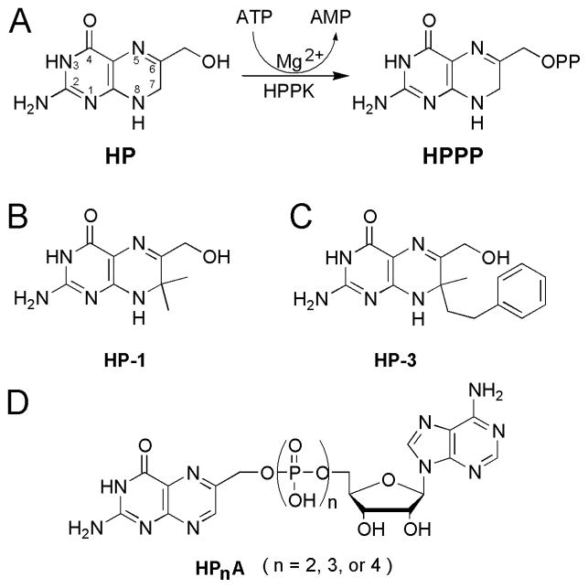
HPPK catalyzes the transfer of pyrophosphate from ATP to 6-hydroxymethyl-7,8-dihydropterin (HP) (Fig. 1A).18 The reaction follows an apparently ordered kinetic mechanism with MgATP binding to the enzyme first followed by rapid addition of HP; the Kd for the binding of MgATP to HPPK is 2.6–4.5 μM, and the Kd for the binding of HP to the HPPK MgATP complex is in the sub-μM range.19–21 In contrast, the Kd for the binding of HP to ligand-free HPPK is in the mM range.22 The cellular ATP concentration is estimated to be ~3 mM.23 Therefore, the enzyme is essentially in the MgATP-bound form, ready for the addition of HP. The products of the reaction are AMP and 6-hydroxymethyl-7,8-dihydropterin pyrophosphate (HPPP, Fig. 1A) and the release of reaction products is rate limiting.21 In-depth structural and mechanistic studies of the enzyme ensured that HPPK is currently the best understood pyrophosphokinase.24,25
Figure 1.
Reaction and molecular structures of previous HPPK inhibitors. (A) Reaction catalyzed by HPPK and chemical structures of substrate HP and product HPPP. (B–D) HPPK inhibitors HP-1,13,15 HP-3,16 and HPnA (n=2,3, or 4).17
Two types of HPPK inhibitors have been developed previously.24 Type 1 inhibitors are HP derivatives, including 6-hydroxymethyl-7,7-dimethyl-7,8-dihydropterin (HP-1, Fig. 1B)13,15 and 6-hydroxymethyl-7-methyl-7-phenethyl-7,8-dihydropterin (HP-3, Fig. 1C).16 Type 2 inhibitors are bisubstrate analogues P1-(6-hydroxymethylpterin)-P2-(5′-adenosyl)diphosphate, P1-(6-hydroxymethylpterin)-P3-(5′-adenosyl)triphosphate, P1-(6-hydroxymethylpterin)-P4-(5′-adenosyl)tetraphosphate (HPnA; n=2, 3, or 4, Fig. 1D).17 However, HP-1 is degraded during prolonged incubation with HPPK,15 the phenethyl group of HP-3 disturbs the geometry of enzyme active site significantly,16,24 which may lead to low binding affinity, and the phosphate bridge of HPnA carries many negative charges,17 which may lead to poor bioavailability. Although none of these compounds are considered useful inhibitors of HPPK,26 they provide valuable information for further development. Here, we present improved bisubstrate analog inhibitors of HPPK, which we have designed on the basis of useful features of aforementioned inhibitors.
2. Results and discussion
2.1. Design
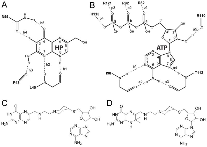
In the catalytic complex of HPPK,16,27,28 pterin is recognized by HPPK via five hydrogen bonds (Fig. 2A); so is adenosine (Fig. 2B). The phosphate bridge, however, is bound optimally only when the phosphoryl transfer reaction is about to occur. One effective approach to enhance the specificity and potency in enzyme inhibition is to design multisubstrate analogs.29,30 Among the three bisubstrate analogs we have previously developed, HP4A is the most potent inhibitor of HPPK (Kd = 0.47 and IC50 = 0.44 μM).17 The overall structure of the protein in the HPPK•MgHP4A complex (PDB entry 1EX8)17 is similar to that observed in the HPPK•MgAMPCPP•HP complex (PDB entry 1Q0N)27 and the pterin and adenosine moieties of HP4A superimpose well with their counterparts in the ternary complex.17 The pterin-protein and adenosine-protein interactions are conserved in the two structures with one exception. In HPPK•MgAMPCPP•HP, N8 of HP is hydrogen bonded to the backbone carbonyl oxygen of L45 (Fig. 2A). In HPPK•MgHP4A, however, this hydrogen bond does not exist because the pterin moiety of HP4A is oxidized (Fig. 1D) and thus N8 cannot serve as a hydrogen bond donor. Under normal conditions, HP is readily oxidized at the 7 and 8 positions, which can be effectively prevented by the introduction of two methyl groups at position 7 as those in HP-1 (Fig. 1B). As expected, the hydrogen bond between N8 of the inhibitor and carbonyl oxygen of L45 has been observed in the crystal structure of HPPK in complex with HP-1 (PDB entry 1CBK).15 Furthermore, in the HPPK•HP-1 structure, the 7,7-dimethyl group of HP-1 interacts favorably with the W89 side chain of HPPK.
Figure 2.
Protein-ligand interaction and inhibitor design. (A, B) Optimized hydrogen bond interactions between HPPK and HP/ATP on the basis of the HPPK•MgAMPCPP•HP (PDB entry 1Q0N), HPPK•MgATP•HP-3 (1DY3), and HPPK•MgAMPCPP•HP-analogs (3IP0) structures. (C, D) New design of bisubstrate analog inhibitors of HPPK.
As aforementioned, the phosphate bridge of HP4A carries several negative charges that may lead to poor bioavailability. Besides, the intact ATP moiety as part of the inhibitor may result in unwanted interaction with other kinases. At this stage of the development, we have modified the HP4A structure in two respects. First, we have replaced the phosphate bridge with a piperidine linkage (Fig. 2C). Second, we have introduced two methyl groups at position 7 of the pterin moiety (Fig. 2D). Using the piperidine, which is seen in other drug molecules, we can eliminate the negative charges carried by the linkage and also diminish unwanted binding of the inhibitor by other kinases. Introducing the 7,7-dimethyl group, we can effectively prevent the hydrogen bond donor N8 from being oxidized and thereby promote the formation of the hydrogen bond between N8 and carbonyl oxygen of L45. The resulting structures fit nicely in the active center of HPPK (not shown).
2.2. Synthesis
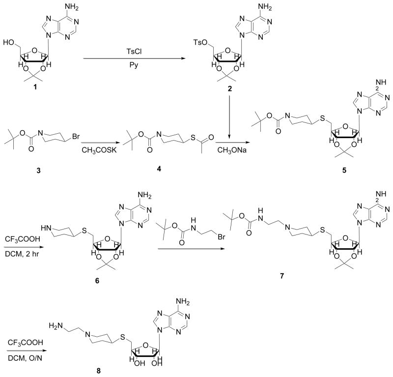
Compounds 7, 8, and 16–19 are new, whereas the rest are either known or commercially available. Using commercially available 2′,3′-isopropylideneadenosine (1, Scheme 1), we followed the Sakami method31 to synthesize 2′,3′-O-isopropylidene-5′-O-toluene-p-sulfonyl adenosine (2), during which an anhydrous pyridine solution of 1 was shaken with p-toluenesulfonyl chloride. The synthetic intermediate 4-acetylsulfanyl-piperidine-1-carboxylic acid tert-butyl ester (4) was synthesized by using the method of Plettenburg et al.32 in which potassium thioacetate and 4-bromo-piperidine (3) were heated in DMF. According to a modified procedure based on an existing protocol,33 4 reacted with sodium methoxide to form the thiol, followed by the reaction with 2 to give 4-[6-(6-amino-purin-9-yl)-2,2-dimethyl-tetrahydro-furo[3,4-d][1,3]dioxol-4-ylmethylsulfanyl]-piperidine-1-carboxylic acid tert-butyl ester (5). Under the TFA/DCM condition, cleavage of the BOC protection group yielded 9-[2,2-dimethyl-6-(piperidin-4-ylsulfanylmethyl)-tetrahydro-furo[3,4-d][1,3]dioxol-4-yl]-9H-purin-6-ylamine (6) and the subsequent reaction of 6 with (2-bromo-ethyl)-carbamic acid tert-butyl ester provided the key intermediate (2-{4-[6-(6-amino-purin-9-yl)-2,2-dimethyl-tetrahydro-furo[3,4-d][1,3]dioxol-4-ylmethylsulfanyl]-piperidin-1-yl}-ethyl)-carbamic acid tert-butyl ester (7). The deprotection of 7 yielded 2-[1-(2-amino-ethyl)-piperidin-4-ylsulfanylmethyl]-5-(6-amino-purin-9-yl)-tetrahydro-furan-3,4-diol (8) that contained an amino group that would be used to link 8 to the pterin moiety.
Scheme 1.
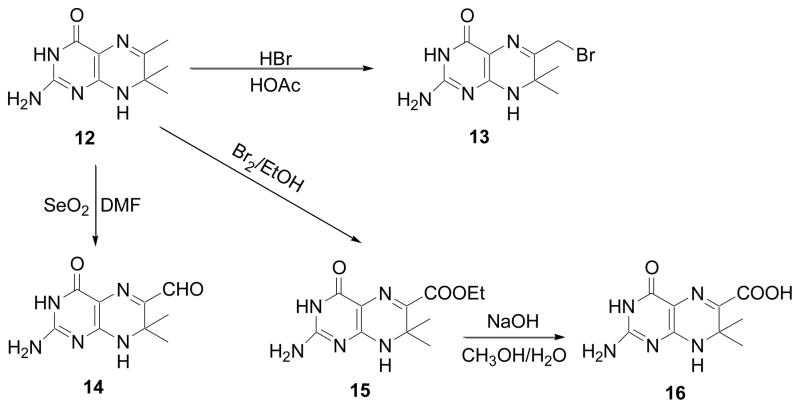
A total of four pterin moieties were synthesized, including 6-bromomethylpterin (11, Scheme 2), 6-bromomethyl-7,7-dimethyl-7,8-dihydropterin (13, Scheme 3), 6-carbaldehyde-7,7-dimethyl-7,8-dihydropterin (14, Scheme 3), and 6-carboxy-7,7-dimethyl-7,8-dihydropterin (16, Scheme 3).
Scheme 2.
Scheme 3.
Compound 11 was synthesized as described.14 Briefly, 2,4-diamino-6-(hydroxymethyl)pteridine hydrochloride (9, Scheme 2) was treated with dibromotriphenylphosphorane in N,N-dimethylacetamide to give 6-bromomethyl-pteridine-2,4-diamine (10). In 48% hydrobromic acid, 10 was converted through hydrolytic deamination to 11 (Scheme 2).
The other three pterin moieties were derived from 2-amino-7,8-dihydro-6,7,7-trimethylpteridin-4(3H)-one (12, Scheme 3) synthesized by a procedure of Al-Hassan et al.14 Subsequently, 12 was converted to 13 by Stuart’s method34 using bromine in acetic acid solution, oxidized by SeO2 in DMF to give 14, or converted to 16 through 2-amino-7,7-dimethyl-4-oxo-3,4,7,8-tetrahydro-pteridine-6-carboxylic acid ethyl ester (15) by heating 12 with bromine in ethanol solution followed by ester bond hydrolysis in base and methanol (Scheme 3). The conditions for the conversion of 12 into 14, and the intermediate (15) for the derivation of 16 from 12 are as recently described.35
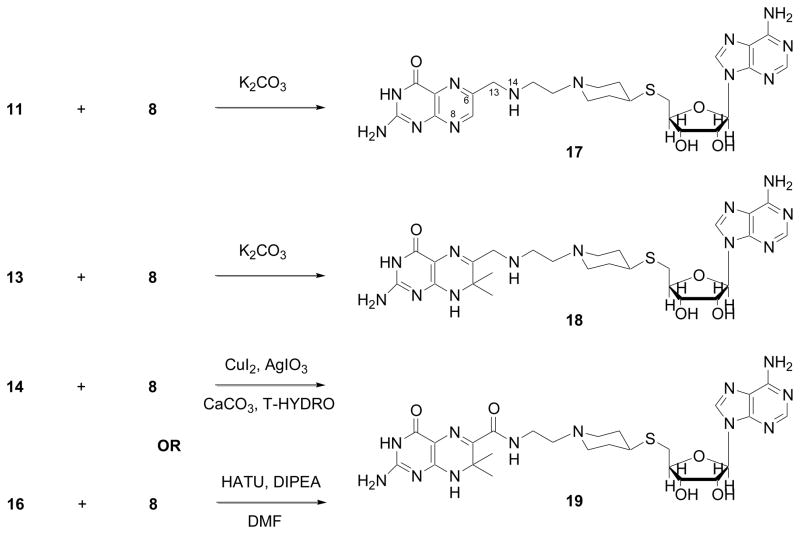
The final step of the synthesis is outlined in Scheme 4, resulting in three final products: 2-amino-6-[(2-{4-[5-(6-amino-purin-9-yl)-3,4-dihydroxy-tetrahydro-furan-2-ylmethylsulfanyl]-piperidin-1-yl}-ethylamino)-methyl]-3H-pteridin-4-one (17), 2-amino-6-[(2-{4-[5-(6-amino-purin-9-yl)-3,4-dihydroxy-tetrahydro-furan-2-ylmethylsulfanyl]-piperidin-1-yl}-ethylamino)-methyl]-7,7-dimethyl-7,8-dihydro-3H pteridin-4-one (18), and 2-amino-7,7-dimethyl-4-oxo-3,4,7,8-tetrahydro-pteridine-6-carboxylic acid (2-{4-[5-(6-amino-purin-9-yl)-3,4-dihydroxy-tetrahydro-furan-2-ylmethylsulfanyl]-piperidin-1-yl}-ethyl)-amide (19). Compound 17 was synthesized by allowing 8 and 11 to react. Compound 18 was derived from 8 and 13 in DMF solution with potassium carbonate. Compound 19 was obtained by two procedures: the procedure developed by Yoo and Li36 using copper-silver catalysis and aqueous tert-butyl hydroperoxide (Method A) and the procedure developed by us35 using the new intermediate 15 (Method B) with a significantly improved yield.
Scheme 4.
2.3. HPPK binding and inhibition
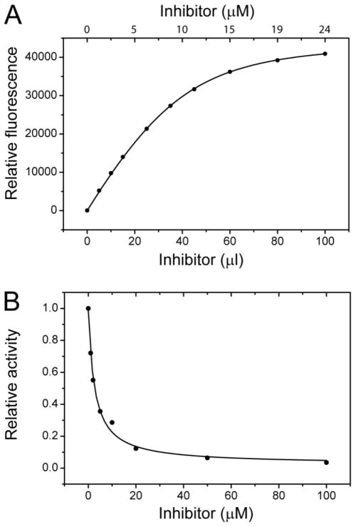
The Kd and IC50 measurements were carried out as described.37,17 The Kd value is > 150 μM for 17 and 2.55 ± 0.15 μM for 19, and the IC50 value is > 100 μM for 17 and 3.16 ± 0.34 μM for 19. Compound 18 was not stable, and therefore, its Kd and IC50 were not determined. These data show that 19 has a 60-fold increased binding affinity and 30-fold increased inhibition ability over 17.
2.4. Crystal structures of compounds 17, 18, and 19 each in complex with HPPK
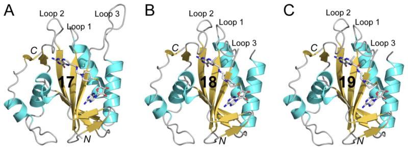
Although 18 was not stable, it was stabilized when bound to HPPK. HPPK in complex with 17, 18, or 19 was crystallized (Table 1) and the crystal structures of all three complexes (HPPK•17, HPPK•18, and HPPK•19) were determined (Table 2). The HPPK•17 structure (Fig. 3A) contains 1 HPPK (residues 1–158), 1 compound 17, 1 ethylene glycol, and 106 water molecules. The HPPK•18 structure (Fig. 3B) contains 1 HPPK (residues 1–82, 87–158), 1 compound 18, 1 acetate ion, and 88 water molecules. The HPPK•19 structure (Fig. 3C) contains 1 HPPK (residues 1–82, 87–158), 1 compound 19, 1 acetate ion, and 91 water molecules. Residues 83–86 in HPPK•18 and HPPK•19 were not observed and presumably disordered.
Table 1.
Crystallization conditions
| HPPK•17 | HPPK•18 | HPPK•19 | |
|---|---|---|---|
|
| |||
| PDB Entry Code | 3UD5 | 3UDE | 3UDV |
| Protein Solution | |||
| HPPK (mg/mL) | 10 | 10 | 10 |
| Compound 17 | Saturated | ||
| Compound 18 | Saturated | ||
| Compound 19 | Saturated | ||
| Tris-HCl [mM (pH)] | 20 (8.0) | 20 (8.0) | 20 (8.0) |
|
| |||
| Reservoir Solution | |||
| PEG 3350 [%(w/v)] | 25 | 25 | 20 |
| CH3·COONH4 (mM) | 200 | 200 | |
| Bis-Tris [mM (pH)] | 100 (6.5) | 100 (8.5) | |
| HEPES [mM (pH)] | 100 (7.5) | ||
|
| |||
| Crystals | |||
| Appear (days) | 7 | 7 | 14 |
| Reach final size (days) | 14 | 14 | 21 |
| Shape | Thin plate | Thin plate | Thin plate |
| Dimension (mm) | 0.15 × 0.10 × 0.005 | 0.10 × 0.05 × 0.005 | 0.15 × 0.10 × 0.005 |
Table 2.
Crystal data, X-ray diffraction, and structures
| HPPK•17 | HPPK•18 | HPPK•19 | |
|---|---|---|---|
|
| |||
| PDB Entry Code | 3UD5 | 3UDE | 3UDV |
| Crystal | |||
| Space group | C2 | P21212 | P21212 |
| Unit cell parameters: a (Å) | 79.98 | 52.91 | 53.00 |
| b (Å) | 52.77 | 70.98 | 70.64 |
| c (Å) | 36.69 | 36.38 | 36.25 |
| β (°) | 102.70 | 90 | 90 |
| Matthews coefficient (Å3/Da) | 2.1 | 1.9 | 1.9 |
|
| |||
| Data | Overall (last shell) | Overall (last shell) | Overall (last shell) |
| Resolution (Å) | 30.00–1.90 (1.97–1.90) | 30.00–1.82 (1.89–1.82) | 30.00–1.79 (1.85–1.79) |
| Unique reflections | 10236 (717) | 11525 (650) | 11716 (658) |
| Redundancy | 6.5 (5.3) | 6.1 (2.5) | 6.0 (2.8) |
| Completeness (%) | 87.0 (61.7) | 89.7 (51.9) | 87.6 (50.4) |
| Rmergea | 0.089 (0.320) | 0.084 (0.561) | 0.074 (0.322) |
| I/σ | 16.2 (3.8) | 16.7 (1.3) | 19.1 (2.3) |
|
| |||
| Refinement | Overall (last shell) | Overall (last shell) | Overall (last shell) |
| Resolution (Å) | 29.84–2.00 (2.13–2.00) | 29.48–1.88 (1.98–1.88) | 29.92–1.88 (1.98–1.88) |
| Unique reflections | 9252 (1203) | 10902 (1180) | 10754 (1150) |
| Completeness (%) | 90.7 (72.0) | 93.5 (73.0) | 92.8 (72.0) |
| Data in the test set | 824 (107) | 949 (104) | 920 (98) |
| R-work | 0.158 (0.164) | 0.178 (0.243) | 0.210 (0.253) |
| R-free | 0.205 (0.262) | 0.237 (0.309) | 0.275 (0.288) |
|
| |||
| Structure | |||
| Protein non-H atoms/B (Å2) | 1448/30.0 | 1389/28.1 | 1399/26.4 |
| Ligand atoms/B (Å2) | 49/44.4 | 47/31.3 | 48/40.7 |
| Water oxygen atoms/B (Å2) | 106/38.3 | 88/36.1 | 91/32.8 |
| Rmsd | |||
| Bond lengths (Å) | 0.012 | 0.012 | 0.013 |
| Bond angles (°) | 1.302 | 1.382 | 1.376 |
| Coordinate error (Å) | 0.54 | 0.50 | 0.48 |
| Ramachandran plotb | |||
| Favored regions (%) | 98.7 | 97.3 | 97.3 |
| Disallowed regions (%) | 0.0 | 0.0 | 0.0 |
Rmerge = Σ|(I-<I>)|/Σ(I), where I is the observed intensity.
Obtained using Ramachandran data by Lovell and coworkers.52
Figure 3.
Schematic illustration of crystal structures. (A) The HPPK•17 complex. (B) The HPPK•18 complex. (C) The HPPK•19 complex. Polypeptide chains are shown as a ribbon diagrams with helices (spirals) in cyan, strands (arrows) in orange, and loops (tubes) in grey. Ligands are shown as sticks in atomic color scheme (C in grey, N in blue, O in red, P in orange, and S in orange).
2.5. Structure-activity relationship
2.5.1. Protein conformation and inhibitor binding
HPPK has three flexible loops (Loop 1, residues 8–15; Loop 2, residues 43–53; Loop 3, residues 82–92), among which Loop 3 undergoes dramatic conformational changes during catalysis.22,38 The catalytic trajectory of HPPK can be described by six consecutive states: ligand-free HPPK, HPPK•MgATP, HPPK•MgATP•HP, HPPK•MgAMP-PP-HP (the transition state), HPPK•AMP•HPPP, and HPPK•HPPP.21,39 In the HPPK•MgATP state, Loop 3 exhibits open conformation as seen in the HPPK•MgADP [Protein Data Bank (PDB) entry 1EQM] and HPPK•MgAMPPCP (PDB entry 1EQ0) structures.38 Loop 3 is also open in the HPPK•AMP•HPPP state (PDB entry 1RAO).39 In the HPPK•MgATP•HP state, in contrast, Loop 3 is closed as seen in the HPPK•MgAMPCPP•HP structure (PDB entry 1Q0N).27 In the HPPK•MgATP state, however, Loop 3 has been seen to have a closed conformation in the HPPK•MgAMPCPP complex (PDB entry 2F65),40 suggesting that Loop 3 in the HPPK•MgATP state is dynamic.25
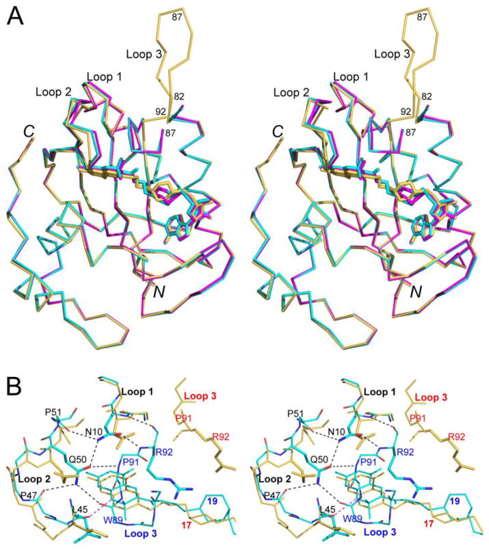
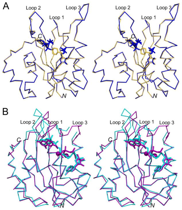
The polypeptide chains in the HPPK•17, HPPK•18, and HPPK•19 structures superimpose well except for Loop 3 (Fig. 4A). The adenosine moieties in the three inhibitors superimpose well; so do the pterin moieties. The linkers in 18 and 19 superimpose well, but the linker in 17 displays a different conformation. In HPPK•17, Loop 3 displays the open conformation (Fig. 5A). During HPPK catalysis, the open Loop 3 facilitates HP binding and product release. For enzyme inhibition, however, the open conformation of Loop 3 has negative impact on binding, which is in agreement with the Kd value for the binding of 17 to HPPK (> 150 μM). As aforementioned, the Kd for the binding of MgATP to HPPK is 2.6–4.5 μM. Therefore, 17 is not a good inhibitor. In contrast, Loop 3 assumes the closed conformation in the HPPK•18 and HPPK•19 structures although residues 83–86 in the loop are disordered (Fig. 4A, 5B). During HPPK catalysis, a closed Loop 3 seals the active center for the reaction to occur. For enzyme inhibition, a closed Loop 3 enhances the affinity for the enzyme and consequently the potency of an inhibitor. Consistently, compound 19 shows a much higher binding affinity (Kd = 2.55 μM). The Kd value of 19 is comparable with that of MgATP, suggesting the compound is a good inhibitor of HPPK.
Figure 4.
Structural comparison in stereo. (A) The HPPK•17 (in orange), HPPK•18 (in magenta), and HPPK•19 (in cyan) structures are superimposed. Proteins are shown as Cα traces and ligands as sticks. (B) Coupled loops in HPPK•19 (in cyan, those in HPPK•18 are not shown for clarity) versus uncoupled loops in HPPK•17 (in orange). Hydrogen bonds are indicated by dashed lines in black, but the N8Pterin···OL45 interaction is highlighted in magenta. Labels are color coded: blue for HPPK•19, red for HPPK•17, and black for the common features in both. For clarity, most side chains that are not involved in loop coupling are not shown.
Figure 5.
Comparison of new with related structures in stereo. (A) The HPPK•17 (in orange, this work) and HPPK•AMP•HPPP (in blue, PDB entry 1RAO) structures are superimposed. (B) The HPPK•19 (in cyan, this work) and HPPK•MgAMPCPP•HP (in purple, PDB entry 1Q0N) structures are superimposed. Proteins are shown as Cα traces and ligands as sticks.
2.5.2. Protein conformation and inhibitor structure
To form the catalytic assembly of HPPK•MgATP•HP in the closed conformation, conserved residues N10 (of Loop 1) and Q50 (of Loop 2) function as the center of a hydrogen bond network with the backbone carbonyl groups of P47 and P51 of Loop 2 and with the backbone amide and/or carbonyl groups of W89, P91, and R92 of Loop 3.27 This hydrogen bond network couples all three loops and helps stabilize the complex and seal the active center. The structures show that such a hydrogen bond network exists in HPPK•18 and HPPK•19, but not in HPPK•17 (Fig. 4B).
The only differences between 18 and 17 are in positions 7 and 8 of the pterin moiety (Scheme 4). In 18, the 7,7-dimethyl group keeps the NH group at position 8 in its reduced form, allowing the formation of the hydrogen bond between N8 and the carbonyl oxygen of L45 (N8Pterin···OL45). In contrast, the dimethyl group is absent from 17 and the pterin moiety is oxidized with an N instead of an NH group at position 8, rendering the formation of the N8Pterin···OL45 hydrogen bond impossible.
The impact of the formation of N8Pterin···OL45 is twofold. First, it is one of the five hydrogen bonds for the recognition of HP by HPPK (Fig. 2A) and therefore contributes to protein-ligand affinity. Second, by anchoring L45 it optimizes the conformation of Loop 2 and Loop-2 residues, especially Q50, favoring the formation of the hydrogen bond network that couples and stabilizes the three loops (Fig. 4B). Taken together, these structures show that the N8Pterin···OL45 hydrogen bond facilitates the formation of the hydrogen bond network that couples the three loops as seen in the closed HPPK•MgATP•HP state. A good inhibitor, such as 19, should not impair the coupling of these loops.
As aforementioned, the 7,7-dimethyl group in HPPK•HP-1 interacts with the side chain of W89.15 This hydrophobic interaction is also observed in HPPK•19, whereas it is not possible in HPPK•17 (Fig. 4B). In addition to the 7,7-dimetyl group, 19 has a new carbonyl group next to the pterin moiety (Scheme 4). It does not, however, affect the conformation of the protein.
2.5.3. Inhibitor structure and stability
Compound 17 is stable, but 18 tends to degrade when not in complex with HPPK. The only difference between 17 and 18 is that the pterin moiety in 17 is oxidized, but it is reduced in 18. Compound 19 is stable. The only difference between 18 and 19 is that the methylene group (–CH2–) at position 13 in 18 becomes a carbonyl group (–CO–) in 19 (Scheme 4).
2.5.4. Partial disorder of Loop 3 and protein-linker interaction
Although 19 is a good inhibitor of HPPK, Loop 3 residues 83–86 in HPPK•19 are disordered (Fig. 4A). Furthermore, all three loops in HPPK•19 display noticeable differences when compared with those in the HPPK•MgAMPCPP•HP structure (Fig. 5B). Such disorder and differences indicate that the protein-linker interactions are not optimized in HPPK•19 although the three loops of the enzyme are coupled and the complex assumes the overall closed conformation. We need to further modify the linker between the pterin and adenosine moieties of 19, which we believe will lead to higher binding affinity. In the HPPK•MgATP•HP state, the phosphate bridge is recognized by conserved HPPK side chains R82, R92, H115, and R121 (Fig. 2B). Therefore, hydrogen bond acceptors should be introduced into the linker.
3. Conclusions
We report a new generation of bisubstrate analog inhibitors of HPPK, compounds 17, 18, and 19 (Scheme 4). In complex with HPPK, these compounds share a common adenosine-protein interaction pattern, but their pterin moieties interact with the protein differently. In HPPK•18 and HPPK•19, the N8Pterin···OL45 hydrogen bond is formed between pterin atom N8 (donor) of the inhibitor and carbonyl oxygen (acceptor) of L45, whereas in HPPK•17, the formation of this hydrogen bond is impossible. The structures show that the formation of N8Pterin···OL45 is sufficient to facilitate the coupling of the three loops of HPPK in the manner as seen in the HPPK•MgAMPCPP•HP complex.27 Without the N8Pterin···OL45 interaction, the HPPK•17 complex exhibits the open conformation without the coupling of the three loops and the binding affinity of 17 is low (Kd > 150 μM). With the formation of N8Pterin···OL45, in contrast, the HPPK•19 complex displays the closed conformation due to the coupling of the three loops and the affinity of 19 is ~60-fold higher. Introducing the 7,7-dimethyl groups into 17 results in 18, which triggers the transition of the open conformation (in HPPK•17) into the closed form (in HPPK•18). Nevertheless, 18 is not stable when it is not in complex with the enzyme. It is the replacement of the methylene group in N=CH–CH2–NH of 18 with the carbonyl group in N=CH–CO–NH of 19 that improves the chemical stability (Scheme 4).
With and without the N8Pterin···OL45 interaction, the binding mode of the linker is also different (Fig. 4A), underscoring the importance of an ordered and closed Loop 3 for optimized protein-inhibitor interaction. For pyrophosphoryl transfer, the phosphate groups of ATP interact with four conserved side chains, R82, R92, H115, and R121 (Fig. 2B). None of these side chains interacts favorably with the linker of any of the three compounds (17, 18, and 19). Further development of bisubstrate analog inhibitors should allow favorable interactions between the linker of inhibitor and the four positively charged side chains of HPPK. Residues R82 and R92 define the boundary of Loop 3. Favored interactions between these two side chains and inhibitor should lead to a completely ordered and closed Loop 3 in the protein-inhibitor complex.
4. Experimental methods
4.1. Modeling
Design and docking of new inhibitors into the active site of HPPK were carried out with program packages CNS and O.41,42 The model complex was subject to geometry optimization using the conjugate gradient method developed by Powell;43 Engh and Huber geometric parameters were used as the basis of the force field.44.
4.2. Chemistry
4.2.1. General methods
All chemicals were purchased from Sigma-Aldrich except that compound 1 was purchased from TCI America. Starting materials and solvents were used without further purification. Anhydrous reactions were conducted under a positive pressure of dry N2. Reactions were monitored by TLC, on Baker-flex Silica Gel IB-F (J. T. Baker). All compounds and intermediates were purified by flash chromatography performed on Teledyne ISCO Combiflash Rf system using RediSep Rf columns. Ion exchange chromatography was performed using strata Scx (50 μm particle size, 70 Å pore) resin cartridges. Preparative high pressure liquid chromatography (HPLC) was conducted using a Waters 600E system using a Waters 2487 dual λ absorbance detector and Phenomenex C18 columns (250 mm × 21.2 mm, 5 μm particle size, 110 Å pore) at a flow rate of 10 mL/min. A binary solvent systems consisting of A = 0.1% aqueous TFA and B = 0.1% TFA in acetonitrile was employed with the gradients as indicated. 1H and 13C NMR data were obtained on a Varian 400 MHz spectrometer and are reported in ppm relative to TMS (tetramethylsilane). Mass spectra were measured with Agilent 1100 series LC/Mass Selective Detector, Agilent 1200 LC/MSD-SL system and Thermoquest Surveyor Finnigan LCQ deca. Chemical purity was determined by HPLC analysis with a Zorbax Eclipse plus C18 column (Narrow Bore RR 2.1 mm × 50 mm, 3.5 micron; flow rate of 0.3 mL/min; solvent, methanol:H2O gradient, 0.1% acetic acid; detection at 260 nm), confirming > 95% purity.
4.2.2. (2-{4-[6-(6-Amino-purin-9-yl)-2,2-dimethyl-tetrahydro-furo[3,4-d][1,3]dioxol-4-ylmethylsulfanyl]-piperidin-1-yl}-ethyl)-carbamic acid tert-butyl ester (7)
To a solution of 9-[2,2-Dimethyl-6-(piperidin-4-ylsulfanylmethyl)-tetrahydro-furo[3,4-d][1,3]dioxol-4-yl]-9H-purin-6-ylamine (6) (4.06 g, 10 mmol, 1 eq) and potassium carbonate (2.76g, 20 mmol, 2 eq) in 100 mL acetonitrile, 2-(Boc-amino)ethyl bromide (1.12g, 5 mmol, 0.5eq) was added and then stirred at 50 °C for a few hours before another dose of 2-(Boc-amino)ethyl bromide (1.12g, 5 mmol, 0.5eq) was added. After the reaction was finished, the mixture was evaporated, diluted with EtOAc (100 mL), and washed with saturated aqueous NaHCO3 (2 × 50 mL) and brine (50 mL). The residue from concentrating the organic layer was purified by silica gel chromatography, eluting with 10% DCM/methanol, to afford 5.43 g (99%) of the title compound as white foam. NMR δH (400 MHz; CD3OD), 1.39 (3 H, s), 1.42–1.48 (13 H, m), 1.58 (3 H, s), 1.72–1.99 (4 H, m), 2.37 (2 H, t), 2.53–2.58 (1 H, m), 2.75–2.82 (2 H, m), 3.17 (2 H, t), 4.33 (1 H, m), 5.07 (1 H, m), 5.58 (1 H, m), 6.18 (1 H, d), 8.23 (1 H, s), 8.28 (1 H, s); δ13C (100 MHz; CD3OD), 158.32 (1C), 157.46 (1C), 154.03 (1C), 150.25 (1C), 142.05 (1C), 120.65 (1C), 115.33 (1C), 91.88 (1C), 88.87 (1C), 85.25 (1C), 85.07 (1C), 80.05 (1C), 58.75 (1C), 54.18 (2C), 42.29 (1C), 38.41 (1C), 33.56 (1C), 33.44 (1C), 33.28 (1C), 28.75 (3C), 27.35 (1C), 25.48 (1C); MS (ESI) calculated for C25H39N7O5S [M+H]+ 550.27, found 550.1.
4.2.3. 2-[1-(2-Amino-ethyl)-piperidin-4-ylsulfanylmethyl]-5-(6-amino-purin-9-yl)-tetrahydro-furan-3,4-diol (8)
To a solution of (2-{4-[6-(6-Amino-purin-9-yl)-2,2-dimethyl-tetrahydro-furo[3,4-d][1,3]dioxol-4-ylmethylsulfanyl]-piperidin-1-yl}-ethyl)-carbamic acid tert-butyl ester (7) (2.75 g, 5 mmol, 1 eq) in 50 mL DCM, 10 ml TFA was added dropwise at −20 °C and then stirred at room temperature overnight. After the reaction was finished, the solvent was removed under vacuum, and the residue was purified by silica gel chromatography, eluting with 15% DCM/methanol, to afford 1.84 g (90%) of the title compound as white foam. NMR δH (400 MHz; CD3OD), 1.46–1.54 (4 H, m), 1.84–1.98 (4 H, m), 2.41 (2 H, t), 2.65–2.82 (3 H, m), 2.89–3.02 (2 H, m), 4.20 (1 H, m), 4.37 (1 H, m), 4.80 (1 H, m), 6.0 (1 H, d), 8.21 (1 H, s), 8.31 (1 H, s); δ13C (100 MHz; CD3OD), 157.40 (1C), 153.97 (1C), 150.73 (1C), 141.55 (1C), 120.61 (1C), 90.19 (1C), 86.00 (1C), 74.72 (1C), 73.97 (1C), 59.76 (1C), 54.32 (2C), 42.64 (1C), 38.66 (1C), 33.67 (2C), 33.13 (1C); MS (ESI) calculated for C17H27N7O3S [M+H]+ 410.27, found 410.1.
4.2.4. 2-Amino-7,7-dimethyl-4-oxo-3,4,7,8-tetrahydro-pteridine-6-carboxylic acid (16)
The intermediate 15 was synthesized as described.35 To a solution of 15 (265mg, 1mmol, 1eq) in methanol (5ml) was added a solution of sodium hydroxide (2M, 2mmol, 2eq). After stirring for two hours, the reaction mixture was acidified to pH = 2 with 1M HCl, the product was precipitated, and the precipitate washed once with water and then dried to obtain 16 (213mg, 0.9mmol, 90%). NMR δH (400 MHz; CD3OD), 1.59 (H, s); δ13C (100 MHz; CD3OD), 166.22 (1C), 161.58 (1C), 157.23 (1C), 156.67 (1C), 142.07 (1C), 101.93 (1C), 36.50 (1C), 29.12 (2C); MS (ESI) calculated for C9H11N5O3 ([M+H]+) 238.09, found 238.10.
4.2.5. 2-Amino-6-[(2-{4-[5-(6-amino-purin-9-yl)-3,4-dihydroxy-tetrahydro-furan-2-ylmethylsulfanyl]-piperidin-1-yl}-ethylamino)-methyl]-3H-pteridin-4-one (17)
To a solution of 8 (100.0 mg, 0.244 mmol, 1 eq) and potassium carbonate (337.9 mg, 2.44 mmol, 10 eq) in 20 mL dimethylacetamide, 11 (81.7 mg, 0.244 mmol, 1eq) was added and stirred at room temperature for 24 hours. It was evaporated under high vacuum and the residue was dissolved in water methanol mixture and purified by HPLC to give 17 (71.0mg, 0.122 mmol, 50%) as a yellowish powder. NMR δH (400 MHz; CD3OD), 1.89 (4 H, m), 2.28–2.32 (4 H, m), 3.05–3.3 (3 H, m), 3.53 (2 H, m), 3.63 (2 H, m), 4.22 (1 H, m), 4.35 (1 H, t, J 5.2), 4.41 (2 H, s), 4.74 (1 H, t, J 5.2), 6.05 (1 H, d, J 4.8), 8.37 (1 H, s), 8.47 (1 H, s), 8.76 (1 H, s); δ13C (100 MHz; DMSO-d6), 160.62 (1C), 158.77 (1C), 158.44 (1C), 158.09 (1C), 154.34 (1C), 149.85 (1C), 148.99 (1C), 141.52 (1C), 141.09 (1C), 127.94 (1C), 119.02 (1C), 87.64 (1C), 84.07 (1C), 72.84 (1C), 72.58 (1C), 51.37 (2C), 48.33 (1C), 41.16 (1C), 40.36 (1C), 37.52 (1C), 32.26 (2C), 31.97 (1C); HRMS (ESI-MS) calculated for C24H32N12O4S (MH+): 585.2463; found: 585.2460.
4.2.6. 2-Amino-6-[(2-{4-[5-(6-amino-purin-9-yl)-3,4-dihydroxy-tetrahydro-furan-2-ylmethylsulfanyl]-piperidin-1-yl}-ethylamino)-methyl]-7,7-dimethyl-7,8-dihydro-3H pteridin-4-one (18)
To a solution of 8 (100.0 mg, 0.244 mmol, 1 eq) and potassium carbonate (337.9 mg, 2.44 mmol, 10 eq) in 20 mL dimethylacetamide, 13 (89.0 mg, 0.244 mmol, 1eq) was added and stirred at room temperature for 24 hours. It was evaporated under high vacuum and the residue was extracted by methanol. It was evaporated again and the residue was used for direct analysis without further purification. MS (ESI) calculated for C26H38N12O4S ([M+H]+) 615.29, found 615.10.
4.2.7. 2-Amino-7,7-dimethyl-4-oxo-3,4,7,8-tetrahydro-pteridine-6-carboxylic acid (2-{4-[5-(6-amino-purin-9-yl)-3,4-dihydroxy-tetrahydro-furan-2-ylmethylsulfanyl]-piperidin-1-yl}-ethyl)-amide (19, Method A)36
Compound 8 (12.3 mg, 0.03 mmol, 1.5 eq) was mixed with CuI (0.0388 mg, 0.0002 mmol, 1.0 mol%), AgIO3 (0.057 mg, 0.0002 mmol, 1.0 mol%), and CaCO3 (2.2 mg, 0.022 mmol, 1.1 eq) in DMF (0.2 mL). Compound 14 (4.5 mg, 0.020 mmol, 1.0 eq) and T-HYDRO® (70 wt% in H2O, 0.00315 mL, 0.022 mmol, 1.1 eq) were added under an inert atmosphere (N2) at room temperature. The reaction was allowed to stir overnight at 40 °C. The crude reaction was purified by HPLC (H2O:Methanol = 2:3) to provide 19 (3.77 mg, 0.006 mmol, 30%) as a pale yellow solid. NMR δH (400 MHz; CD3OD), 1.58 (6 H, s), 1.76–2.28 (8 H, m), 2.85–3.06 (3 H, m), 3.25 (2H, m) 3.61 (2 H, m), 4.22 (1 H, m), 4.33 (1 H, m), 4.74 (1 H, m), 6.05 (1 H, d, J 4.8), 8.37 (1 H, s), 8.46 (1 H, s); δ13C (100 MHz; DMSO-d6), 164.22 (1C), 157.79 (1C), 155.09 (1C), 155.00 (1C), 148.84 (1C), 143.01 (1C), 141.49 (1C), 118.96 (1C), 117.62 (1C), 114.69 (1C), 100.66 (1C), 87.64 (1C), 84.18 (1C), 72.96 (1C), 72.53 (1C), 55.31 (1C), 53.27 (2C), 51.90 (1C), 37.83 (1C), 33.44 (2C), 31.97 (1C), 29.85 (1C), 27.71 (2C); HRMS (ESI-MS) calculated for C26H36N12O5S (MH+): 629.2725; found: 629.2708.
4.2.8. Compound 19 (Method B)
To a solution of 16 (190 mg, 0.8 mmol, 1eq), O-(7-azabenzotriazol-1-yl)-1,1,3,3-tetramethyluronium hexafluorophosphate (HATU) (334.6, 0.88 mmol, 1.1eq), and compound 8 (327.2 mg, 0.8 mmol, 1eq) in anhydrous DMF (100 mL) was added DIPEA (4.18 uL, 2.4 mmol, 3q). After 18 h, the solvent was evaporated under high vacuum, the reaction residue was purified by HPLC (H2O:Methanol = 2:3) to provide 19 (377 mg, 0.6 mmol, 75%) as a pale yellow solid. NMR and HRMS (ESI-MS) data are found in 4.2.7.
4.3. Fluorometric titration
The dissociation constants of the inhibitors were measured as described37 for the fluorometric measurement of the dissociation constant of Ant-ATP with modifications. Briefly, the inhibitors were dissolved in dimethylsulfoxide. Dimethylsulfoxide concentrations were kept within 1.7% during the titration experiments and control experiments showed that dimethylsulfoxide at these concentrations had no effects on activity (substrate binding and catalysis) of the enzyme. The excitation and emission wavelengths were 420 and 450 nm, respectively, for 17, but 450 and 480, respectively, for 19. The excitation and emission slits were 1 and 4 nm, respectively. The titration was performed by adding aliquots of a 500 μM 19 stock solution to an HPPK solution. The initial HPPK concentration and volume were 10 μM and 2 ml, respectively. The Kd values were obtained by nonlinear least-squares regression regression of the data to equation 1 as described37
| (Eq. 1) |
where ΔFobs and ΔFmol are observed and molar fluorescence changes caused by binding, Et is the total concentration of HPPK, and Lt is the total concentration of the inhibitor. As an example, the fluorometric titration of HPPK with compound 19 is shown in Fig. 6A.
Figure 6.
Kd and IC50 measurements. (A) Fluorometric titration of HPPK with compound 19. (B) Inhibition of HPPK by 19.
4.4. Enzyme inhibition assay
IC50 measurements were carried out as described17 except for the concentrations of reaction mixtures, which contained 1 nM E. coli HPPK, 2 μM ATP, 1 μM HP, 5 mM MgCl2, 25 mM DTT, and a trace amount of [α-32P]-ATP (~1 μCi) in 100 mM Tris, pH 8.3. IC50 values were obtained by fitting the data to a logistic equation by nonlinear least-squares regression of the data to equation 2 as described45
| (Eq. 2) |
where v is the reaction rate, vmin the minimum reaction rate, vmax the maximum reaction rate, and [I] the concentration of the inhibitor. The inhibition of HPPK by compound 19 is shown in Fig. 6B.
4.5. Crystallization, X-ray diffraction, structure solution, and refinement
Crystals were grown in sitting drops at 19±1 °C. Crystallization conditions are summarized in Table 1. A Hydra II Plus crystallization robot (Matrix Technologies, Hudson, New Hampshire, USA) and Crystal Screen kits from Hampton Research (Laguna Niguel, California, USA) were used. X-ray diffraction data were collected at 100K with an MARCCD detector mounted at the synchrotron Beamline 22 at the Advanced Photon Source, Argonne National Laboratory. Data processing was carried out with the HKL2000 program suite.46 The structure was solved by Fourier synthesis starting with a homologous structure: PDB entry 1EQM for HPPK•17, 3ILJ for HPPK•18, and 3UDE for HPPK•19. Multiple conformations of amino acid residues, ligands, and solvent molecules were removed from the starting models. Structure solution and refinement were done with PHENIX.47 All graphics work, including model building and rebuilding, was performed with COOT.48 The structures were verified with annealed omit maps and the geometry was assessed using PROCHECK49 and WHAT IF.50 The statistics of X-ray diffraction data and structures are summarized in Table 2. Illustrations were prepared with PyMOL.51
Supplementary Material
Acknowledgments
This research was supported by NIH grant GM51901 (H.Y.), NIAID Trans NIH/FDA Intramural Biodefense Program Y3-RC-8007-01 (X.J.), and the Intramural Research Program of the NIH, National Cancer Institute, Center for Cancer Research. Mass spectrometry experiments were conducted on an Agilent 1100 series LC/Mass Selective Detector maintained by the Biophysics Resource in the Structural Biophysics Laboratory, an Agilent 1200 LC/MSD-SL system in the Chemical Biology Laboratory, and a Thermoquest Surveyor Finnigan LCQ deca maintained by the Comparative Carcinogenesis Laboratory of National Cancer Institute at Frederick. X-ray diffraction data were collected at the Southeast Regional Collaborative Access Team (SER-CAT) 22-ID and 22-BM beamlines at the Advanced Photon Source (APS), Argonne National Laboratory (ANL).
Abbreviations
- AMPCPP
α,β-methyleneadenosine 5′-triphosphate
- HP
6-hydroxymethyl-7,8-dihydropterin
- HP-1
6-hydroxymethyl-7,7-dimethyl-7,8-dihydropterin
- HP-3
6-hydroxymethyl-7-methyl-7-phenethyl-7,8-dihydropterin
- HP2A
P1-(6-hydroxymethylpterin)-P2-(5′-adenosyl)diphosphate
- HP3A
P1-(6-hydroxymethylpterin)-P3-(5′-adenosyl)triphosphate
- HP4A
P1-(6-hydroxymethylpterin)-P4-(5′-adenosyl)tetraphosphate
- HPPK
6-hydroxymethyl-7,8-dihydropterin pyrophosphokinase
- HPPP
6-hydroxymethyl-7,8-dihydropterin pyrophosphate
- PDB
Protein Data Bank
Footnotes
The coordinates and structure factors have been deposited in the PDB under entry codes 3UD5 (HPPK•17), 3UDE (HPPK•18), and 3UDV (HPPK•19).
Supplementary data (the 1H and 13C spectra of compounds 7, 8, 16, 17, and 19) associated with this article can be found, in the online version, at…
Publisher's Disclaimer: This is a PDF file of an unedited manuscript that has been accepted for publication. As a service to our customers we are providing this early version of the manuscript. The manuscript will undergo copyediting, typesetting, and review of the resulting proof before it is published in its final citable form. Please note that during the production process errors may be discovered which could affect the content, and all legal disclaimers that apply to the journal pertain.
References
- 1.Blakley RL, Benkovic SJ. In: Folates and Pterins. Blakley RL, Benkovic SJ, editors. John Wiley & Sons, Inc; New York: 1984. [Google Scholar]
- 2.Hitchings GH, Burchall JJ. Adv Enzymol Relat Areas Mol Biol. 1965;27:417–468. doi: 10.1002/9780470122723.ch9. [DOI] [PubMed] [Google Scholar]
- 3.Cohen ML. Science. 1992;257:1050–1055. doi: 10.1126/science.257.5073.1050. [DOI] [PubMed] [Google Scholar]
- 4.Neu HC. Science. 1992;257:1064–1073. doi: 10.1126/science.257.5073.1064. [DOI] [PubMed] [Google Scholar]
- 5.Kunin CM. Ann Intern Med. 1993;118:557–561. doi: 10.7326/0003-4819-118-7-199304010-00011. [DOI] [PubMed] [Google Scholar]
- 6.Levy SB. Adv Exp Med Biol. 1995;390:1–13. doi: 10.1007/978-1-4757-9203-4_1. [DOI] [PubMed] [Google Scholar]
- 7.Murray BE. Adv Intern Med. 1997;42:339–367. [PubMed] [Google Scholar]
- 8.Bermingham A, Derrick JP. Bioessays. 2002;24:637–648. doi: 10.1002/bies.10114. [DOI] [PubMed] [Google Scholar]
- 9.Walsh C. Nat Rev Microbiol. 2003;1:65–70. doi: 10.1038/nrmicro727. [DOI] [PubMed] [Google Scholar]
- 10.Hughes DTD. Sulphonamides Antibiotic and Chemotherapy. 7. Churchill Livingstone; New York: 1997. [Google Scholar]
- 11.Hughes DTD. In: Antibiotics and Chemotherapy; O’Grady F, Lambert HP, Finch RG, Greenwood D, editors. Churchill Livingstone; New York: 1997. pp. 346–356. [Google Scholar]
- 12.Zinner SH, Mayer KH. In: Principles and Practice of Infectious Diseases; Mandell GL, Bennett JE, Dolin R, editors. Churchill Livingstone; Philadelphia: 2005. pp. 440–451. [Google Scholar]
- 13.Wood HCS. In: Chemistry and Biology of Pteridines; Pfleiderer W, editor. Walter de Gruyter; Berlin-New York: 1975. [Google Scholar]
- 14.Al-Hassan SS, Cameron RJ, Curran AWC, Lyall WJS, Nicholson SH, Robinson DR, Stuart A, Suckling CJ, Stirling I, Wood HCS. J Chem Soc, Perkin Trans. 1985;1:1645–1659. [Google Scholar]
- 15.Hennig M, Dale GE, D’Arcy A, Danel F, Fischer S, Gray CP, Jolidon S, Muller F, Page MG, Pattison P, Oefner C. J Mol Biol. 1999;287:211–219. doi: 10.1006/jmbi.1999.2623. [DOI] [PubMed] [Google Scholar]
- 16.Stammers DK, Achari A, Somers DO, Bryant PK, Rosemond J, Scott DL, Champness JN. FEBS Lett. 1999;456:49–53. doi: 10.1016/s0014-5793(99)00860-1. [DOI] [PubMed] [Google Scholar]
- 17.Shi G, Blaszczyk J, Ji X, Yan H. J Med Chem. 2001;44:1364–1371. doi: 10.1021/jm0004493. [DOI] [PubMed] [Google Scholar]
- 18.Shiota T. In: Chemistry and Biochemistry of Folates; Blakley RT, Benkovic SJ, editors. John Wiley & Sons; New York: 1984. pp. 121–134. [Google Scholar]
- 19.Shi G, Gong Y, Savchenko A, Zeikus JG, Xiao B, Ji X, Yan H. Biochim Biophys Acta. 2000;1478:289–299. doi: 10.1016/s0167-4838(00)00043-1. [DOI] [PubMed] [Google Scholar]
- 20.Bermingham A, Bottomley JR, Primrose WU, Derrick JP. J Biol Chem. 2000;275:17962–17967. doi: 10.1074/jbc.M000331200. [DOI] [PubMed] [Google Scholar]
- 21.Li Y, Gong Y, Shi G, Blaszczyk J, Ji X, Yan H. Biochemistry. 2002;41:8777–8783. doi: 10.1021/bi025968h. [DOI] [PubMed] [Google Scholar]
- 22.Xiao B, Shi G, Chen X, Yan H, Ji X. Structure. 1999;7:489–496. doi: 10.1016/s0969-2126(99)80065-3. [DOI] [PubMed] [Google Scholar]
- 23.Neuhard J, Nygaard P. In: Escherichia coli and Salmonella typhimurium: Cellular and Molecular Biology; Neidhardt FC, Ingraham JL, Low KB, Magasanik B, Schaechter M, Umbarger HE, editors. ASM Press; Washington, D. C: 1987. pp. 445–473. [Google Scholar]
- 24.Derrick JP. In: Folic Acid and Folates; Litwack G, editor. Academic Press; Oxford, UK: 2008. pp. 411–433. [Google Scholar]
- 25.Yan H, Ji X. Protein Pept Lett. 2011;18:328–335. doi: 10.2174/092986611794654003. [DOI] [PMC free article] [PubMed] [Google Scholar]
- 26.Kompis IM, Islam K, Then RL. Chem Rev. 2005;105:593–620. doi: 10.1021/cr0301144. [DOI] [PubMed] [Google Scholar]
- 27.Blaszczyk J, Shi G, Yan H, Ji X. Structure. 2000;8:1049–1058. doi: 10.1016/s0969-2126(00)00502-5. [DOI] [PubMed] [Google Scholar]
- 28.Blaszczyk J, Li Y, Shi G, Yan H, Ji X. Biochemistry. 2003;42:1573–1580. doi: 10.1021/bi0267994. [DOI] [PubMed] [Google Scholar]
- 29.Broom AD. J Med Chem. 1989;32:2–7. doi: 10.1021/jm00121a001. [DOI] [PubMed] [Google Scholar]
- 30.Radzicka A, Wolfenden R. Methods Enzymol. 1995;249:284–312. doi: 10.1016/0076-6879(95)49039-6. [DOI] [PubMed] [Google Scholar]
- 31.Sakami W. Biochemical Prepparations. 1961;8:5–8. [Google Scholar]
- 32.Plettenburg O, Hofmeister A, Goerlitzer J, LÖHn M. WO/2008/077,552. WO Patent. 2008
- 33.Isakovic L, Saavedra OM, Llewellyn DB, Claridge S, Zhan L, Bernstein N, Vaisburg A, Elowe N, Petschner AJ, Rahil J. Bioorg Med Chem Lett. 2009;19:2742–2746. doi: 10.1016/j.bmcl.2009.03.132. [DOI] [PubMed] [Google Scholar]
- 34.Stuart A. 4,036,961. US Patent. 1977
- 35.Shi G, Ji X. Tetrahedron Lett. 2011;52:6174–6176. doi: 10.1016/j.tetlet.2011.09.047. [DOI] [PMC free article] [PubMed] [Google Scholar]
- 36.Yoo WJ, Li CJ. J Am Chem Soc. 2006;128:13064–13065. doi: 10.1021/ja064315b. [DOI] [PubMed] [Google Scholar]
- 37.Li Y, Wu Y, Blaszczyk J, Ji X, Yan H. Biochemistry. 2003;42:1581–1588. doi: 10.1021/bi026800z. [DOI] [PubMed] [Google Scholar]
- 38.Xiao B, Shi G, Gao J, Blaszczyk J, Liu Q, Ji X, Yan H. J Biol Chem. 2001;276:40274–40281. doi: 10.1074/jbc.M103837200. [DOI] [PubMed] [Google Scholar]
- 39.Blaszczyk J, Shi G, Li Y, Yan H, Ji X. Structure (Camb) 2004;12:467–475. doi: 10.1016/j.str.2004.02.003. [DOI] [PubMed] [Google Scholar]
- 40.Li G, Felczak K, Shi G, Yan H. Biochemistry. 2006;45:12573–12581. doi: 10.1021/bi061057m. [DOI] [PubMed] [Google Scholar]
- 41.Brünger AT, Adams PD, Clore GM, DeLano WL, Gros P, Grosse-Kunstleve RW, Jiang JS, Kuszewski J, Nilges M, Pannu NS, Read RJ, Rice LM, Simonson T, Warren GL. Acta Crystallogr D. 1998;54:905–921. doi: 10.1107/s0907444998003254. [DOI] [PubMed] [Google Scholar]
- 42.Jones TA, Kjeldgaard M. Methods Enzymol. 1997;277:173–208. doi: 10.1016/s0076-6879(97)77012-5. [DOI] [PubMed] [Google Scholar]
- 43.Powell MJD. Math Prog. 1977;12:241–254. [Google Scholar]
- 44.Engh RA, Huber R. Acta Crystallogr A. 1991;47:392–400. [Google Scholar]
- 45.Graeser D, Neubig RR. In: Signal transduction; Milligan G, editor. IRL Press; Oxford: 1992. pp. 1–30. [Google Scholar]
- 46.Otwinowski Z, Minor W. Methods Enzymol. 1997;276:307–326. doi: 10.1016/S0076-6879(97)76066-X. [DOI] [PubMed] [Google Scholar]
- 47.Adams PD, Grosse-Kunstleve RW, Hung LW, Ioerger TR, McCoy AJ, Moriarty NW, Read RJ, Sacchettini JC, Sauter NK, Terwilliger TC. Acta Crystallogr D. 2002;58:1948–54. doi: 10.1107/s0907444902016657. [DOI] [PubMed] [Google Scholar]
- 48.Emsley P, Cowtan K. Acta Crystallogr D. 2004;60:2126–32. doi: 10.1107/S0907444904019158. [DOI] [PubMed] [Google Scholar]
- 49.Laskowski RA, MacArthur MW, Moss DS, Thornton JM. J Appl Crystallogr. 1993;26:283–291. [Google Scholar]
- 50.Vriend G. J Mol Graph. 1990;8:52–56. 29. doi: 10.1016/0263-7855(90)80070-v. [DOI] [PubMed] [Google Scholar]
- 51.DeLano WL. Delano Scientific. San Carlos, CA: 2002. [Google Scholar]
- 52.Lovell SC, Davis IW, Arendall WB, 3rd, de Bakker PI, Word JM, Prisant MG, Richardson JS, Richardson DC. Proteins: Struct Funct Genet. 2003;50:437–450. doi: 10.1002/prot.10286. [DOI] [PubMed] [Google Scholar]
Associated Data
This section collects any data citations, data availability statements, or supplementary materials included in this article.


