Abstract
In contrast to previous electron capture dissociation (ECD) studies, we find that electron transfer dissociation (ETD) of Cu(II)-peptide complexes can generate c- and z- type product ions when the peptide has a sufficient number of strongly coordinating residues. Double-resonance experiments, ion-molecule reactions, and collision-induced dissociation (CID) prove that the c and z product ions are formed via typical radical pathways without the associated reduction of Cu(II), despite the high second ionization energy of Cu. A positive correlation between the number of Cu(II) binding groups in the peptide sequence and the extent of c and z ion formation was also observed. This trend is rationalized by considering that the recombination energy of Cu(II) can be lowered by strong binding ligands to an extent that enables electron transfer to non-Cu sites (e.g. protonation sites) to compete with Cu(II) reduction, thereby generating c/z ions in a manner similar to that observed for protonated (i.e. non-metalated) peptides.
Introduction
Electron capture dissociation (ECD) [1] and electron transfer dissociation (ETD) [2] have shown great utility for sequencing peptides and proteins, especially those with labile post-translational modifications. The vast majority of peptides and proteins that are subjected to ECD or ETD are protonated, and sequence informative c′ and z• ions are typically formed. Recently, the effect of different metal ions on the dissociation patterns of peptides subjected to ECD or ETD have been investigated to improve the structural information available from these techniques or to gain deeper insight into mechanistic aspects of these dissociation techniques. For example, in studying alkali-metalated peptides Williams and co-workers found that ECD of Li- or Cs-adducted peptides gave product ions that are analogous to those generated by protonated peptides, whereas ions with multiple alkali metal ions and protons result in dissociations near protonated sites [3]. Chan and co-workers reported that different alkaline-earth metal ions complexed with synthetic Gly-rich peptides produce both metalated and non-metalated c′ and z• ions after ECD [4]. This group also found that peptides adducted with Group IIB metal ions (Zn, Cd, and Hg) give different ECD spectra, depending on the metal that is bound to the peptide [5]. ECD of Zn-bound peptide ions generate typical c′ and z• ions, while Hg-bound peptide ions produce peptide radical cations (M+•) and associated small neutral losses. In the latter case, electron capture by Hg(II) followed by spontaneous electron transfer from the neutral peptide to Hg(I) was proposed to be responsible for the observed peptide radical cation formation. ECD of alkaline-earth metal ion bound substance P (SubP) has been studied by Håkansson and co-workers [6], and they observed protonated c′ ions and their complementary metal-containing (z• + M – H)+ ions, indicating that the metal ions are bound close to the C-terminus and that ECD can provide such information.
The influence of divalent transition metal ions on the ECD dissociation patterns of peptide ions has also been investigated. Håkansson and co-workers studied the ECD behavior of SubP complexed with first-row transition metal ions and observed metal-dependent dissociation behavior [6]. Mn(II)-, Fe(II)-, and Zn(II)-bound peptide ions were found to generate protonated c′ ions and complementary metal-containing (z• + M – H)+ ions in a manner similar to that of the alkaline-earth metal ions. In contrast, ECD of Co(II)- and Ni(II)-bound SubP predominately generated cleavages of the C-terminal Met side chain. Differences in the dissociation patterns of a series of first-row transition metals were ascribed to differences in their second ionization energies. Van der Burgt et al. studied the ECD behavior of transition metal ion complexes of the peptides oxytocin (OT1) and vasopressin (VP1), which contain a disulfide-bonded tocin ring [7]. Like in previous studies, the identity of the metal ion influenced the dissociation patterns of the OT1 and VP1 complexes, and for Ni(II) and Zn(II) complexes c and z product ions were formed as well as b- and y product ions. Interestingly, the authors also found that the type of product ions changed somewhat when the tocin ring was opened. Generally speaking, the ECD behavior of peptides complexed by Mn(II), Fe(II), Co(II), Ni(II), and Zn(II) is different that the ECD behavior of protonated peptides; however, typical c- and z-type product ions are still formed, even if accompanied by the formation of b- and y-type product ions.
In contrast to peptide complexes of the other first-row transition metal ions, the ECD behavior of Cu(II)-bound peptides is unique with regards to the type of product ions formed. Several studies report that ECD results in the formation of few or no c- and z-type product ions for Cu(II)-peptide complexes [6–8]. Instead, CID-type product ions (i.e. a-, b-, and y-type ions) are formed. For example, ECD of [SubP+Cu+H]3+ [6], [VP+Cu]2+, and [OT+Cu]2+ [7, 8] resulted in the formation of b- and y-type ions almost exclusively. The absence of c and z ions during ECD of Cu(II)-peptide complexes has been rationalized by the ease of Cu reduction during electron capture, thereby preventing the formation of aminoketyl radicals that can lead to c- and z-type product ions. Electron capture by Cu(II) is thought to produce vibrationally excited Cu(I)-peptide ions that can then dissociate into b- and y-type product ions [6, 8].
In contrast to previous studies, we demonstrate here that Cu(II)-peptide complexes can generate typical ECD/ETD product ions (i.e. c- and z-type ions). Using a series of peptides with different numbers of Cu(II)-binding groups, we investigate the effect of Cu’s coordination sphere on the degree of c and z ion formation. We find that electron transfer to non-Cu sites and thus the formation of c and z ions is possible when a sufficient number of strong Cu(II)-binding amino acids are present in the peptide sequence.
Experimental
Materials and Sample Preparation
Methanol, glacial acetic acid, and copper(II) acetate were purchased from Sigma Aldrich (St. Louis, MO). The peptides β-amyloid 1-16 (Aβ1-16, DAEFRHDSGYEVHHQK), β-amyloid 10-20 (Aβ10-20, YEVHHQKLVFF), β-amyloid 22-25 (Aβ22-35, EDVGSNKGAIIGLM), angiotensin 1-13 (Agt1-13, DRVYIHPFHLVIH), angiotensin I (AgtI, DRVYIHPFHL), angiotensin II (AgtII, DRVYIHPF), Angiotensin III (AgtIII, RVYIHPF), brain derived acidic fibroblast growth factor (102–111) (aFGF, HAEKHWFVGL), myelin proteolipid protein (139–151) (MPP, HSLGKWLGHPDKF), neuromedin C (NMC, GNHWAVGHLM-NH2), adrenocorticotropic hormone (ACTH, SYSMEHFRWGKPVG), neurokinin B (DMHDFFVGLM-NH2), substance P (SubP, RPKPQQFFGLM-NH2), bradykinin (RPPGFSPFR), and oxytocin (OT, CYIQNCPLG-NH2) were all purchased from the American Peptide Company (Sunnyvale, CA). The N-terminus of AgtI was acetylated by reacting the peptide with sulfo N-hydroxysulfosuccinimide (NHSA) at a 1:5 peptide:NHSA ratio for 3 min at a pH of 7.0.
Cu(II)-peptide complexes were formed by mixing 1 μM of the peptide of interest with copper(II) acetate at the appropriate concentration (i.e. between 2 μM and 20 μM) in 50:50 methanol/water to generate significant ion signals during electrospray ionization.
Mass Spectrometry
All ETD and CID experiments were performed on a Bruker AmaZon quadrupole ion trap mass spectrometer (Bruker Daltonics, Billerica, MA). ETD reagent anions from fluoranthene were generated in a chemical ionization source (reagent gas: CH4) and injected into the ion trap to react with the stored ions for 75 – 90 ms. Double resonance ETD experiments were performed by applying a supplementary waveform during the ETD reaction to eject the charge-reduced product ion as it was formed. The charge-reduced ion was resonantly ejected by applying a resonance excitation voltage of between 2.5 and 3.5 V, depending upon the peptide studied. The excitation voltage was chosen to be between the minimum voltage required for complete ion ejection and the maximum voltage that could be applied without affecting the abundance of other ETD fragment ions that had similar m/z ratios. The minimum and maximum excitation voltages were determined by two sets of control experiments for each peptide studied. In the first set of control experiments, the minimum value was obtained by gradually increasing the resonance excitation voltage after isolation of the charge reduced species until it was completely ejected from the ion trap. The maximum voltage was obtained by monitoring the ion abundances of ETD fragments that were within a m/z ratio of ±100 while increasing the resonance ejection voltage applied to the charge reduced ion until the signals of other ETD fragments were affected.
Ion-molecule reactions between ETD-generated product ions and O2 were conducted inside the mass spectrometer to help identify radical-containing product ions. Mass-selected product ions were isolated and reacted in the ion trap with background O2 for up to 4 s.
Nomenclature
The nomenclature used here for Cu(II)-peptide product ions is based on the nomenclature proposed by Zubarev and coworkers [9], in which ′ denotes addition of an H atom to the radical product ion that would result from direct homolytic cleavage of a N-Cα bond. Conventional c- and z- type ions produced by ECD/ETD are denoted as c′ and z•. For reasons of clarity, the prime forms are not indicated explicitly except where the differentiation of a radical form and H atom adduct is needed. This holds both for c and z ions and a, b, and y product ions. In this work, ETD product ions still containing Cu are indicated with an asterisk (*). For charge-reduced ions of Cu(II)-peptide complexes, [M+Cu+nH](n+1)+/(n+1)+ •, “(n+1)+” indicates electron transfer to Cu(II) center and “(n+1)+ •” indicates electron transfer to protonation site during ETD.
In the analysis of the ETD spectra, the resulting product ions are divided into four categories: 1) c- and z-type ions, representing typical ETD product ions; 2) a-, b-, and y-type ions; 3) charge reduced (CR) ions that result from electron transfer to a precursor ion but no dissociation; and 4) ions arising from neutral losses (NL). The percent partitioning between the four categories is given in equations 1 through 4.
| (1) |
| (2) |
| (3) |
| (4) |
The total percentage of precursor ions that undergo dissociation (% Efficiency) is given by equation 5:
| (5) |
CID spectra of the charge-reduced ion for each Cu(II)-peptide complex (CR CID) was also obtained. The percentage of a, b, and y ions generated in each CR CID spectrum is given by equation 6. The result was then used to estimate the percentage of CR Cu-peptide complexes that contained Cu(I) (equation 7).
| (6) |
| (7) |
Results and discussion
The production of c/z ions
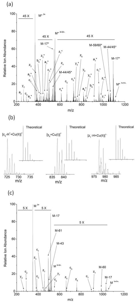
Unlike previous ECD studies of Cu(II)-peptide complexes, we find that many Cu(II)-peptide complexes can produce typical ECD/ETD type product ions, namely c- and z-type ions, upon ETD (Figure 1). The ETD spectrum (Figure 1a and b) of [AgtII+Cu(II)+H]3+, for example, shows numerous c and z ions, with and without Cu, although the overall product ion abundances are lower than that observed for the triply protonated form of this peptide (Figure 1c). Commonly observed neutral losses are also observed during ETD of [AgtII+Cu(II)+H]3+, including losses of H•, NH3, and CO2. In contrast to the ETD spectrum of the triply protonated peptide, a-, b- and y-type product ions are also present but to a much lower extent than c and z product ions. Overall, we find that about 5% of the product ions, including the charge-reduced species (i.e. [AgtII+Cu+H]2+/2+•]), are c and z ions, while only about 2% are a, b, and y ions. Similar or higher percentages of c and z product ions are found upon ETD of a wide range of triply-charged Cu(II)-bound peptides (Table 1).
Figure 1.
(a) ETD spectrum of [AgtII+Cu(II)+H]3+; (b) expanded regions of the ETD spectrum of [AgtII+Cu(II)+H]3+, showing the isotope distributions of selected ions; and (c) ETD spectrum of [AgtII+3H]3+.
Table 1.
ETD results for selected triply-charged Cu(II)-peptide complexes.
| Peptide | % Efficiencya | % c/zb | % a/b/yc | % CR with Cu(I)d | % NLe |
|---|---|---|---|---|---|
| Aβ1-16 | 15.4 ± 0.6 | 9.4 ± 0.2 | 1.0 ± 0.1 | 58.8 ± 0.4 | 5.0 ± 0.3 |
| Neurokinin B | 30 ± 1 | 11 ± 2 | 4.0 ± 0.2 | 63 ± 1 | 15.0 ± 0.8 |
| Agt1-13 | 18.6 ± 0.2 | 12.1 ± 0.3 | 1.1 ± 0.1 | 57.0 ± 0.3 | 5.5 ± 0.1 |
| NMC | 19.4 ± 0.6 | 12.4 ± 0.5 | 1.5 ± 0.2 | 66.5 ± 0.4 | 4.9 ± 0.1 |
| ACTH | 14 ± 2 | 11 ± 2 | 1.0 ± 0.1 | 66 ± 2 | 2.0 ± 0.2 |
| AgtI | 12 ± 2 | 8 ± 2 | 1.1 ± 0.3 | 61 ± 1 | 2.7 ± 0.4 |
| aFGF | 13 ± 3 | 8 ± 3 | 1.9 ± 0.2 | 63 ± 4 | 2.6 ± 0.5 |
| MPP | 16 ± 2 | 8 ± 2 | 3.7 ± 0.5 | 64 ± 2 | 4.2 ± 0.5 |
| Aβ22-35 | 15 ± 1 | 7.1 ± 0.4 | 1.7 ± 0.4 | 64.3 ± 0.5 | 5.8 ± 0.1 |
| Aβ10-20 | 7.6 ± 0.4 | 4.5 ± 0.3 | 1.4 ± 0.2 | 65 ± 1 | 1.7 ± 0.3 |
| AgtII | 13 ± 1 | 5.2 ± 0.2 | 2.4 ± 0.3 | 67 ± 1 | 4.6 ± 0.6 |
| SubP | 11 ± 2 | 5.3 ± 0.2 | 0.2 ± 0.1 | 82 ± 2 | 5.4 ± 0.4 |
| AgtIII | 7.0 ± 0.4 | 1.5 ± 0.3 | 2.7 ± 0.2 | 78.1 ± 0.6 | 2.5 ± 0.5 |
| Bradykinin | 8.8 ± 0.4 | 3.9 ± 0.1 | 1.2 ± 0.1 | 80.3 ± 0.1 | 3.5 ± 0.1 |
| OT | 46.7 ± 0.3 | 0.7 ± 0.2 | 17.5 ± 0.2 | 47.4 ± 0.5 | 26.3 ± 0.2 |
Calculated using equation 5 in the experimental section
Calculated using equation 1 in the experimental section
Calculated using equation 2 in the experimental section
Calculated using equation 7 in the experimental section
Calculated using equation 4 in the experimental section
The production of c- and z-type product ions suggests that in some cases the initial electron transfer event does not result in the reduction of Cu(II) to Cu(I) but rather in the production of aminoketyl radicals that subsequently dissociate to the observed c and z ions. An alternate explanation for the production of the c and z ions is double electron transfer in which a first electron transfer event reduces Cu(II) to Cu(I) and a second electron transfer generates c and z product ions via backbone cleavage at N-Cα bonds of the Cu(I)-peptide complex. For all the experiments described in this study, though, short ETD reaction times were employed to minimize the possibility of double electron transfer events. Indeed, the relative abundances of doubly charge-reduced species were always below 3%, indicating minimal double electron transfer had occurred. To further confirm that the observed c and z ions are generated directly from the precursor ions rather than by a secondary electron transfer event, “double resonance” experiments were performed. In these experiments, the product ion corresponding to the charge-reduced precursor ion (i.e. [M+Cu+H]2+/2+•) was resonantly ejected from the ion trap during ETD. If the c and z ions were produced via a second electron transfer event, then one would expect these product ions to disappear during the double resonance experiments. A comparison of ETD spectra with and without resonance ejection of the charged-reduced precursor ion, however, indicates that the dissociation patterns are very similar with regard to the number and types of c/z and a/b/y product ions (see Figure S1 in the Supplemental Information for an example with [Agt1-13+Cu(II)+H]3+). The similarity between these two spectra indicates that the c and z ions are being formed directly from the precursor ion via a single electron transfer event. This similarity is somewhat surprising, though, considering the double resonance experiments performed on ECD generated ions by O’Connor and co-workers [10]. In their work, they found notable differences between regular ECD spectra and ECD spectra with double resonance signals applied that were explained by the formation of long-lived and short-lived dissociation channels. One might expect most of the product ions formed from Cu(II)-bound peptides to be long-lived and therefore lead to different spectra when a double resonance signal is applied. In our experiments, however, we do not see major differences, but we are unsure if this is due to differences in ETD and ECD or differences in the dissociation behavior of protonated and Cu(II)-bound peptides.
If the c- and z-type product ions are being generated via a single electron transfer event and thus typical aminoketyl radicals are formed, then the product ions containing copper should have the metal in the +2 oxidation state. Three sets of experimental data were used to further explore the nature of these Cu-containing product ions. First, close examination of the product ion m/z ratios can confirm the oxidation state of Cu. For example, Figure 1b shows the isotope distributions of the c5*, y6*, and z7* product ions, which are consistent with the theoretical isotope distributions of [c5-H++Cu(II)]+, [y6+Cu(I)]+, and [z7•-H++Cu(II)]+ •, respectively. These data indicate that the c and z ions contain Cu(II), while the y ion contains Cu(I). Many of the zn*, however, do not exactly fit the theoretical isotope distributions of [zn•-H++Cu(II)]+ •. As is commonly seen in the ECD and ETD spectra of peptides, H• addition or loss can occur for z• product ions [11–12]. The addition of H• to z• radicals is thought to be correlated with the lifetime of the [c′+z•] complex, the identity of the amino acid adjacent to the cleavage site, and may be influenced by the relatively low precursor ion internal energies in the quadrupole ion trap [13]. These nominally zn′ product ions could be bound to Cu(II) (i.e. [zn•-H++H•+Cu(II)]+ or to Cu(I) [i.e. zn•+Cu(I)]+ •. To determine if [zn•+Cu(I)]+ • species are the major contributors, we considered the theoretical isotope distributions of [zn•-H++Cu(II)]+ • and [zn•+Cu(I)]+ • for each product ion and found that almost all the z-type product ions have isotope distributions that are more consistent with [zn•-H++Cu(II)]+ • than [zn•+Cu(I)]+ •. Taken as a whole, the m/z ratios of the product ions indicate that the Cu-containing c and z ions for all the peptides have the metal in an oxidation state of +2, whereas the a, b, and y ions have the product ions in an oxidation state of +1.
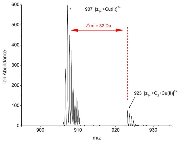
In a second set of experiments, gas-phase ion/molecule reactions between z* ions and O2 inside the mass spectrometer were carried out to further prove that the Cu-containing z ions had carbon-centered radicals and thus were produced by the initial formation of an aminoketyl radical. Studies have shown that the radical character of z ions enables them to add O2 (i.e. mass increase of 32 Da) upon storage in a quadrupole ion trap, and this mass addition permits the distinction between radical ions and even-electron species [14]. Most of the z ions with and without Cu were reacted with O2, and in the vast majority of the cases, an addition of 32 Da was seen (see Figure 2 as an example with the z14•*2+ ion of [Aβ1-16+Cu(II)+H]3+). The reactivity of the z ions with O2 strongly supports the notion that carbon-centered radicals are formed upon ETD without the associated reduction of Cu(II). It could be argued that Cu(I)-containing peptide ions could also react with O2 in the gas phase because proteins and small complexes with Cu(I) are known to bind O2 in solution. Two lines of evidence suggest that O2 addition to Cu(I) complexes are very unlikely. First, ion-molecule reactions between O2 and non-radical yn* ions or charged-reduced precursor ions (e.g. [M+Cu(I)+H]2+) do not lead to the addition of O2, even for reactions as long as 4 s (see Figure S2 in the Supplemental Information). Second, based on experimental studies by Karlin and co-workers, the affinities of even the most avid O2 binding Cu(I) complexes, which have ΔH values around −50 kJ/mol and ΔS values around −130 J/K-mol in non-coordinating solvents [15], are too low to be observed at room temperature under our experimental conditions (see Supplemental Information for calculations to support this claim).
Figure 2.
Mass spectrum that is obtained after the gas-phase reaction between O2 and the z14•*2+ ion of [Aβ1-16+Cu(II)+H]3+.
Finally, a third set of experiments involving CID of Cu-bound and Cu-free z ions formed by ETD indicate that z ions are radical species rather than ones in which Cu(II) has been reduced. CID of ETD-generated z• ions is known to give rise to side-chain losses and backbone cleavages via radical driven processes [16]. CID of selected Cu-containing z ions indicate that such product ions are indeed observed. As an example, Figure S3 in the Supplemental Information shows the CID spectrum of z8•* that is generated after ETD of [MPP+Cu(II)+H]3+. In this spectrum, several product ions arising from radical-driven processes are observed, including a side chain loss from Leu.
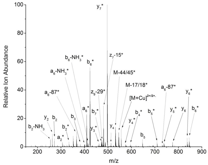
From these three sets of experimental results, we conclude the following. First, the observed c and z ions are formed via direct electron transfer to sites other than Cu(II) to generate regular ETD-type product ions. Second, the observed a, b, and y ions contain Cu(I), suggesting that they are formed via initial electron transfer to Cu(II), which then results in vibrational excitation and subsequent dissociation of the peptide ions, as proposed previously. These observations then raise the question about copper’s oxidation state in the charged-reduced precursor ions (i.e. [M+Cu+H]2+/2+•), which are the most abundant product ions in almost all the ETD spectra. CID of the charge reduced species results mainly in the formation of b- and y-type product ions, implying that the charged-reduced species contain mainly Cu(I). For example, the CID spectrum of the charge reduced species of [AgtII+Cu(II)+H]3+ after ETD shows an almost complete series of b and y ions with [y7+Cu(I)+H++]2+ as the most abundant product ion (Figure 3). Radical ions (e.g. z* with side chain losses as indicated in Figure 3) of lower abundance are detected as well, but these are always minor product ion channels. In fact, the a, b, and y ions account for about 75% of the product ion intensity in the CID spectrum of the charge-reduced species of [AgtII+Cu(II)+H]3+.
Figure 3.
CID spectrum of the charge reduced species of [AgtII+Cu(II)+H]3+ after ETD.
The effect of the Cu’s coordination sphere
One key difference between the Cu(II)-binding peptides studied here and those studied in previous ECD experiments on Cu(II)-binding peptides is the presence of amino acids, particularly His residues, with high affinity for Cu(II). Binding of Cu(II) by these His residues likely tunes the electronic character of Cu(II) in a way that makes electron transfer to non-Cu sites (i.e. protonated sites) possible, thereby enabling the formation of typical ETD product ions with Cu(II). To test this idea, the ETD behavior of a series of peptides with decreasing numbers of coordinating functionality was investigated. Assuming that His, Glu, and Asp residues along with the N- and C-termini are the most likely Cu(II) binding groups, the peptides Agt1-13 (DRVYIHPFHLVIH), AgtI (DRVYIHPFHL), AgtII (DRVYIHPF), and AgtIII (RVYIHPF), have 6, 5, 4, and 3 coordinating groups. Upon ETD of the [M+Cu(II)+H]3+ ion of these peptides, the percentage of c and z ions are found to decrease as the number of coordinating groups decrease. The measured percentages are about 12%, 8%, 5%, and 2% for DRVYIHPFHLVIH, DRVYIHPFHL, DRVYIHPF, and RVYIHPF, respectively. Similarly, the percentage of the charge-reduced species (i.e. [M+Cu+H]2+/2+•) that contain Cu(I) are found to be 57%, 61%, 67%, and 78% for the same series of peptides (see experimental section for how these percentages are calculated). These two sets of results indicate that the peptides with more coordinating functionality give rise to a greater number of c and z ions and less charge-reduced ions containing Cu(I). As a further test of this idea, when the free N-terminus of DRVYIHPFHL is acetylated, thereby removing the N-terminal amine as a Cu(II) binding group, the percentage of c/z ions decreases to 6%, and the percentage of charge-reduced species containing Cu(I) increase to 64%.
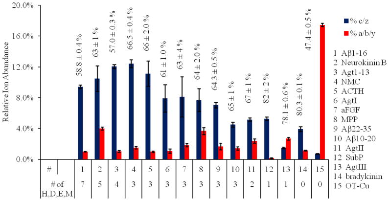
When ETD data from a wider collection of peptides are considered (Figure 4), we find a positive correlation between the number of binding residues and the percentage of c and z ions that are formed. Also, as the number of binding residues decreases, the percentage of charge-reduced ions with Cu(I) roughly increases. The number of binding residues indicated in Figure 4 is the total of His, Asp, Glu, and Met residues in each peptide (no Cys residues with free thiols were present in the peptides studied here). These residues were chosen because they represent the residues most commonly found in the coordination sphere of Cu(II)-bound proteins [17].
Figure 4.
Summary of the percentages of c and z ions, a, b, and y ions, and charge-reduced ions with Cu(I) (as determined by equation 7) after ETD of the [M+Cu(II)+H]3+ ions of 15 different peptides. The percentages of charged-reduced ions are indicated above the bars on the graph. The # of H, D, E, and M correspond to the total number of these residues in the given peptide.
| (8) |
| (9) |
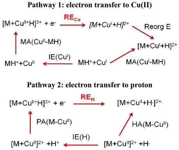
We propose that the observed effect of the coordination sphere on the degree of c and z formation (Figure 4) is related to the effect that the peptide binding groups have on Cu(II)’s recombination energy. Strong binding groups are able to lower Cu(II)’s recombination energy to an extent that electron transfer to other sites (i.e. protonated side chains) can be competitive with electron transfer to Cu(II). A similar use of recombination energies was used previously by Williams and co-workers to explain the ECD behavior of protonated and alkali-metalated peptides [3]. Recombination energy (RE) is a measure of the thermodynamic favorability of electron transfer to a given site, and several factors influence this value for Cu(II) or a protonated site in a given peptide. RE values for Cu(II) and a protonated site in a peptide can be estimated using the thermochemical cycles shown in Scheme 1. The RE for Cu(II) bound to the peptide (RECu, pathway 1) is influenced by the ionization energy (IE) of Cu(I), the binding affinity of Cu(II) to the protonated peptide [MA(CuII-MH)], the binding affinity of Cu(I) to the protonated peptide [MA(CuI-MH)], and the energy (ReorgE) required for the peptide to reorganize its structure to accommodate the preferred binding geometry of Cu(I) (i.e. typically tetrahedral [18]) from the preferred binding geometry of Cu(II) (i.e. typically square planar [18]). The RE for a protonated site in the peptide (REH, pathway 2) depends on the IE of the hydrogen atom, the proton affinity (PA) of the Cu(II) bound peptide [PA(CuII-M)], and the hydrogen atom affinity (HA) of the Cu(II) bound peptide [HA(CuII-M)]. From the thermochemical cycle depicted in Scheme 1, one can ascribe the trend observed for Cu(II)-peptide complexes (Figure 4) to the increased value of MA(CuII-MH) relative to MA(CuI-MH). A high binding energy to Cu(II) will increase MA(CuII-MH) so that the RECu is lowered to a value closer to REH, thereby making electron transfer to sites other than Cu(II) feasible.
Scheme 1.
Thermochemical cycles of the two competing pathways during ETD of [M+Cu(II)+H]3+. Pathway 1: electron transfer to Cu(II). Pathway 2: electron transfer to a protonated site.
This model can be semi-quantitatively evaluated by considering the relative binding energies of the peptides to Cu(II) and Cu(I). Unfortunately, no experimental data exists for the Cu binding energies of the peptides used in this study; however, binding energies of Cu and other metals to small molecule models (e.g. imidazole) of amino acid side chains have been measured or calculated. Rodgers and co-workers have measured the binding energies of Cu(I) to imidazole and have found the values to be 287.5 kJ/mol (2.98 eV), 257.5 kJ/mol (2.67 eV), and 79.8 kJ/mol (0.83 eV) for one, two, and three bound imidazoles, respectively [19]. This leads to a total binding energy of 624.8 kJ/mol or 6.48 eV for Cu(I). To our knowledge, no comparable measurements or calculations have been made for imidazole binding to Cu(II), but values have been calculated for imidazoles binding to Zn(II). These binding energies for Zn(II) are 754.0 kJ/mol (7.82 eV), 529.7 kJ/mol (5.49 eV), and 263.6 kJ/mol (2.73 eV) for one, two, and three bound imidazoles, respectively [20]. This leads to a total binding energy of 1547.7 kJ/mol or 16.0 eV. Other computational studies of divalent metal complexes with similar ligand sets indicate that Cu(II) tends to bind ligands with 1.2 to 1.4 times greater affinity than Zn(II) [21], so an estimated value of about 21 eV is probably better. If these values are plugged in equation 8 along with the IE of Cu(I), which is 20.3 eV, a RECu value of approximately 6 eV is obtained, ignoring contributions from the ReorgE. It should be noted that the RECu value would be even lower with more than three functional groups bound to Cu and would also be lower if the peptide has to reorganize its structure to accommodate the preferred binding geometry of Cu(I).
As Scheme 1 shows, REH will be influenced by the proton and hydrogen atom affinities of the Cu(II)-peptide complex. No PA values exist for Cu(II)-peptide complexes, but this value can be estimated by using the average PA values of Arg and Lys which is about 1023.5 kJ/mol (10.6 eV). Even though peptides are known to have higher PA values than their most basic residue [22], the dipositive charge of Cu(II) bound to the peptide is likely to lower the overall PA of the peptide, making our estimated PA reasonable. Hydrogen atom affinities for peptides have been determined to be approximately 0.6 eV [23]. Plugging these values into equation 9, we find that REH could be approximately 3.6 eV. While lower than the estimate for RECu, we still find that values are comparable enough that electron transfer to a protonated site could compete with electron transfer to Cu(II). When one further considers that ReorgE will likely be significant during Cu(II) reduction (but will be quite small for H+ to H), the processes would be even more competitive; indeed ReorgE for typical small copper complexes undergoing reduction are in the range of 2.5 eV [24]. Thus, this model roughly predicts that the right collection of coordinating residues with sufficient binding energies will enable Cu(II)-peptide complexes to undergo typical ETD dissociation processes to produce c and z ions without associated Cu(II) reduction. Indeed, the data in Figure 4 confirms this prediction.
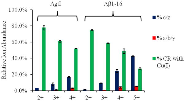
The effect of charge states
This model also predicts that a lower peptide PA will cause electron transfer to protonated sites to compete with electron transfer to Cu(II). Lower PA values are expected for peptides with a lower number of basic residues and a higher number of extra protons, as each additional proton will decrease the PA of the Cu(II)-peptide complex. If we consider Cu(II)-peptide complexes with an increasing number of protons, we find that the total abundances of the c and z ions increase, while the ion abundances of the charge-reduced species with Cu(I) decreases (Figure 5). It is also important to point out that peptides without any basic residues tend to have the greatest percentage of c and z ion formation for a given number of coordinating residues. Most notable is Neuromedin C (NMC in Figure 4), which has two His residues but no Arg or Lys residues. Interestingly, substance P and bradykinin have higher than expected c/z ion formation given their very low number of good binding residues and high number of basic residues. Because these peptides do not have good coordinating residues, it is quite possible that the basic Lys and Arg residues bind to Cu in the gas phase. Cu(II) binding via these residues would then substantially lower the PA of these peptides, thereby enabling more effective electron transfer to the Cu(II) center instead of protonated sites.
Figure 5.
Summary of the percentages of c and z ions, a, b, and y ions, and charge-reduced ions with Cu(I) (as determined by equation 7) after ETD of the [M+Cu(II)+nH](n+2)+ ions of AgtI and Aβ1-16, where n=0, 1, 2 and 3 (i.e. charge states of +2, +3, +4 for AgtI, and +2, +3, +4, +5 for Aβ1-16).
It should be noted that the use of thermochemical cycles to describe the results in this work do not account for the possibility of initial electron transfer to excited electronic states as shown in recent work by Tureček and co-workers [26]. Unfortunately, the peptides in our study are too large for extensive electronic structure calculations. Even so, if electron transfer to excited states is a common occurrence for Cu(II) complexes during ETD, we predict that coordination by strong ligands such as His will still influence the energy levels of these excited states in a way that makes electron transfer to non-Cu sites competitive. Future work will investigate this possibility with smaller peptides that are more amenable to detailed electronic structure calculations. In addition, the effect of reorganization energy will also be evaluated in future studies using metal complexes with measured or calculated reorganization energies.
Conclusions
When peptides have a sufficient number of coordinating residues (i.e. His, Asp, Glu, and Met), ETD of their Cu(II) complexes generate abundant c- and z- type product ions, presumably via typical aminoketyl radical formation. The resulting Cu-containing c and z product ions are bound by Cu(II) rather than Cu(I), suggesting that the initial electron transfer event does not reduce Cu(II) despite the high ionization energy of Cu. From the ETD behavior of a series of Cu(II)-bound peptides containing different numbers of binding residues (i.e. His, Asp, Glu, and Met), we found a positive correlation between the number of strong binding residues and the relative abundance of c and z ions. This trend can be rationalized by the effect that these binding groups have on Cu’s recombination energy relative to the recombination energy of protonated sites in these peptides. Strong binding ligands are able to lower Cu(II)’s recombination energy so that electron transfer to non-Cu sites is competitive, thereby allowing c and z product ions to be formed in a manner similar that observed for protonated (i.e. non-metalated) peptides. In addition, the relatively high reorganization energy associated with the conformational changes necessary to accommodate the preferred coordination geometry of Cu(I) (i.e. tetrahedral) relative to Cu(II) (i.e. square planar) may also further tune Cu(II)’s recombination energy. This latter effect will be studied in future work.
Supplementary Material
Acknowledgments
This work was supported by the National Institute of Health (R01 GM 075092). The authors thank Dr. Desmond A. Kaplan from Bruker Daltonics for help in implementing the ion-molecule reactions with O2.
References
- 1.Zubarev RA, Kelleher NL, McLafferty FW. Electron Capture Dissociation of Multiply Charged Protein Cations. A Nonergodic Process. J Am Chem Soc. 1998;120:3265–3266. [Google Scholar]
- 2.Syka JEP, Coon JJ, Schroeder MJ, Shabanowitz J, Hunt DF. Peptide and Protein Sequence Analysis by Electron Transfer Dissociation Mass Spectrometry. Proc Natl Acad Sci. 2004;101:9528–9533. doi: 10.1073/pnas.0402700101. [DOI] [PMC free article] [PubMed] [Google Scholar]
- 3.Iavarone AT, Paech K, Williams ER. Effects of Charge State and Cationizing Agent on the Electron Capture Dissociation of a Peptide. Anal Chem. 2004;76:2231–2238. doi: 10.1021/ac035431p. [DOI] [PMC free article] [PubMed] [Google Scholar]
- 4.Fung YME, Liu HC, Chan TWD. Electron Capture Dissociation of Peptides Metalated with Alkaline-Earth Metal Ions. J Am Soc Mass Spectrom. 2006;17:757–771. doi: 10.1016/j.jasms.2006.01.014. [DOI] [PubMed] [Google Scholar]
- 5.Chen XF, Chan WYK, Wong PS, Yeung HS, Chan TWD. Formation of Peptide Radical Cations (M+ ) in Electron Capture Dissociation of Peptides Adducted with Group IIB Metal Ions. J Am Soc Mass Spectrom. 2011;22:233–244. doi: 10.1007/s13361-010-0035-2. [DOI] [PubMed] [Google Scholar]
- 6.Liu HC, Håkansson K. Divalent Metal Ion-Peptide Interactions Probed by Electron Capture Dissociation of Trications. J Am Soc Mass Spectrom. 2006;17:1731–1741. doi: 10.1016/j.jasms.2006.07.027. [DOI] [PubMed] [Google Scholar]
- 7.Van der Burgt YEM, Palmblad M, Dalebout H, Heeren RMA, Deelder AM. Electron Capture Dissociation of Peptide Hormone Changes upon Opening of the Tocin Ring and Complexation with Transition Metal Cations. Rapid Commun Mass Spectrom. 2009;23:31–38. doi: 10.1002/rcm.3849. [DOI] [PubMed] [Google Scholar]
- 8.Kleinnijenhuis AJ, Mihalca R, Heeren RMA, Heck AJR. Atypical Behavior in the Electron Capture Induced Dissociation of Biologically Relevant Transition Metal Ion Complexes of the Peptide Hormone Oxytocin. Int J Mass Spectrom. 2006;253:217–224. [Google Scholar]
- 9.Kjeldsen F, Haselmann KF, Budnik BA, Jensen F, Zubarev RA. Dissociative Capture of Hot Electrons by Polypeptide Polycations: An Efficient Process Accompanied by Secondary Fragmentation. Chem Phys Lett. 2002;356:201–206. [Google Scholar]
- 10.Lin C, Cournoyer JJ, O’Connor PB. Use of a Double Resonance Electron Transfer Capture Dissociation Experiment to Probe Fragment Intermediate Lifetimes. J Am Soc Mass Spectrom. 2006;17:1605–1615. doi: 10.1016/j.jasms.2006.07.007. [DOI] [PubMed] [Google Scholar]
- 11.Savitiski MM, Kjeldsen F, Nielsen ML, Zubarev RA. Hydrogen Rearrangement to and from Radical z Fragments in Electron Capture Dissociation of Peptides. J Am Soc Mass Spectrom. 2007;18:113–120. doi: 10.1016/j.jasms.2006.09.008. [DOI] [PubMed] [Google Scholar]
- 12.O’Connor PB, Lin C, Cournoyer JJ, Pittman JL, Belyayev M, Budnik BA. Long-Lived Electron Capture Dissociation Product Ions Experience Radical Migration via Hydrogen Abstraction. J Am Soc Mass Spectrom. 2006;17:576–585. doi: 10.1016/j.jasms.2005.12.015. [DOI] [PubMed] [Google Scholar]
- 13.Ben Hamidane H, Chiappe D, Hartmer R, Vorobyev A, Moniatte M, Tysbin YO. Electron Capture and Transfer Dissociation: Peptide Structure Analysis at Different Ion Internal Energy Levels. J Am Soc Mass Spectrom. 2009;20:567–575. doi: 10.1016/j.jasms.2008.11.016. [DOI] [PubMed] [Google Scholar]
- 14.Xia Y, Chrisman PA, Pitteri SJ, Erickson DE, Mcluckey SA. Ion/Molecule Reactions of Cation Radicals Formed from Protonated Polypeptide via Gas-Phase Ion/Ion Electron Transfer. J Am Chem Soc. 2006;128:11792–11798. doi: 10.1021/ja063248i. [DOI] [PubMed] [Google Scholar]
- 15.Fry HC, Scaltrito DV, Karlin KD, Meyer GJ. The Rate of O2 and CO Binding to a Copper Complex, Determined by a “Flash-and-Trap” Technique, Exceeds that for Hemes. J Am Chem Soc. 2003;125:11866–11871. doi: 10.1021/ja034911v. [DOI] [PubMed] [Google Scholar]
- 16.Han HL, Xia Y, Mcluckey SA. Ion Trap Collisional Activation of c and z•Ions Formed via Gas-Phase Ion/Ion Electron-Transfer Dissociation. J Proteome Res. 2007;6:3062–3069. doi: 10.1021/pr070177t. [DOI] [PMC free article] [PubMed] [Google Scholar]
- 17.Zheng H, Chruszcz M, Lasota P, Lebioda L, Minor W. Data Mining of Metal Environments Present in Protein Structures. J Inorg Biochem. 2008;102:1765–1776. doi: 10.1016/j.jinorgbio.2008.05.006. [DOI] [PMC free article] [PubMed] [Google Scholar]
- 18.Cotton FA, Wilkinson G. Advanced Inorganic Chemistry. 4. John Wiley & Sons; pp. 800–818. [Google Scholar]
- 19.Rannulu NS, Rodgers MT. Solvation of Copper Ions by Imidazole: Structures and Sequential Binding Energies of Cu+ (Imidazole)x, x = 1 – 4. Competition Between Ion Solvation and Hydrogen Bonding. Phys Chem Chem Phy. 2005;7:1014–1025. doi: 10.1039/b418141g. [DOI] [PubMed] [Google Scholar]
- 20.Peschke M, Blades AT, Kebarle P. Metalloion-Ligand Binding Energies and Biological Function of Metalloenzymes Such as Carbonic Anhydrase. A Study Based on ab Initio Calculations and Experimental Ion-Ligand Equilibria in the Gas Phase. J Am Chem Soc. 2000;122:1492–1505. [Google Scholar]
- 21.Rulíšek L, Havlas Z. Theoretical Studies of Metal Ion Selectivity. 1 DFT Calculations of Interaction Energies of Amino Acid Side Chains with Selected Transition Metal Ions (Co2+, Ni2+, Cu2+, Zn2+, Cd2+, and Hg2+) J Am Chem Soc. 2000;122:10428–10439. [Google Scholar]
- 22.Harrison AG. The Gas-Phase Basicities and Proton Affinities of Amino Acids and Peptides. Mass Spectrom Rev. 1997;16:201–217. [Google Scholar]
- 23.Zubarev RA, Haselmann KF, Budnik B, Kjeldsen F, Jensen F. Account: Towards an Understanding of the Mechanism of Electron-Capture Dissociation: a Historical Perspective and Modern Ideas. Eur J Mass Spectrom. 2002;8:337–349. [Google Scholar]
- 24.Winkler JR, WittungStafshede P, Leckner J, Malmstrom BG, Gary HB. Effects of Folding on Metalloprotein Active Sites. Proc Natl Acad Sci. 1997;94:4246–4249. doi: 10.1073/pnas.94.9.4246. [DOI] [PMC free article] [PubMed] [Google Scholar]
- 25.Wu ZZ, Fernandez-Lima FA, Russell DH. Amino Acid Influence on Copper Binding to Peptides: Cystein Versus Arginine. J Am Soc Mass Spectrom. 2010;21:522–533. doi: 10.1016/j.jasms.2009.12.020. [DOI] [PubMed] [Google Scholar]
- 26.Tureček F, Jones JW, Holm AIS, Panja S, Nielsen SB, Hvelplund P. Transition Metals Are Electron Traps. I Structures, Energies, Electron Capture and Electron-Transfer-Induced Dissociations of Ternary Copper-Peptide Complexes in the Gas Phase. J Mass Spectrom. 2009;44:707–724. doi: 10.1002/jms.1546. [DOI] [PubMed] [Google Scholar]
Associated Data
This section collects any data citations, data availability statements, or supplementary materials included in this article.


