Abstract
N-linked glycans are composed of three major types: high-mannose (Man), hybrid or complex. The functional role of hybrid- and complex-type N-glycans in Newcastle disease virus (NDV) infection and fusion was examined in N-acetylglucosaminyltransferase I (GnT I)-deficient Lec1 cells, a mutant Chinese hamster ovary (CHO) cell incapable of synthesizing hybrid- and complex-type N-glycans. We used recombinant NDV expressing green fluorescence protein or red fluorescence protein to monitor NDV infection, syncytium formation and viral yield. Flow cytometry showed that CHO-K1 and Lec1 cells had essentially the same degree of NDV infection. In contrast, Lec2 cells were found to be resistant to NDV infection. Compared with CHO-K1 cells, Lec1 cells were shown to more sensitive to fusion induced by NDV. Viral attachment was found to be comparable in both lines. We found that there were no significant differences in the yield of progeny virus produced by both CHO-K1 and Lec1 cells. Quantitative analysis revealed that NDV infection and fusion in Lec1 cells were also inhibited by treatment with sialidase. Pretreatment of Lec1 cells with Galanthus nivalis agglutinin specific for terminal α1-3-linked Man prior to inoculation with NDV rendered Lec1 cells less sensitive to cell-to-cell fusion compared with mock-treated Lec1 cells. Treatment of CHO-K1 and Lec1 cells with tunicamycin, an inhibitor of N-glycosylation, significantly blocked fusion and infection. In conclusion, our results suggest that hybrid- and complex-type N-glycans are not required for NDV infection and fusion. We propose that high-Man-type N-glycans could play an important role in the cell-to-cell fusion induced by NDV.
Keywords: fusion, high-mannose-type, hybrid- and complex-type, N-glycan, Newcastle disease virus
Introduction
Newcastle disease viruses (NDVs) are non-segmented, single-stranded RNA viruses that belong to the genus Avulavirus in the family Paramyxoviridae (Mayo 2002). As with other viruses, sialic acid-containing glycans play a key role in the initial steps of NDV infectious life cycle (Olofsson and Bergstrom 2005; Isa et al. 2006; Villar and Barroso 2006). The entry of NDV into the host cell can be broken down into two steps: attachment to the cellular receptors and membrane fusion (Villar and Barroso 2006). Hemagglutinin–neuraminidase (HN) protein binds to sialylated glycans on the cell surface and fusion (F) protein is responsible for the fusion between the viral envelope and the plasma membrane (Lamb and Kolakofsky 2001). However, there are details of the mechanism that remain to be fully clarified. Recently, Ferreira et al. (2004) reported that gangliosides and N-linked glycoproteins serve as NDV receptors.
N-glycan is covalently attached to protein at asparagine (Asn) residues by an N-glycosidic bond. In eukaryotes cells, there is a wide diversity of N-glycan structures. Manα1-6(Manα1-3)Manβ1-4GlcNAcβ1-4GlcNAcβ1-Asn-X-Ser/Thr is a common core sugar sequence of all N-glycans, which are divided into three types: high-mannose (Man), in which only Man residues are attached to the core; hybrid, which have two branches from the core, one that terminates in Man and one that terminates in a sugar of the complex type (Yamashita et al. 1978); and complex, such as tri- and tetraantennary glycopeptides containing outer chains of sialic acid, galactose (Gal), N-acetylglucosamine (GlcNAc) residues and an α-linked Man residue substituted at positions C-2 and C-6 (Tabas and Kornfeld 1978). The biosynthesis of a complex N-linked glycan is a complex and high-ordered process, which is initiated in the endoplasmic reticulum (ER) and completed in the Golgi apparatus. In the ER, the Glc3Man9GlcNAc2 sequence is initially assembled on the lipid dolichol carrier and then transferred to the Asn residues of the nascent peptide or protein as the core oligosaccharide unit. Subsequent to the transfer, the glycan undergoes a series of modifications before it reaches its maturity in the Golgi apparatus. In the Golgi complex, the conversion of high-Man-type glycan to hybrid-type glycan is initiated by the action of N-acetylglucosaminyltransferase I (GnT I), which transfers GlcNAc, in a β1-2 linkage, to the 3-arm Man residue of the oligo-Man substrate, Man5GlcNAc2. Hybrid-type glycan is further remodeled by a series of enzymes to yield complex-type N-glycans. Incomplete processing of N-linked glycan results in the production of high-Man glycans, which terminate in Man (Tabas et al. 1978). Many inhibitors have been identified that interfere with glycoprotein biosynthesis and processing of N-glycans. For example, tunicamycin is an N-glycosylation inhibitor which blocks the first step in the lipid-linked pathway by specifically suppressing the transfer of GlcNAc to dolichol monophosphate, therefore significantly reducing the formation of GlcNAc-pyrophosphoryl-dolichol (Duksin and Mahoney 1982).
Lec1 Chinese hamster ovary (CHO) mutants were isolated for sensitivity to Galanthus nivalis agglutinin (GNA) which specifically binds the terminal α1-3-Man of high-Man glycans but not hybrid glycans (Shibuya et al. 1988; Hester and Wright 1996) and resistance to the leucoagglutinin from Phaseolus vulgaris (L-PHA; Stanley, Caillibot, et al. 1975; Stanley, Narasimhan, et al. 1975), which binds to certain branched, complex-type N-glycans containing the pentasaccharide sequence Galβ1-4GlcNAcβ1-2(Galβ1-4GlcNAcβ1-6)Manα1-R. Lec1 cells have been characterized as having an insertion or deletion within the Mannosyl-α1,3-glycoprotein-β1,2-N-acetylglucosaminyltransferase 1 (Mgat1) gene open reading frame (ORF), resulting in an absence of GnT I activity and do not synthesize complex and hybrid N-glycans (Chen and Stanley 2003; North et al. 2010) and accumulation of abundant Man5GlcNAc2 N-glycans (North et al. 2010). In addition, an Lec2 cell line is also a mutant CHO cell line defective in the Cytidine monophosphate (CMP)-sialic acid transporter (SAT), which is responsible for the translocation of CMP-sialic acid into the lumen of the Golgi apparatus (Eckhardt et al. 1998). As such, the loss of CMP-SAT activity results in significantly reduced levels of sialylation of glycoproteins and gangliosides (North et al. 2010).
In this study, we have engineered a recombinant NDV carrying a red fluorescence protein (RFP) reporter inserted into the region between the P/V and the M genes via reverse genetics. We used this recombinant NDV expressing RFP, and another recombinant NDV expressing green fluorescence protein (GFP; Liu et al. 2007), to study viral infection and fusion in host cells. To elucidate the role of complex N-linked glycans in NDV infection and fusion, we used Lec1 cells which are deficient in terminal N-linked glycosylation, with no effect on the synthesis of O-glycan and glycolipid (Sarkar et al. 1991; Puthalakath et al. 1996; North et al. 2010). Our results clearly indicate that NDV undergoes efficient infection and replication and enhanced fusion in Lec1 cells, supporting that hybrid and complex N-linked glycans are not functionally essential for infection and cell-to-cell fusion induced by NDV. In addition, we propose that high-Man N-linked glycans on the surface of Lec1 cells could play an important role in the cell–cell fusion induced by NDV.
Results
Generation of recombinant NDV carrying an RFP reporter
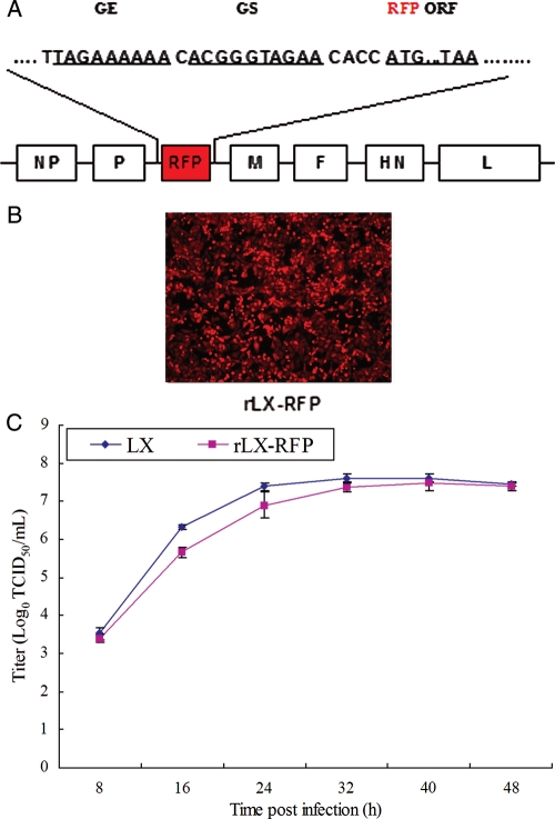
RFP autofluorescence is a convenient marker for identification and quantitation of infected cells; thus, we generated a recombinant NDV carrying an RFP reporter via reverse genetics (Peeters et al. 1999; Romer-Oberdorfer et al. 1999). The RFP transcription cassette containing the RFP ORF, flanked by NDV gene-start (GS) and gene-end (GE) sequence motifs, was inserted into in the downstream non-coding region of the P/V gene of the full-length complementary DNA (cDNA) clone of the NDV Laoxi (LX) strain (Figure 1A) following the rule of six (Peeters et al. 2000). For virus recovery, the full-length pLX-RFP plasmid was then cotransfected with the helper plasmids as described previously (Peeters et al. 2000). The resultant recombinant virus, which was designated rLX-RFP, was readily rescued and found to replicate efficiently in titers similar to those of the wild-type NDV LX strain [5 × 10⁸ plaque forming unit (PFU)/mL] in 9-day-old embryonated eggs (Figure 1C). To determine whether RFP was expressed in infected cells, CHO-K1 cells were infected with the rLX-RFP at a multiplicity of infection (MOI) of 0.1. At 12 hpi (hour post-infection), RFP expression was observed in cells (Figure 1B). The results showed that rLX-RFP could efficiently infect and express RFP in CHO-K1 cells.
Fig. 1.
Generation of recombinant NDV expressing an added RFP reporter gene (rLX-RFP). (A) Schematic representation of rLX-RFP construct. The coding sequence of RFP was inserted into the downstream non-coding region of the P/V gene. NDV GE, IG, GS signals and RFP gene were inserted into the nucleotide position 3143 of the NDV genome. (B) CHO-K1 cells were infected with rLX-RFP at an MOI of 0.1. Original magnification, ×10. (C) Multistep growth curve for LX and rLX-RFP in SPF chicken embryos. Embryonated chicken eggs were inoculated with 2 × 10³ TCID50 LX and rLX-RFP. Allantoic fluid was harvested at 8 h intervals for 48 h, and virus titers were determined in triplicate by TCID50 in CEF cells.
Lec1 cells are deficient in terminal N-linked glycosylation
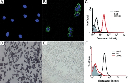
Lec1 cells, a mutant CHO cell line, are incapable of synthesizing hybrid and complex N-glycans due to a defect the GnT I gene. Therefore, we used the lectin L-PHA, which binds to branched, complex glycan structures, such as tri- and tetraantennary glycopeptides containing outer Gal residues and an α-linked Man residue substituted at positions C-2 and C-6, to detect terminal N-glycans on CHO-K1 cells and on Lec1 cells. Confocal fluorescence microscopy and flow cytometry showed that CHO-K1 cells bound high levels of L-PHA, whereas Lec1 cells did not bind L-PHA (Figure 2A–C). Additionally, the lectin GNA was used to identify the terminal α1-3-linked Man residues (Shibuya et al. 1988). In the immunohistochemical staining assay and flow cytometry, we found that Lec1 cells showed a strong reaction with GNA. In contrast, CHO-K1 cells failed to bind GNA (Figure 2D–F). These data confirmed that we had authentic Lec1 cells.
Fig. 2.
Characterization of Lec1 cells phenotype. CHO-K1 (B) and Lec1 (A) cells were fixed in 2% paraformaldehyde, blocked in TBS containing 10% goat serum, and then incubated with FITC-labeled lectin L-PHA, with nuclei counterstained with DAPI. Cells were visualized under Leica confocal fluorescence microscope (TCS SP2). CHO-K1 (red line) and Lec1 cells (black line) were incubated with FITC-labeled L-PHA for 45 min at 4°C and analyzed by flow cytometry. Cells analyzed in the absence of FITC-labeled L-PHA are shown in the filled light-gray trace (C). CHO-K1 (E) and Lec1 (D) cells were fixed in 2% paraformaldehyde, blocked in TBS containing 10% goat serum, and then incubated with DIG-labeled lectin GNA at 37°C. After 45 min incubation, cells were incubated with AP-conjugated anti-DIG for 45 min at 37°C and then treated with NBT/BCIP for visualization under Leical fluorescence microscope. CHO-K1 (red line) and Lec1 cells (black line) were incubated with the DIG-labeled lectin GNA for 45 min at 4°C and incubated with FITC-conjugated anti-DIG for 45 min at 37°C and then analyzed by flow cytometry. Cells analyzed in the absence of lectin are shown in the filled light-gray trace (F).
Lec1 cells express equivalent levels of cell-surface sialic acids to CHO-K1 cells
To evaluate the levels of α2,3- or α2,6-linked sialic acids on the surface, We used Maackia amurensis agglutinin (MAA; specific for the α2-3 linkage) and Sambucus nigra agglutinin (SNA; specific for the α2-6 linkage) to carry out a lectin-binding assay. In fluorescence-activated cell sorting analysis (FACS), we found that Lec1 cells have overall α2-3 sialic acid levels equal to those of CHO-K1 cells (as shown in Figure 3). In contrast, both cells failed to SNA specific for α2,6-linked sialic acid (data not shown).
Fig. 3.
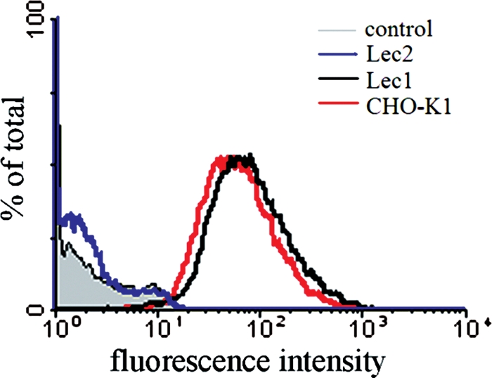
Lec1 cells express equivalent levels of cell-surface sialic acid to CHO-K1 cells. CHO-K1 (red line), Lec1 (black line) and Lec2 (blue line) cells were treated with the DIG-labeled lectin MAA for 45 min at 4°C, and then incubated with FITC-conjugated anti-DIG for 30 min at 4°C and analyzed by flow cytometry. Cells analyzed in the absence of DIG-labeled lectin are shown in the filled light-gray trace.
NDV binds efficiently to Lec1 cells
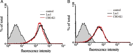
To investigate the effect of the absence of hybrid or complex N-linked glycans on attachment of NDV to the cells, we performed virus-binding assays using Lec1 and CHO-K1 cells. Cells were exposed to rZJ1 at an MOI of 50 or 10 on ice, to allow virus binding, but not internalization. Virus binding was analyzed by flow cytometry after 90-min incubation on ice. Both Lec1 and CHO-K1 cells (Figure 4A and B) showed comparable high levels of virus binding, suggesting that the attachment of NDV is essentially not affected in Lec1 cells.
Fig. 4.
Both Lec1 and CHO cells show comparable high levels of virus binding. CHO-K1 (red line) and Lec1 (black line) cells were exposed to rZJ1 at an MOI of 50 or 10 (A and B) on ice to allow virus binding. After 90-min incubation on ice, cells were incubated with the monoclonal antibody to NDV HN protein (6B1) for 45 min at 4°C, and then incubated with the FITC-labeled antibody for 45 min at 4°C. Virus binding was analyzed by flow cytometry. Cells analyzed in the absence of monoclonal antibody to NDV HN protein are shown in the filled light-gray trace.
Lec1 cells are markedly infectable with NDV
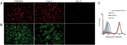
To determine whether hybrid or complex N-linked glycans were dispensable for NDV infection, we tested the infection with rZJ1-GFP and rLX-RFP. Particularly, rZJ1-GFP was chosen as a good model, because ZJ-1/00(Go) was a member of genotype VIId NDV, which is the predominant genotype circulating worldwide and contains only virulent viruses. In these experiments, Lec1 cells were readily and efficiently infected with two NDV strains used, which was similar to CHO-K1 cells.
To compare the susceptibility of different cell lines to NDV infection quantitatively, we performed the flow cytometry assay calculating the numbers of cells infected in each cell line by the same virus dosage. In this assay, we chose avirulent rLX-RFP, because it did not form syncytia without addition of exogenous l-(tosylamido-2-phenyl) ethyl chloromethyl ketone (TPCK)-treated trypsin. The results revealed that Lec1 cells were readily infected with NDV, albeit the susceptibility of Lec1 cells to NDV infection was somewhat lower than that of CHO-K1 cells (Figure 5). Furthermore, Lec2 cells were found to become resistant to NDV infection (Figure 5). Therefore, we conclude that the sensitivity of Lec1 cells to virus infection and fusion appears to be a feature of NDV.
Fig. 5.
Analysis of three CHO cell lines for the susceptibility to NDV infection. CHO-K1, Lec1 and Lec2 cells were infected with NDV (rZJ1-GFP or rLX-RFP) at an MOI of 1 for 16 h (viral replication was revealed by GFP or RFP). At 16 hpi, infected cells were visualized for GFP (shown in B) or RFP (shown in A) expression under the inverted fluorescence microscope. At 16 hpi, CHO-K1 (black line), Lec1 (red line) and Lec2 (blue line) cells were also subjected to flow cytometric analysis for quantitating NDV infection (rLX-RFP), as shown in C. Mock-infected CHO-K1 cells were shown in the filled light gray trace.
NDV infection leads to syncytia formation in Lec1 cells to a markedly greater extent than in CHO-K1 cells
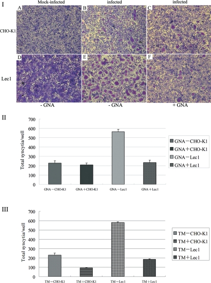
To overcome the addition of TPCK-treated trypsin to activate the precursor F0, we chose fully velogenic rZJ1-GFP to perform syncytium assays rather than lentogenic rLX-RFP. The relative sensitivity of CHO-K1 and Lec1 cells to cell-to-cell fusion induced by rZJ1-GFP was shown in Figure 6I. The results revealed that the cell-to-cell fusion of Lec1 cells was significantly above that of CHO-K1 cells 12 hpi. In contrast, Lec2 cells were resistant to fusion induced by rZJ1-GFP (data not shown). Our results indicated that NDV was able to propagate in Lec1 cells and induced much more syncytia formation, supporting that the lack of hybrid and complex N-linked glycans does not reduce the sensitivity of Lec1 cells to NDV-induced fusion.
Fig. 6.
NDV-induced syncytium formation in CHO-K1 and Lec1 cells. (A) CHO-K1 and Lec1 cells were pretreated or non-treated with GNA for 1 h at 37°C prior to viral infection, and then infected with NDV (rZJ1-GFP) at an MOI of 1. At 16 hpi, cells were fixed with methanol for 10 min and stained with Giemsa. Syncytia formation was shown in (I). The number of syncytia (cells containing more than three nuclei) was counted in 10 random areas of the well (data are mean ± SD of three independent experiments), as shown in (II). (B) CHO-K1 and Lec1 cells were pretreated or non-treated with tunicamycin (TM) for 24 h at 37°C prior to viral infection, and then infected with NDV (rZJ1-GFP) at an MOI of 1. At 16 hpi, cells were fixed with methanol for 10 min and stained with Giemsa. The number of syncytia (cells containing more than three nuclei) was counted in 10 random areas of the well (data are mean ± SD of three independent experiments), as shown in (III).
Terminal α1-3-linked Man residues of high-Man N-glycans are critical for cell-to-cell fusion induced by NDV
The sensitivity of Lec1 cells to NDV-induced fusion led to consider the possible effect of high-Man N-glycans on enhanced cell-to-cell fusion. CHO-K1 and Lec1 cells were incubated with GNA and then subjected to NDV infection at an MOI of 1. The extent of cell–cell fusion was quantified by syncytium assays at 24 hpi. As shown in Figure 6I and II, we observed that GNA treatment caused a marked reduction in cell–cell fusion in Lec1 cells, while the fusion was essentially unaffected in CHO-K1 cells. The data suggested that the increase in high-Man N-glycans in Lec1 correlates closely with enhanced cell–cell fusion induced by NDV.
Tunicamycin pretreatment blocks NDV infection and fusion
To further analyze the importance of high-Man N-glycans in the cell-to-cell fusion induced by NDV, we treated CHO-K1 and Lec1 cells with tunicamycin. Then, cells pretreated with tunicamycin were infected with NDV at an MOI of 1 and the extent of syncytium formation between NDV-infected cells was analyzed. The data were shown in Figure 6III. As can be seen, treatment of Lec1 cells with 0.5 μg/mL of tunicamycin for 24 h before infection markedly inhibited the extent of cell-to-cell fusion. Therefore, we suggest that high-Man N-glycans in the surface of Lec1 cells play an important role in enhanced cell–cell fusion induced by NDV.
Pretreatment of Lec1 cells with sialidase blocks NDV infection and fusion
In order to examine the importance of sialic acid for the initiation of NDV infection, Lec1 cells were treated with NA from Vibrio cholerae prior to infection with rZJ1-GFP. Infection was evaluated by observation of GFP under the fluorescence microscopy. As shown in Figure 7, in cells infected with rZJ1-GFP at an MOI of 1 following pre-treatment with 200 mU NA, we observed a distinct reduction in the number of infected cells. These results revealed that neuraminidase (NA) treatment rendered Lec1 cells resistant to infection by the NDV-ZJ1 strain, similar to Lec2 cells. These findings strongly suggested that NDV requires a greater amount of sialic acids on the cell surface to initiate an infection.
Fig. 7.
Effect of sialidase treatment on NDV infection and fusion in Lec1 cells. Lec1 cells were incubated in the presence of 200 mU/mL sialidase from V. cholerae for 3 h at 37°C prior to infection with NDV (rZJ1-GFP or rLX-RFP) at an MOI of 1. (A) At 16 hpi, infected cells were visualized for GFP expression under the inverted fluorescence microscope. (B) At 16 hpi, sialidase-treated (red line) and non-treated (black line) Lec1 cells were also subjected to flow cytometric analysis for quantitating NDV infection (rLX-RFP). Mock-infected Lec1 cells were shown in the filled light gray trace.
Kinetics of rZJ1-GFP infection in different cells
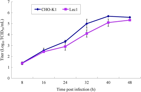
To test whether the absence of hybrid or complex N-linked glycans in Lec1 cells could reduce the yield of infectious virus progeny, we carried out a multicycle growth in CHO-K1 and Lec1 cells. The cells were infected with rZJ1-GFP at an MOI of 0.01. At different time points post-infection, viral titers in the supernatants were determined by GFP autofluorescence, highly correlated with the immunofluorescence assay (IFA) titer (Engel-Herbert et al. 2003). rZJ1-GFP displayed a growth pattern in Lec1 cells similar to in CHO-K1 cells, as shown in Figure 8. This result revealed that no significant differences in the yield of progeny virus were shown in both Lec1 and CHO-K1 cells, suggesting that hybrid and complex N-linked glycans are not essential for NDV infection and replication.
Fig. 8.
Multistep growth curve of NDV infection in CHO-K1 and Lec1 cells. CHO-K1 and Lec1 cells were infected with rZJ1-GFP at an MOI of 0.01, and at the assigned time points post-infection, nearly 300 μL of supernatant was harvested and replenished with the same amount of fresh medium. The virus titers in the collected supernatant were quantitated in triplicate by TCID50 in CEF cells.
Discussion
In this report, we show that the susceptibility of L-PHA-resistant Lec1 cells, which lack hybrid or complex N-linked glycans, to NDV infection was comparable with that of CHO-K1 cells. Our findings are consistent with the previous findings that a ricin-resistant CHO-15B isolated from CHO-K1 cells (Gottlieb et al. 1974, 1975; Brandli 1991), which lack GnT I activity, did not differ from the wild-type CHO cells in its interactions with NDV (Polos and Gallaher 1979). Virus attachment and lectin-binding assay may account for the observed sensitivity of NDV to Lec1 cells. In fact, virus binding was quantitatively almost equivalent with both Lec1 and CHO-K1 cells. This is correlated with the results that the quantity of sialic acids on the surface of Lec1 cells was comparable with that of CHO-K1 cells, which is consistent with those of Chu and Whittaker (2004). In addition, we also found that the yield of progeny virus from Lec1 cells was comparable with that obtained from CHO-K1 cells. Therefore, as may be deduced from our data, it seems that, like vesicular stomatitis virus (Schlesinger et al. 1975; Chu and Whittaker 2004), the alterations in terminal N-glycosylation which result from the mutation(s) in GnT I do not significantly affect the replication and infection of NDV, suggesting that hybrid or complex N-glycans of glycoprotein are not absolutely a requirement for NDV infection.
Furthermore, we observed that Lec1 cells were more sensitive to cell-to-cell fusion induced by NDV than that of the wild-type CHO-K1 cells, albeit the sensitivity of Lec1 cells to NDV infection was comparable with that of CHO-K1 cells. Lec1 cells were infected by NDV mainly via substantial syncytia formation in Lec1 cells (cell-to-cell fusion), whereas NDV first infected a single CHO-K1 cell and then syncytia were formed in CHO-K1 cells. However, Polos and Gallaher (1979) discovered that ricin-resistant CHO-15B cells were resistant to cell fusion and lysis induced by NDV. The discrepancy may be attributed to the different NDV strains and CHO cell mutants used in both works.
The observed enhancement of cell-to-cell fusion induced by NDV was not due to virus binding. Because the results described above showed that the binding of NDV to Lec1 cells was quantitatively similar to that of CHO-K1 cells. In light of these results, we propose that high-Man N-linked glycans could play a critical role in the cell-to-cell fusion induced by NDV. To support this hypothesis, it has been reported that the plant lectin GNA specifically binds the terminal α1-3-linked Man of high-Man-type N-linked glycans but not hybrid-type N-glycans (Shibuya et al. 1988) and Man5GlcNAc2 N-glycans were the predominant high-Man N-glycans in Lec1 cells (North et al. 2010). From our studies, the sensitivity of GNA-treated Lec1 cells to cell-to-cell fusion induced by NDV was less than that of mock-treated Lec1 cells. On the other hand, the treatment with tunicamycin, which suppresses N-glycan expression globally in eukaryotes by blocking the transfer of GlcNAc-1-phosphate from Uridine diphosphate (UDP)-GlcNAc to dolichol monophosphate and reducing the formation of dolichol-pyrophosphoryl GlcNAc, markedly inhibited syncytium formation between NDV-infected Lec1 cells. Taken together, this would explain why Lec1 cells are more sensitive to fusion induced by NDV than that of CHO-K1 cells.
A recent report proposed that there could be two different phases in NDV binding: an early ganglioside-dependent phase, and a later N-glycoprotein-dependent phase (Ferreira et al. 2004). In this multistep attachment model, the interaction of HN protein with gangliosides would trigger a partial conformational change in HN, rendering it suitable to interact with second receptors: N-glycoproteins; subsequently, the interaction of HN with N-glycoproteins would elicit another conformational change in HN protein that would induce a conformational change in F protein, resulting in complete virus-cell fusion (Ferreira et al. 2004). As deduced from this hypothesis, the change of oligosaccharide sequences of N-glycoproteins which might affect a membrane rearrangement has an influence on the interaction of HN with N-glycoproteins that, in turn, might affect the conformational change in F protein. Cell-to-cell fusion was decreased or enhanced by the presence on the cell surface of ligands, such as lectins (e.g. GNA), or by the absence of hybrid-and complex-type N-glycans at the cell surface, as in the Lec1 cell system.
Based on these results, we speculate that the mechanism of enhanced sensitivity to NDV-induced fusion in Lec1 cells may be attributed to the change of virus–cell interaction subsequent to viral binding.
In conclusion, our results clearly show that hybrid- and complex-type N-glycans are not essential for NDV infection and cell-to-cell fusion. We also propose that high-Man-type N-glycans could play a critical role in the cell-to-cell fusion induced by NDV. These observations continue to add to our knowledge of the complex mechanism of cell–NDV interactions.
Materials and methods
Cells
CHO-K1 (CCL-61; Puck et al. 1958) and Lec1 (CRL-1735; Stanley, Caillibot, et al. 1975; Stanley, Narasimhan, et al. 1975) cells were purchased from cell resource center of Shanghai Institutes for Biological Sciences of the Chinese Academy of Sciences. Lec2 cells (CRL-1736; Stanley and Siminovitch 1977; Deutscher et al. 1984) were a kind gift from Dr P. Stanley (Albert Einstein College of Medicine). CHO-K1, Lec1 and Lec2 cells were cultured in α-minimal essential medium (αMEM) supplemented with 10% fetal bovine serum (FBS), and 1% of an antibiotic stock containing penicillin (100 units/mL), streptomycin (100 μg/mL) and fungizone (2.5 μg/mL). Primary chicken embryo fibroblast (CEF) cells were prepared from 9-day-old specific-pathogen-free (SPF) chicken embryos and grown in Dulbecco's modification of Eagle's medium supplemented with 10% FBS.
Viruses
A recombinant ZJ1-NDV strain expressing GFP, rZJ1-GFP, was generated by Liu et al. (2007) using the reverse genetics approach. LX strain, rZJ1-GFP and rZJ1 were propagated in 9-day-old SPF chicken embryos.
Construction and generation of a recombinant NDV expressing RFP
To construct rLX-RFP (based on the NDV LX strain), a 678 bp cDNA encoding the RFP flanking NDV GE, intergenic (IG), GS sequences (Figure 1A), was modified by fusion polymerase chain reaction (PCR) with three pairs of primers (PU: 5′-GG GCATGATGAAGATCCTGGACCCTGGTTGTG-3′; PD: 5′-G GTGTTCTACCCGTGTTTTTTCTAAGAGGAGCTTGGTGC AGATACCGTG-3′; RFPU: 5′-TTAGAAAAAACACGGGTAGAACACCATGGCCTCCTCCGAGGACGTC-3′; RFPD: 5′-TGTTGGACCTTGGGTTTGCAGCTACAGGAACAGGTGGTG GC-3′; MU: 5′-CTGTAGCTGCAAACCCAAGGTCCAAC-3′; MD: 5′-GCTCAACAAGATCTCCGACATTTGGTACACTC C-3′). The resultant PCR product was digested with Age I and Avr II and inserted into the Age I-to-Avr II window of a subclone, nucleotides 2311–8157 in the antigenomic cDNA of NDV strain LX. This subclone encompassed the downstream sequence of the P gene, and the upstream sequence of the M gene (Figure 1A). The RFP-coding sequence was inserted into nucleotide position 3143 in the antigenomic cDNA because it allowed flanking a set of GE, IG and GS. This subclone was digested with Sac II and Spe I, and then assembled into the full-length antigenomic cDNA to result in the full-length clone pLX-RFP. The total number of added nucleotides was 702, thus maintaining a final genome length that was an even multiple of six, as required for efficient RNA replication (Kolakofsky et al. 2005). rLX-RFP was rescued by cotransfecting BSR T7/5 cells with the antigenomic plasmid pLX-RFP, N, P, and L support plasmids of ZJ1 strain as previously described (Liu et al. 2007).
Virus infection
CHO-K1, Lec1 or Lec2 cells, which were grown to confluency in 35 mm tissue culture dishes, were washed three times with phosphate-buffered saline (PBS) and inoculated with rZJ1-GFP or rLX-RFP at an MOI of 1 in serum-free αMEM. After absorption at 37°C for 1 h, the cells were washed and further incubated in αMEM supplemented with 1% FBS. Cells were constantly observed for the cytopathic effect (CPE) and RFP or GFP expression at 16 hpi. Photomicrographs of RFP- or GFP-expressing cells were acquired using a Leica DMI 3000B inverted fluorescence microscope. RFP fluorescence expressed by rLX-RFP was quantitatively measured by flow cytometry.
Sialidase treatment assays
Lec1 cells were treated with NA type III from V. cholerae (Roche Diagnostics, Indianapolis, IN) to determine the role of cell-surface sialylated glycans in NDV infection as described previously (Shen et al. 2011). Briefly, the monolayers of Lec1 cells in 35 mm tissue culture dishes were incubated with 200 mU/mL NA in serum-free αMEM at 37°C for 3 h. Cells were then washed three times and subjected to NDV infection at an MOI of 1. Infected cells were visualized for GFP or RFP expression at 16 hpi. RFP fluorescence expressed by rLX-RFP was quantitatively measured by flow cytometry.
Flow cytometry
For flow cytometry preparation, cells were digested with typsin from the dish, washed in PBS, fixed in 2% paraformaldehyde. To quantitate virus infection, infected cells were analyzed on BD FACSAria cytometry as previously described (Chu and Whittaker 2004). Lectin-binding assays used fluorescein isothiocyanate (FITC)-labeled L-PHA (Vector Laboratories, Burlingame, CA), and digoxigenin (DIG)-labeled MAA, SNA and GNA (Roche Diagnostics, Indianapolis, IN). DIG-labeled lectins were localized with FITC-conjugated anti-DIG antibody (Roche Diagnostics, Indianapolis, IN).
Lectin-binding assays
To examine hybrid- or complex-type N-glycans on the CHO-K1 and Lec1 cells, cells were fixed in 2% paraformaldehyde at room temperature for 20 min, blocked in Tris-buffered saline (TBS) containing 10% goat serum and then incubated with FITC-labeled lectin L-PHA (10 μg/mL). Cell nuclei were couterstained with DAPI [2-(4-amidinophenyl)-6-indolecarbamidine dihydrochloride; Sigma-Aldrich, St. Louis, MO]. Fluorescent images were imaged with a Leica confocal fluorescence microscope (Leica model TCS SP2), using × 63 objectives.
To determine the level of α1-3-linked Man on the cell surface, we used DIG-labeled GNA (2 μg/mL) specific for terminal α1-3-linked Man. Immunohistochemical staining was performed using DIG Glycan Differentiation Kit (Roche Diagnostics, Indianapolis, IN).
To investigate the role of high-Man N-linked in the virus infection and fusion, cells were pretreated with GNA (500 μg/mL) for 1 h at 37°C prior to viral infection and syncytia assays.
Treatment of Lec1 cells with N-glycosylation inhibitor
N-glycosylation inhibitor assay was performed as described previously (Wu et al. 2006). CHO-K1 and Lec1 in 12-well plates were grown to confluence and incubated in the presence of 0.5 μg/mL tunicamycin for 24 h at 37°C. After the treatment with tunicamycin (Sigma-Aldrich, St. Louis, MO), the cells were washed twice with fresh serum-free αMEM and then infected with NDV at an MOI of 1. After 1 h infection, the medium was removed and the cells were washed and incubated for 16 h before a syncytia assay.
Virus-binding assays
CHO-K1, Lec1 and Lec2 cells were exposed to rZJ1 at an MOI of 50 or 10 on ice to allow virus binding. After 90 min incubation on ice, cells were incubated with the monoclonal antibody to NDV HN protein (6B1) (Hu et al. 2010) for 45 min at 4°C and then incubated with the FITC-labeled antibody for 45 min at 4°C. Virus binding was analyzed by flow cytometry.
Syncytia assays
Analysis of syncytium formation was carried out as described previously (San Roman et al. 2002). CHO-K1 and Lec1 cells were plated on a 12-well plate for 24 h before infection. rZJ1-GFP at an MOI of 1 was left to be adsorbed for 1 h at 37°C; thereafter, the inoculum was removed and the cells were incubated in complete αMEM for 16 h. The cells were then fixed with methanol for 10 min, stained with Giemsa, and the number of syncytia (cells containing more than three nuclei) was counted in 10 random areas of the well.
Virus growth kinetics
CHO-K1 and Lec1 cells were grown to confluency in 35 mm dishes. After the removal of the growth media, the cells were washed twice with serum-free αMEM and rZJ1-GFP was added to each dish at an MOI of 0.01. After incubation for 1 h at 37°C with frequent shaking, the virus inoculum was removed and 3 mL of αMEM containing 1% fetal calf serum. The cells were constantly observed for the CPE. At the assigned time, nearly 300 μL of supernatant was harvested and replenished with the same amount of fresh medium. Collected supernatants were stored in aliquots at −70°C for virus titration. Virus titers were determined by inoculation of 10-fold serial virus dilutions into CEF cells and counting by GFP autofluorescence under fluorescence microscopy as described previously (Engel-Herbert et al. 2003; Zhang et al. 2005). Average virus titers (expressed as log10 TCID50/mL) were calculated using the Reed and Muench method (Reed and Muench 1938).
Funding
This work was supported by the National Natural Science Foundation of China (key program, grant number: 30630048), the earmarked fund for Modern Agro-industry Technology Research System (grant number: nycytx-41-G07), the Priority Academic Program Development of Jiangsu Higher Education Institutions, and Natural science fund for colleges and universities in Jiangsu Province (grant number: 07KJB230140).
Abbreviations
αMEM, α-modified minimal essential medium; Asn, asparagine; cDNA, complementary DNA; CEF, Chicken embryo fibroblast; CHO, Chinese hamster ovary; CMP, Cytidine monophosphate; CPE, cytopathic effect; DAPI, 2-(4-amidinophenyl)-6-indolecarbamidine dihydrochloride; DIG, digoxigenin; ER, endoplasmic reticulum; F protein, fusion protein; FBS, fetal bovine serum; FITC, fluorescein isothiocyanate; Gal, galactose; GE, gene end; GFP, green fluorescence protein; GlcNAc, N-acetylglucosamine; GNA, Galanthus nivalis agglutinin; GnT I, N-acetylgluco saminyltransferase I; GS, gene start; HN, hemagglutinin–neuraminidase; hpi, hour post-infection; IFA, immunofluorescence assay; IG, intergenic; L-PHA, Phaseolus vulgaris leucoagglutinin; LX, Laoxi; MAA, Maackia amurensis agglutinin; Man, mannose; Mgat1, Mannosyl-α1,3-glycoprotein-β1,2-N-acetylglucosaminyltransferase 1; MOI, multiplicity of infection; NA, neuraminidase; NDV, Newcastle disease virus; ORF, open reading frame; P, phosphoprotein; PBS, phosphate-buffered saline; PCR, polymerase chain reaction; PFU, plaque forming unit; RFP, red fluorescence protein; SAT, sialic acid transporter; SNA, Sambucus nigra agglutinin; SPF, specific-pathogen-free; TBS, Tris-buffered saline; TCID, tissue culture infectious dose; TPCK, l-(tosylamido-2-phenyl) ethyl chloromethyl ketone; UDP, Uridine diphosphate.
Acknowledgements
We are indebted to Yuliang Liu for language correction and proofreading the manuscript.
References
- Brandli AW. Mammalian glycosylation mutants as tools for the analysis and reconstitution of protein transport. Biochem J. 1991;276(Pt 1):1–12. doi: 10.1042/bj2760001. [DOI] [PMC free article] [PubMed] [Google Scholar]
- Chen W, Stanley P. Five Lec1 CHO cell mutants have distinct Mgat1 gene mutations that encode truncated N-acetylglucosaminyltransferase I. Glycobiology. 2003;13:43–50. doi: 10.1093/glycob/cwg003. doi:10.1093/glycob/cwg003. [DOI] [PubMed] [Google Scholar]
- Chu VC, Whittaker GR. Influenza virus entry and infection require host cell N-linked glycoprotein. Proc Natl Acad Sci USA. 2004;101:18153–18158. doi: 10.1073/pnas.0405172102. doi:10.1073/pnas.0405172102. [DOI] [PMC free article] [PubMed] [Google Scholar]
- Deutscher SL, Nuwayhid N, Stanley P, Briles EI, Hirschberg CB. Translocation across Golgi vesicle membranes: A CHO glycosylation mutant deficient in CMP-sialic acid transport. Cell. 1984;39:295–299. doi: 10.1016/0092-8674(84)90007-2. doi:10.1016/0092-8674(84)90007-2. [DOI] [PubMed] [Google Scholar]
- Duksin D, Mahoney WC. Relationship of the structure and biological activity of the natural homologues of tunicamycin. J Biol Chem. 1982;257:3105–3109. [PubMed] [Google Scholar]
- Eckhardt M, Gotza B, Gerardy-Schahn R. Mutants of the CMP-sialic acid transporter causing the Lec2 phenotype. J Biol Chem. 1998;273:20189–20195. doi: 10.1074/jbc.273.32.20189. doi:10.1074/jbc.273.32.20189. [DOI] [PubMed] [Google Scholar]
- Engel-Herbert I, Werner O, Teifke JP, Mebatsion T, Mettenleiter TC, Romer-Oberdorfer A. Characterization of a recombinant Newcastle disease virus expressing the green fluorescent protein. J Virol Methods. 2003;108:19–28. doi: 10.1016/s0166-0934(02)00247-1. doi:10.1016/S0166-0934(02)00247-1. [DOI] [PubMed] [Google Scholar]
- Ferreira L, Villar E, Munoz-Barroso I. Gangliosides and N-glycoproteins function as Newcastle disease virus receptors. Int J Biochem Cell Biol. 2004;36:2344–2356. doi: 10.1016/j.biocel.2004.05.011. doi:10.1016/j.biocel.2004.05.011. [DOI] [PubMed] [Google Scholar]
- Gottlieb C, Baenziger J, Kornfeld S. Deficient uridine diphosphate-N-acetylglucosamine: Glycoprotein Nacetylglucosaminyltransferase activity in a clone of Chinese hamster ovary cells with altered surface glycoproteins. J Biol Chem. 1975;250:3303–3309. [PubMed] [Google Scholar]
- Gottlieb C, Skinner AM, Kornfeld S. Isolation of a clone of Chinese hamster ovary cells deficient in plant lectin-binding sites. Proc Natl Acad Sci USA. 1974;71:1078–1082. doi: 10.1073/pnas.71.4.1078. doi:10.1073/pnas.71.4.1078. [DOI] [PMC free article] [PubMed] [Google Scholar]
- Hester G, Wright CS. The mannose-specific bulb lectin from Galanthus nivalis (snowdrop) binds mono- and dimannosides at distinct sites. Structure analysis of refined complexes at 2.3 A and 3.0 A resolution. J Mol Biol. 1996;262:516–531. doi: 10.1006/jmbi.1996.0532. doi:10.1006/jmbi.1996.0532. [DOI] [PubMed] [Google Scholar]
- Hu SL, Wang TY, Liu YL, Meng C, Wang XQ, Wu YT, Liu XF. Identification of a variable epitope on the Newcastle disease virus hemagglutinin-neuraminidase protein. Vet Microbiol. 2010;140:92–97. doi: 10.1016/j.vetmic.2009.07.029. doi:10.1016/j.vetmic.2009.07.029. [DOI] [PubMed] [Google Scholar]
- Isa P, Arias CF, Lopez S. Role of sialic acids in rotavirus infection. Glycoconj J. 2006;23:27–37. doi: 10.1007/s10719-006-5435-y. doi:10.1007/s10719-006-5435-y. [DOI] [PMC free article] [PubMed] [Google Scholar]
- Kolakofsky D, Roux L, Garcin D, Ruigrok RW. Paramyxovirus mRNA editing, the “rule of six” and error catastrophe: A hypothesis. J Gen Virol. 2005;86:1869–1877. doi: 10.1099/vir.0.80986-0. doi:10.1099/vir.0.80986-0. [DOI] [PubMed] [Google Scholar]
- Lamb RA, Kolakofsky D. 2001. Paramyxoviridae: The viruses and their replication. In Fields BN, Knippe DM, & Kato A (Eds.), Fundamental virology (pp. 1305–1340). New York: Lippincot-Raven. [Google Scholar]
- Liu YL, Hu SL, Zhang YM, Sun SJ, Romer-Oberdorfer A, Veits J, Wu YT, Wan HQ, Liu XF. Generation of a velogenic Newcastle disease virus from cDNA and expression of the green fluorescent protein. Arch Virol. 2007;152:1241–1249. doi: 10.1007/s00705-007-0961-x. doi:10.1007/s00705-007-0961-x. [DOI] [PubMed] [Google Scholar]
- Mayo MA. Virus taxonomy - Houston 2002. Arch Virol. 2002;147:1071–1076. doi: 10.1007/s007050200036. doi:10.1007/s007050200036. [DOI] [PubMed] [Google Scholar]
- North SJ, Huang HH, Sundaram S, Jang-Lee J, Etienne AT, Trollope A, Chalabi S, Dell A, Stanley P, Haslam SM. Glycomics profiling of Chinese hamster ovary cell glycosylation mutants reveals N-glycans of a novel size and complexity. J Biol Chem. 2010;285:5759–5775. doi: 10.1074/jbc.M109.068353. doi:10.1074/jbc.M109.068353. [DOI] [PMC free article] [PubMed] [Google Scholar]
- Olofsson S, Bergstrom T. Glycoconjugate glycans as viral receptors. Ann Med. 2005;37:154–172. doi: 10.1080/07853890510007340. doi:10.1080/07853890510007340. [DOI] [PubMed] [Google Scholar]
- Peeters BP, de Leeuw OS, Koch G, Gielkens AL. Rescue of Newcastle disease virus from cloned cDNA: Evidence that cleavability of the fusion protein is a major determinant for virulence. J Virol. 1999;73:5001–5009. doi: 10.1128/jvi.73.6.5001-5009.1999. [DOI] [PMC free article] [PubMed] [Google Scholar]
- Peeters BP, Gruijthuijsen YK, de Leeuw OS, Gielkens AL. Genome replication of Newcastle disease virus: Involvement of the rule-of-six. Arch Virol. 2000;145:1829–1845. doi: 10.1007/s007050070059. doi:10.1007/s007050070059. [DOI] [PubMed] [Google Scholar]
- Polos PG, Gallaher WR. Insensitivity of a ricin-resistant mutant of Chinese hamster ovary cells to fusion induced by Newcastle disease virus. J Virol. 1979;30:69–75. doi: 10.1128/jvi.30.1.69-75.1979. [DOI] [PMC free article] [PubMed] [Google Scholar]
- Puck TT, Cieciura SJ, Robinson A. Genetics of somatic mammalian cells. III. Long-term cultivation of euploid cells from human and animal subjects. J Exp Med. 1958;108:945–956. doi: 10.1084/jem.108.6.945. doi:10.1084/jem.108.6.945. [DOI] [PMC free article] [PubMed] [Google Scholar]
- Puthalakath H, Burke J, Gleeson PA. Glycosylation defect in Lec1 Chinese hamster ovary mutant is due to a point mutation in N-acetylglucosaminyltransferase I gene. J Biol Chem. 1996;271:27818–27822. doi: 10.1074/jbc.271.44.27818. doi:10.1074/jbc.271.44.27818. [DOI] [PubMed] [Google Scholar]
- Reed LJ, Muench HA. A simple method of estimating fifty per cent endpoints. Am J Hypertens. 1938;27:493–497. [Google Scholar]
- Romer-Oberdorfer A, Mundt E, Mebatsion T, Buchholz UJ, Mettenleiter TC. Generation of recombinant lentogenic Newcastle disease virus from cDNA. J Gen Virol. 1999;80(Pt 11):2987–2995. doi: 10.1099/0022-1317-80-11-2987. [DOI] [PubMed] [Google Scholar]
- San Roman K, Villar E, Munoz-Barroso I. Mode of action of two inhibitory peptides from heptad repeat domains of the fusion protein of Newcastle disease virus. Int J Biochem Cell Biol. 2002;34:1207–1220. doi: 10.1016/s1357-2725(02)00045-6. doi:10.1016/S1357-2725(02)00045-6. [DOI] [PubMed] [Google Scholar]
- Sarkar M, Hull E, Nishikawa Y, Simpson RJ, Moritz RL, Dunn R, Schachter H. Molecular cloning and expression of cDNA encoding the enzyme that controls conversion of high-mannose to hybrid and complex N-glycans: UDP-N-acetylglucosamine: Alpha-3-D-mannoside beta-1,2-N-acetylglucosaminyltransferase I. Proc Natl Acad Sci USA. 1991;88:234–238. doi: 10.1073/pnas.88.1.234. doi:10.1073/pnas.88.1.234. [DOI] [PMC free article] [PubMed] [Google Scholar]
- Schlesinger S, Gottlieb C, Feil P, Gelb N, Kornfeld S. Growth of enveloped RNA viruses in a line of Chinese hamster ovary cells with deficient N-acetylglucosaminyltransferase activity. J Virol. 1975;17:239–246. doi: 10.1128/jvi.17.1.239-246.1976. [DOI] [PMC free article] [PubMed] [Google Scholar]
- Shen S, Bryant KD, Brown SM, Randell SH, Asokan A. Terminal N-linked galactose is the primary receptor for adeno-associated virus 9. J Biol Chem. 2011;286:13532–13540. doi: 10.1074/jbc.M110.210922. doi:10.1074/jbc.M110.210922. [DOI] [PMC free article] [PubMed] [Google Scholar]
- Shibuya N, Goldstein IJ, Van Damme EJ, Peumans WJ. Binding properties of a mannose-specific lectin from the snowdrop (Galanthus nivalis) bulb. J Biol Chem. 1988;263:728–734. [PubMed] [Google Scholar]
- Stanley P, Caillibot V, Siminovitch L. Selection and characterization of eight phenotypically distinct lines of lectin-resistant Chinese hamster ovary cell. Cell. 1975;6:121–128. doi: 10.1016/0092-8674(75)90002-1. doi:10.1016/0092-8674(75)90002-1. [DOI] [PubMed] [Google Scholar]
- Stanley P, Narasimhan S, Siminovitch L, Schachter H. Chinese hamster ovary cells selected for resistance to the cytotoxicity of phytohemagglutinin are deficient in a UDP-N-acetylglucosamine–glycoprotein N-acetylglucosaminyltransferase activity. Proc Natl Acad Sci USA. 1975;72:3323–3327. doi: 10.1073/pnas.72.9.3323. doi:10.1073/pnas.72.9.3323. [DOI] [PMC free article] [PubMed] [Google Scholar]
- Stanley P, Siminovitch L. Complementation between mutants of CHO cells resistant to a variety of plant lectins. Somatic Cell Genet. 1977;3:391–405. doi: 10.1007/BF01542968. doi:10.1007/BF01542968. [DOI] [PubMed] [Google Scholar]
- Tabas I, Kornfeld S. The synthesis of complex-type oligosaccharides. III. Identification of an alpha-D-mannosidase activity involved in a late stage of processing of complex-type oligosaccharides. J Biol Chem. 1978;253:7779–7786. [PubMed] [Google Scholar]
- Tabas I, Schlesinger S, Kornfeld S. Processing of high mannose oligosaccharides to form complex type oligosaccharides on the newly synthesized polypeptides of the vesicular stomatitis virus G protein and the IgG heavy chain. J Biol Chem. 1978;253:716–722. [PubMed] [Google Scholar]
- Villar E, Barroso IM. Role of sialic acid-containing molecules in paramyxovirus entry into the host cell: A minireview. Glycoconj J. 2006;23:5–17. doi: 10.1007/s10719-006-5433-0. doi:10.1007/s10719-006-5433-0. [DOI] [PubMed] [Google Scholar]
- Wu Z, Miller E, Agbandje-McKenna M, Samulski RJ. Alpha2,3 and alpha2,6 N-linked sialic acids facilitate efficient binding and transduction by adeno-associated virus types 1 and 6. J Virol. 2006;80:9093–9103. doi: 10.1128/JVI.00895-06. doi:10.1128/JVI.00895-06. [DOI] [PMC free article] [PubMed] [Google Scholar]
- Yamashita K, Tachibana Y, Kobata A. The structures of the galactose-containing sugar chains of ovalbumin. J Biol Chem. 1978;253:3862–3869. [PubMed] [Google Scholar]
- Zhang L, Bukreyev A, Thompson CI, Watson B, Peeples ME, Collins PL, Pickles RJ. Infection of ciliated cells by human parainfluenza virus type 3 in an in vitro model of human airway epithelium. J Virol. 2005;79:1113–1124. doi: 10.1128/JVI.79.2.1113-1124.2005. doi:10.1128/JVI.79.2.1113-1124.2005. [DOI] [PMC free article] [PubMed] [Google Scholar]


