Abstract
Stomata play an important role in plant innate immunity by limiting pathogen entry into leaves but molecular mechanisms regulating stomatal closure upon pathogen perception are not well understood. Here we show that the Arabidopsis thaliana L-type lectin receptor kinase-V.5 (LecRK-V.5) negatively regulates stomatal immunity. Loss of LecRK-V.5 function increased resistance to surface inoculation with virulent bacteria Pseudomonas syringae pv tomato DC3000. Levels of resistance were not affected after infiltration-inoculation, suggesting that LecRK-V.5 functions at an early defense stage. By contrast, lines overexpressing LecRK-V.5 were more susceptible to Pst DC3000. Enhanced resistance in lecrk-V.5 mutants was correlated with constitutive stomatal closure, while increased susceptibility phenotypes in overexpression lines were associated with early stomatal reopening. Lines overexpressing LecRK-V.5 also demonstrated a defective stomatal closure after pathogen-associated molecular pattern (PAMP) treatments. LecRK-V.5 is rapidly expressed in stomatal guard cells after bacterial inoculation or treatment with the bacterial PAMP flagellin. In addition, lecrk-V.5 mutants guard cells exhibited constitutive accumulation of reactive oxygen species (ROS) and inhibition of ROS production opened stomata of lecrk-V.5. LecRK-V.5 is also shown to interfere with abscisic acid-mediated stomatal closure signaling upstream of ROS production. These results provide genetic evidences that LecRK-V.5 negatively regulates stomatal immunity upstream of ROS biosynthesis. Our data reveal that plants have evolved mechanisms to reverse bacteria-mediated stomatal closure to prevent long-term effect on CO2 uptake and photosynthesis.
Author Summary
During their lifetime, plants face numerous pathogenic microbes. Plants recognize microbial pathogens via plant receptors and recognition leads to the activation of a general defense response. Some foliar pathogens such as bacteria enter plant leaves through natural surface openings such as stomata. To restrict bacterial entry, plants close stomata upon contact with bacteria. A better understanding of stomatal immunity may lead to development of crops with improved disease resistance. Here, we used the model plant Arabidopsis thaliana to study activation of defense responses after infection by Pseudomonas syringae pv. tomato (Pst) DC3000 bacteria. We found that a gene not previously known to function in the defense response, LecRK-V.5, is modulating Arabidopsis resistance. By studying plants with mutations in or overexpressing this gene, we show that LecRK-V.5 negatively regulates plant stomatal immunity to Pst DC3000. In addition, LecRK-V.5 is rapidly expressed at stomata upon activation of the general defense response. Plants with mutations in LecRK-V.5 also demonstrated constitutive accumulation of reactive oxygen species in stomatal guard cells. We conclude that LecRK-V.5 is a protein that negatively regulates closure of stomata upon bacterial infection.
Introduction
Plants are continuously exposed to a variety of microorganisms and have elaborated defense mechanisms to successfully avoid infection by limiting pathogen invasion and multiplication. The earliest event in plant defense response is recognition of microbial molecular signatures called pathogen- or microbe-associated molecular patterns (PAMPs or MAMPs) by pattern recognition receptors (PRRs) located at the plasma membrane [1]. One of the best characterized PRR is the Arabidopsis thaliana receptor kinase Flagellin Insensitive 2 (FLS2) that recognizes and interacts with the peptide flg22, the biologically active epitope of the bacterial PAMP flagellin. PAMPs perception initiates a variety of basal defense response referred to as PAMP-triggered immunity (PTI), which mostly includes reactive oxygen species (ROS) production, increase in Ca2+ influx, activation of mitogen-activated protein kinase (MAPK) cascades, transcriptional activation, callose deposition and stomatal closure [2].
Stomata are microscopic pores surrounded by a pair of guard cells and located at the leaf epidermis. They control CO2 uptake for photosynthesis, water loss during transpiration and play a crucial role in biotic and abiotic stress tolerance [3]. Stomata are critical during the plant innate immune response [4], [5]. Bacteria such as Pseudomonas syringae pv tomato (Pst) strain DC3000 induce stomatal closure in Arabidopsis within 1 to 2 h post inoculation. However, Pst DC3000 is able to reopen stomata 3 to 4 h after infection through the action of coronatine (COR) [4]. COR acts downstream of ROS accumulation and reverses the inhibitory effects of flg22 on both K+ (in) currents and stomatal opening [6]. Both salicylic acid (SA) and abscisic acid (ABA) synthesis and signaling pathways are required during bacterial- and PAMP-induced stomatal closure in Arabidopsis [4], [7]. Several studies suggest that PAMP-induced stomatal closure share common signaling pathway with the ABA-induced stomatal closure [4], [6], [8]. PAMP-induced stomatal closure requires the synthesis of H2O2 [9]. In Arabidopsis, ABA- and flg22-induced ROS is dependent on the NADPH oxidase Rboh [10], [11], [12] and Rboh is specifically required for flg22- and bacterial-induced stomatal closure [13].
The Arabidopis lectin receptor kinase LecRK-V.5 (also known as LecRK1 or LecRK-a1) belongs to a multigenic family comprising 45 members [14], [15], [16]. LecRK-V.5 protein is likely localized at the plasma membrane and the kinase domain can be phosphorylated on serine residues [14], [17]. LecRK-V.5 is up-regulated in senescing leaves, and is induced by wounding and oligogalacturonic acid treatments [18]. Although a significant number of LecRKs demonstrate an increased expression upon pathogen or elicitor treatments [16], only a few reports demonstrated a function for a LecRK in plant-pathogen interactions [19], [20], [21]. In this study, we show that lecrk-V.5 is a key regulatory gene in stomatal immunity. In particular, our data suggest that LecRK-V.5 negatively regulates bacterial- and PAMP-triggered stomatal closure upstream of ROS production.
Results
LecRK-V.5 negatively regulates disease resistance to bacteria
To identify novel players in the Arabidopsis defense response, a reverse genetic approach was undertaken with the PAMP- and bacteria-responsive LEGUME-LIKE LECTIN RECEPTOR KINASE-V.5 (LecRK-V.5, At3g59700) [16]. Towards this goal, we isolated lecrk-V.5-1, a transcriptional knockout Ds transposon insertion line and lecrk-V.5-2, a T-DNA insertion line producing a truncated transcript (Figure 1A, B). Arabidopsis mutants were dip-inoculated with the virulent bacteria Pst DC3000 and disease progression was evaluated. lecrk-V.5-1 and lecrk-V.5-2 mutants developed less disease symptoms and lower bacterial titers than wild-type (WT) controls (Figure 1C, D). To confirm that the mutation in LecRK-V.5 is responsible for the enhanced Pst DC3000 resistance observed in the lecrk-V.5-1 mutant, 35S::LecRK-V.5 (CL-1 for complemented line 1) and ProLecRK-V.5::LecRK-V.5-HA (CL-2 for complemented line 2) constructs were produced for complementation analysis. LecRK-V.5 was up-regulated about 75 times in CL-1 while CL-2 showed a WT level of LecRK-V.5 expression (Figure S1). The mutant lecrk-V.5-1 transformed with both constructs demonstrated WT susceptibility to Pst DC3000 dip-inoculation (Figure 1C, D). To further ascertain whether LecRK-V.5 is involved in bacterial resistance, we generated transgenic Arabidopsis Col-0 plants harboring the 35S::LecRK-V.5 (OE-1) and 35S::HA-LecRK-V.5 (OE-2) constructs. Both lines demonstrated a strong up-regulation of LecRK-V.5 characterized by expression levels about 250 times higher than WT (Figure S1). Such overexpression lines demonstrated higher Pst DC3000 titer levels than WT controls (Figure 1E). Collectively, these data indicate that LecRK-V.5 negatively regulates Arabidopsis resistance to Pst DC3000.
Figure 1. LecRK-V.5 negatively regulates disease resistance to bacteria.
(A) Insertional mutation sites in two lecrk-V.5 mutant lines. Ds transposon (lecrk-V.5-1) and T-DNA (lecrk-V.5-2) insertion sites are shown. Filled box represents exon and arrows denote the different positions of the primers used in the RT-PCR experiments in (B). The relative position of lectin, transmembrane (TM) and kinase domains in LecRK-V.5 predicted protein structure is indicated. (B) RT-PCR analysis of LecRK-V.5 transcripts in WT and lecrk-V.5 mutants. EF-1 was used as a control. (C) Disease symptoms assessed 3 days after dip-inoculation with 1×10⁷ cfu.ml−1 Pst DC3000 in WT (Ler), lecrk-V.5-1 and two complemented lines (CL-1 and CL-2). (D) Bacterial growth at 3 days post-inoculation (1×10⁷ cfu.ml−1 Pst DC3000 (Pst)) in WT (Col-0 and Ler), lecrk-V.5 mutants and two complemented lines (CL-1 and CL-2). (E) Bacterial titers evaluated 3 days after dip-inoculation with 1×10⁷ cfu.ml−1 Pst DC3000 (Pst) in lines overexpressing LecRK-V.5 (OE-1 and OE-2). For (D) and (E), data represent average ± SD. Statistical differences between WT controls and mutants or transgenics are detected with a t test (P<0.01, n = 6). All experiments were repeated at least three times with similar results.
Phenotypes of lecrk-V.5 mutants and lines over-expressing LecRK-V.5 are associated with stomatal immunity
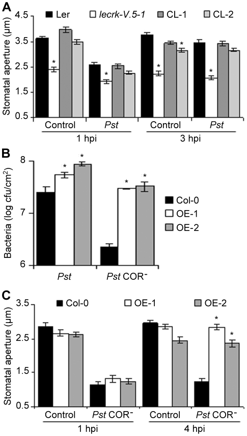
Although lecrk-V.5-1 and lecrk-V.5-2 were more resistant to Pst DC3000 after dip-inoculation (Figure 1C, D), both mutants demonstrated WT susceptibility levels after infiltration-inoculation (Figure S2). Since Arabidopsis restricts bacterial invasion through stomatal closure [4], [7], we hypothesized that lecrk-V.5 mutants increased resistance to surface-inoculation with bacteria is due to their ability to prevent bacterial entry inside leaves via stomata. To determine LecRK-V.5 possible function in stomatal immunity, we examined stomatal aperture of lecrk-V.5 mutants upon Pst DC3000 inoculation. Stomata of buffer-treated epidermal peels of lecrk-V.5 mutants were closed at levels similar to those observed in WT controls at 1 hpi with bacteria (Figure 2A and Figure S3A). In addition, the COR-dependent stomatal reopening that occurred in WT at 3 hpi was not observed in lecrk-V.5 mutants (Figure 2A and Figure S3A). Complemented lines demonstrated a WT stomatal aperture in response to Pst DC3000 (Figure 2A), suggesting that mutations in LecRK-V.5 caused the stomatal response phenotype observed in lecrk-V.5 mutants. We also analyzed stomatal aperture in WT and lines overexpressing LecRK-V.5 at 1, 1.5 and 3 hpi with Pst DC3000. Although stomatal closure in OE-1 and OE-2 was observed at 1 hpi with Pst DC3000, stomata reopened in overexpression lines at 1.5 hpi, a time point where WT stomata were still closed (Figure S3B). An early stomatal reopening may explain the increased susceptibility of OE-1 and OE-2 lines to bacteria.
Figure 2. Altered stomatal immunity responses in lecrk-V.5-1 and in lines over-expressing LecRK-V.5.
(A) Stomatal apertures in epidermal peels exposed to MES buffer (control) or 1×10⁸ cfu.ml−1 Pst DC3000 (Pst) for 1 or 3 hrs. (B) Bacterial growth assessed 3 days after dip-inoculation with 1×10⁷ cfu.ml−1 Pst DC3000 (Pst) or Pst DC3000 COR− (Pst COR−) in WT Col-0 and overexpression lines (OE-1 and OE-2). Values are the means ± SD. Statistical differences between WT controls and OE lines are detected with a t test (P<0.01, n = 6). (C) Stomatal aperture in WT Col-0 and overexpression lines (OE-1 and OE-2) after 1 and 4 hours incubation in MES buffer (Control) or 1×10⁸ cfu.ml−1 Pst DC3000 COR− (Pst COR−). For (A) and (C), results are shown as mean of ≥60 stomata measurements ± SE. Asterisks indicate significant differences between WT and mutant/CL/OE lines based on a t test (P<0.001). hpi, hour post inoculation. All experiments were repeated at least three times with similar results.
WT Col-0 Arabidopsis are resistant to surface inoculation with COR-deficient mutants of Pst DC3000 (Pst DC3000 COR−), presumably because their stomata do not reopen upon infection [4], [22]. To further assess the possible role of LecRK-V.5 in stomatal immunity, lines overexpressing LecRK-V.5 (OE-1 and OE-2) were dip-inoculated with Pst DC3000 COR− bacteria and disease development was evaluated 3 days later. The defective virulence of Pst DC3000 COR− observed in Col-0 was almost fully rescued in lines overexpressing LecRK-V.5 (Figure 2B). We also analyzed stomatal aperture in WT and overexpression lines after Pst DC3000 COR− inoculation. Lines overexpressing LecRK-V.5 reopened stomata as early as 4 hpi (Figure 2C). Since Arabidopsis WT stomata do not reopen upon infection with Pst DC3000 COR− (Figure 2C) [4], [22], stomatal reopening in OE-1 and OE-2 lines may explain their increased susceptibility to Pst DC3000 COR− (Figure 2B). Taken together these data suggest a role for LecRK-V.5 in mediating stomatal movement in response to Pst DC3000 bacteria in Arabidopsis.
A negative role for LecRK-V.5 in PAMP-induced stomatal closure
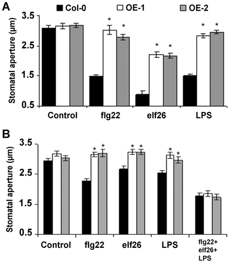
To further define the role of LecRK-V.5 during stomatal immunity, we tested the ability of lines overexpressing LecRK-V.5 to respond to PAMP-mediated stomatal closure. Epidermal peels were incubated with the PAMPs flg22, the active elongation factor Tu epitope elf26 and lipopolysaccharides (LPS), which promote stomatal closure [4], [9]. Stomatal closure was strongly reduced in OE-1 and OE-2 lines after PAMP treatments at concentrations that are usually used to induce stomatal closure in WT controls (Figure 3A) [4]. Treatments with higher concentrations of flg22 or elf26 partially restored OE-1 and OE-2 sensitivity to these PAMPs (Figure S4A, B). By contrast, these transgenics demonstrated a defective stomatal response to LPS concentrations up to 200 ng.µL−1 (Figure S4C). Surprisingly, stomata of lines overexpressing LecRK-V.5 responded similarly to WT control to low concentrations of these PAMPs when all 3 PAMPs were applied together (Figure 3B). This observation may explain the observed stomatal closure of OE-1 and OE-2 after Pst DC3000 inoculation (Figure S3B).
Figure 3. Altered PAMP-induced stomatal closure in lines overexpressing LecRK-V.5.
(A) Stomatal apertures in epidermal peels of WT Col-0 and overexpression lines (OE-1 and OE-2) after 3 hrs of incubation with MES buffer (Control), 5 µM flg22, 5 µM elf26 or 100 ng.µL−1 LPS. (B) Stomatal apertures were measured on epidermal peels incubated in MES buffer (Control), 1 µM flg22, 1 µM elf26, 10 ng.µL−1 LPS or 1 µM flg22, 1 µM elf26 and 10 ng.µL−1 LPS together. Results are shown as mean of ≥60 stomata measurements ± SE. Asterisks indicate significant differences between WT and OE lines based on a t test (P<0.001). All experiments were repeated at least three times with similar results. hpi, hour post inoculation.
lecrk-V.5 mutants demonstrate normal apoplastic PTI responses
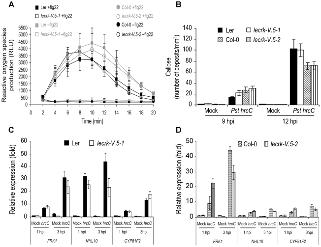
Stomatal closure is only one aspect of the PTI response. To determine whether apoplastic PTI responses were constitutively elevated in lecrk-V.5 mutants, we analyzed the production of H2O2 as an early response to PAMPs in lecrk-V.5-1 and lecrk-V.5-2 leaves in response to flg22 [23]. ROS production after flg22 treatment was at a WT level in both lecrk-V.5 mutants (Figure 4A). We also examined callose deposition [24] and accumulation of the PTI-responsive FRK1, NHL10 and CYP81F2 [25] after inoculation with the Type III secretion system deficient bacterial mutant Pst DC3000 hrcC (CB200) [26]. Callose deposition and PTI-gene expression levels were not constitutively elevated in lecrk-V.5 mutants and inoculation with Pst DC3000 hrcC induced WT callose deposition (Figure 4B) and PTI-gene expression levels (Figure 4C, D). These data suggest that enhanced resistance of lecrk-V.5 mutants to Pst DC3000 is not correlated with a constitutively activated apoplastic PTI or boosted ROS production, increased callose deposition or PTI-responsive gene expression levels upon PTI activation in the leaves.
Figure 4. Apoplastic PTI responses in lecrk-V.5 mutants.
(A) Production of reactive oxygen species in Arabidopsis leaves after treatment with 1 µM flg22 as relative light units (RLU). Values represent averages ± SE (n = 6). (B) Callose deposition in WT or lecrk-V.5 mutants leaves infiltrated with 10 mM MgSO4 (Mock) or Pst DC3000 hrcC (Pst hrcC). Data represent the number of callose deposits per square millimeter ± SD. Differences were not significantly different to WT based on a t test (P<0.01). (C, D) FRK1, NHL10, CYP81F2 expression levels in WT or lecrk-V.5 mutant seedlings soaked in 10 mM MgSO4 (Mock) or 1×10⁷ cfu.ml−1 Pst DC3000 hrcC (hrcC). Transcript levels were determined by qRT-PCR and normalized to both EF-1 and UBQ10. Bars indicate SD (n = 9). All experiments were repeated 3 times with similar results.
LecRK-V.5 is localized at the plasma membrane and expressed in stomatal guard cells
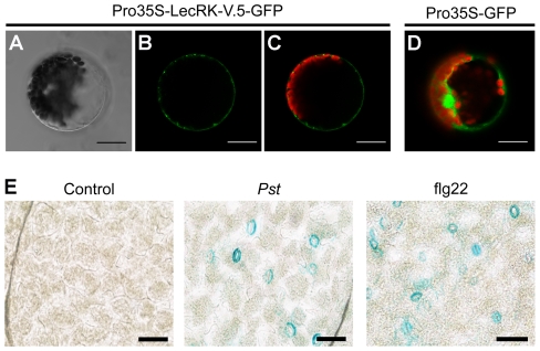
To determine the subcellular localization of LecRK-V.5, the LecRK-V.5-GFP fusion protein driven by the cauliflower mosaic virus 35S promoter was transiently expressed in Arabidopsis mesophyll protoplasts. Confocal imaging indicates that the fluorescence signal is confined to a ring external to the chloroplast signal, while the control protoplasts expressing GFP alone showed a nuclear and cytoplasmic GFP localization (Figure 5A–D). Since LecRK-V.5 was previously detected in plasma membrane fraction [17], our data confirm that LecRK-V.5 is localized at the plasma membrane.
Figure 5. LecRK-V.5 is localized at the plasma membrane and is expressed at guard cells upon PTI activation.
(A, B and C) Subcellular localization of LecRK-V.5-GFP fusion protein in Arabidopsis mesophyll protoplasts. LecRK-V.5-GFP expression was driven by the cauliflower mosaic virus 35S promoter and transiently expressed in Arabidopsis mesophyll protoplasts. The bright-field image (A), images of the GFP fluorescence (green) only (B), and the overlap image of the GFP (green) and chlorophyll (red) fluorescence (C) are presented. (D) Photo of a control protoplast expressing GFP alone. Bars = 20 µm. (E) The activity of LecRK-V.5 promoter was detected by GUS staining in guard cell 15 min after inoculation with 1×10⁸ cfu.ml−1 Pst DC3000 (Pst) or 1 µM flg22 treatment. Bar represents 20 µm.
We then asked whether LecRK-V.5 expression is localized at stomatal guard cells upon stomatal immunity activation. LecRK-V.5 promoter GUS analyses indicated that LecRK-V.5 is induced specifically in stomatal guard cells 15 min after Pst DC3000 inoculation or flg22 treatment (Figure 5E). This expression pattern suggests a role for LecRK-V.5 in stomatal movement upon pathogen infection.
LecRK-V.5 regulates stomatal closure upstream of COR site of action
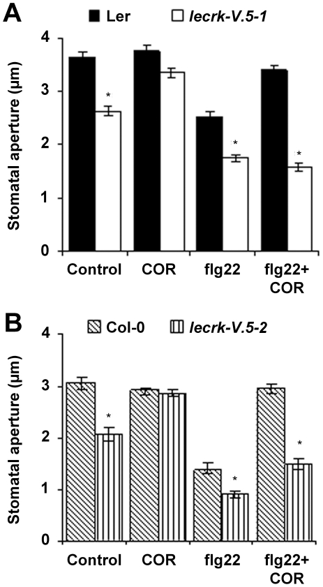
COR counteracts PAMP-induced stomatal closure and reverses flg22 inhibition of inward K+ channels downstream of ROS production [4], [6]. To determine whether LecRK-V.5 regulates stomatal closure upstream or downstream of COR site of action, we evaluated stomatal apertures in epidermal peels of lecrk-V.5 mutants treated with COR. Treatments with COR only opened closed stomata in lecrk-V.5-1 and lecrk-V.5-2 (Figure 6A, B), suggesting that LecRK-V.5 negatively regulates PAMP-induced stomatal closure upstream of COR site of action. Although Pst DC3000 bacteria produce COR [4], inoculation with Pst DC3000 did not open stomata of lecrk-V.5 mutants (Figure 2A). Epidermal peels were thus treated with flg22 alone or flg22 together with COR. The PAMP flg22 induced stomatal closure in WT plants and further closure in both lecrk-V.5 mutants (Figure 6A, B). However, COR did not counteract flg22-mediated stomatal closure in lecrk-V.5 mutants while stomata of flg22-treated WT controls did reopen after COR treatment (Figure 6A, B). Together, these data suggest that mutations in LecRK-V.5 inhibit the COR-dependent reopening of stomata during PAMP-induced stomatal closure.
Figure 6. Effects of COR on flg22-mediated stomatal closure in lecrk-V.5 mutants.
(A, B) Stomatal aperture in epidermal peels of WT (Ler and Col-0) and lecrk-V.5 mutants exposed to MES buffer (Control), 0.5 ng.µL−1 COR, 5 µM flg22 or 5 µM flg22 and 0.5 ng.µL−1 COR together (flg22+COR) for 3 hrs. Results are shown as mean of ≥60 stomata measurements ± SE. Asterisks indicate significant differences between WT and mutants based on a t test (P<0.001). All experiments were repeated at least three times with similar results.
Both lecrk-V.5 mutants accumulate high levels of ROS in guard cells
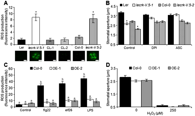
To further clarify the role of LecRK-V.5 in the stomatal response, ROS levels in lecrk-V.5-1 and lecrk-V.5-2 guard cells were analyzed with the fluorescent dye 2′,7′-dichlorofluorescein diacetate (H2DCFDA) [27], [28]. Microscopy and fluorescence emission analyses revealed a higher level of ROS in guard cells of both lecrk-V.5 mutants (Figure 7A). WT controls and complemented lines demonstrated similar levels of ROS production (Figure 7A), suggesting that mutations in LecRK-V.5 caused ROS accumulation observed in lecrk-V.5 mutants. Diphenylene iodium chloride (DPI), an inhibitor of NADPH oxidases known to inhibit ABA-induced stomatal closure [28], was used to test a possible role for NADPH oxidases in lecrk-V.5 stomatal phenotype. DPI treatments induced stomatal opening in lecrk-V.5-1 and lecrk-V.5-2 (Figure 7B). To further evaluate the role of ROS, plants were treated with ascorbic acid (ASC), a chemical known to reduce ROS levels [27]. Treatment with ASC also opened constitutively closed stomata in lecrk-V.5 mutants (Figure 7B). These results suggest that over-accumulation of ROS is responsible for the constitutive stomatal closure observed in lecrk-V.5 mutants. Treatments with PAMPs such as flg22, elf26 or LPS boosted guard cell ROS production at WT levels in lecrk-V.5-1 (Figure S5). By contrast, treatments with these PAMPs increased guard cell ROS levels in WT, but no increase of ROS production was observed in Arabidopsis overexpressing LecRK-V.5 (Figure 7C). Furthermore, H2O2-induced stomatal closure was normal in overexpression lines (Figure 7D) suggesting that LecRK-V.5 does not influence stomatal closure signaling downstream of ROS biosynthesis. LecRK-V.5 may thus function upstream of ROS production in guard cell movement.
Figure 7. Role of LecRK-V.5 in ROS-mediated stomatal closure.
(A) ROS detected by H2DCFDA fluorescence in non-treated WT (Ler and Col-0), lecrk-V.5 mutants and two complemented line (CL-1 and CL-2) guard cells. A representative stoma is shown. Bars represent 7.5 µm. (B) Stomatal aperture in epidermal peels of WT (Ler and Col-0) and lecrk-V.5 mutants exposed to MES buffer (Control), 20 µM DPI or 1 mM ASC for 3 hrs. (C) ROS detected by H2DCFDA fluorescence in guard cells of WT Col-0 and lines overexpressing LecRK-V.5 (OE-1 and OE-2) after treatments with MES buffer (Control), 5 µM flg22, 5 µM elf26 or 100 ng.µL−1 LPS. (D) Stomatal aperture in epidermal peels of WT Col-0 and overexpression line OE-1 and OE-2 after 3 h incubation with H2O2. For all experiments, results are shown as mean ± SE. In (A), (B) and (D) asterisks indicate a significant difference to WT control based on a t test analysis (n≥60; P<0.001). In (C), different letters indicate statistically significant differences compared with the non-treated WT Col-0 (Fisher's Least Significant Difference test; n≥60; P<0.05). All experiments were repeated at least three times with similar results.
LecRK-V.5 role in stomatal immunity is mechanistically linked to ABA signaling
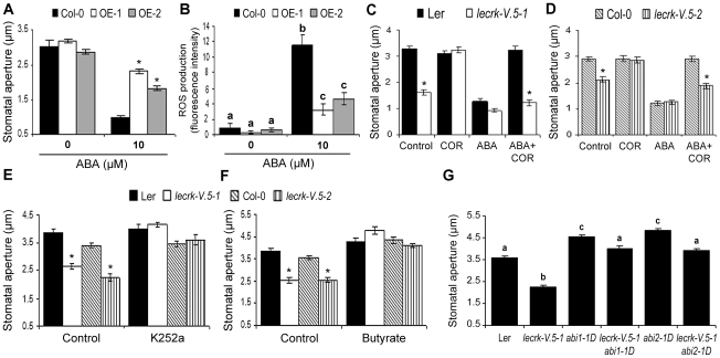
Innate immunity-mediated stomatal closure depends on ABA signaling [4], [6], [22]. To evaluate whether LecRK-V.5 plays a role in the ABA-mediated stomatal closure, stomatal apertures after treatment with ABA were assessed in lines overexpressing LecRK-V.5. Stomata of such lines were greatly compromised in their ability to respond to ABA (Figure 8A). Furthermore, overexpression lines demonstrated reduced ROS production after ABA treatment (Figure 8B). COR inhibits ABA-induced stomatal closure [4], [6]. We thus evaluated the possibility that lecrk-V.5 mutants were resistant to COR-inhibition of stomatal closure upon ABA treatments. Similarly to PAMP-induced stomatal closure (Figure 6A, B), COR treatments did not open ABA-treated lecrk-V.5-1 and lecrk-V.5-2 stomata while reopening in WT controls was observed (Figure 8C, D). To further evaluate the role of LecRK-V.5 in ABA-mediated stomatal closure, we manipulated the ABA-mediated regulation of guard cell pH. Butyrate which causes an acidification of cytoplasm, and K252a, a protein kinase inhibitor, are able to suppress ABA-induced alkalization occurring upstream of ROS production in guard cells [29]. Both compounds opened constitutively closed lecrk-V.5-1 and lecrk-V.5-2 stomata (Figure 8E, F). Double mutants generated by crossing lecrk-V.5-1 with ABA-insensitive mutants abi1-1D and abi2-1D [30], [31] demonstrated a WT stomatal aperture further indicating that LecRK-V.5 functions in ABA signaling (Figure 8G). In addition to ABA, the plant hormones jasmonate (JA) and SA play a positive role in stomatal closure [7], [29], [32]. Both OE-1 and OE-2 lines demonstrated WT levels of stomatal closure in response to these two phytohormones (Figure S6). Collectively, these results suggest that LecRK-V.5 interferes with ABA- but not with JA- or SA-mediated stomatal closure signaling.
Figure 8. LecRK-V.5 role in stomatal immunity is mechanistically linked to ABA signaling.
(A) Stomatal apertures in epidermal peels of WT Col-0 or LecRK-V.5 overexpression transgenics OE-1 and OE-2 were measured after 3 hrs incubation in ABA. (B) H2DCFDA-detected ROS production after ABA treatments. (C, D) Stomatal apertures in epidermal peels of WT (Ler and Col-0) and lecrk-V.5 mutants exposed to MES buffer (Control), 0.5 ng.µL−1 COR, 10 µM ABA, or 10 µM ABA together with 0.5 ng.µL−1 COR (ABA+COR) for 3 hrs. (E, F) Arabidopsis WT (Ler and Col-0) or lecrk-V.5 mutants epidermal peels were floated in MES buffer (Control) or 1 µM K252a (E) or 0.5 mM butyrate (F) for 3 hrs before stomatal aperture measurement. (G) Stomatal aperture in lecrk-V.5-1 abi1-1D and lecrk-V.5-1 abi2-1D double mutants. For B and G, different letters indicate statistically significant differences compared with WT (Fisher's Least Significant Difference test; n≥60; P<0.05). For A, C, D, E and F, results are shown as mean ± SE (n≥60) and asterisks indicates significant differences between WT and mutant/OE based on a t test (P<0.001). All experiments were repeated at least three times with similar results.
Discussion
As a part of the plant innate immune system, stomata play an active role in limiting bacterial entry into plant tissues and subsequent disease symptoms [4], [5]. A rapid stomatal closure occurs upon bacterial challenge and some pathogens have evolved virulence factors such as COR for Pst DC3000 to overcome plant stomatal immunity [4], [33], [34]. Plants lacking a functional LecRK-V.5 displayed enhanced resistance to Pst DC3000 surface-inoculation, but were susceptible as WT after bacteria-infiltration, an inoculation method that bypasses the epidermal barrier. These results suggest that disruption of LecRK-V.5 affects early Arabidopsis defenses by restricting bacterial entry into leaves. Differences between surface- and infiltration-inoculation were also observed in Arabidopsis mutants defective in bacteria-induced stomatal closure [4], [22], [35], [36]. Since lecrk-V.5 mutants demonstrated WT apoplastic PTI (Figure 4), closed stomata and inhibition of COR-dependent stomatal reopening is the most straightforward explanation for the enhanced resistance phenotype of lecrk-V.5 mutants to Pst DC3000 surface inoculation. LecRK-V.5 overexpression lines were more susceptible than WT plants to bacteria, notably to Pst DC3000 COR− dip-inoculation (Figure 2B). Enhanced susceptibility to WT Pst DC3000 was correlated with an earlier reopening of stomata, further pointing for a role of LecRK-V.5 in stomatal immunity. The reopening of stomata after inoculation with Pst DC3000 COR− suggests the existence of a plant stomatal reopening mechanism positively modulated by LecRK-V.5. Taken together these data suggest that LecRK-V.5 negatively regulates Arabidopsis resistance to bacteria through fine-tuning of stomatal immunity.
Treatments with PAMPs such as flg22, elf26, or LPS induce rapid stomatal closure and the fls2 mutant is defective in flg22-induced stomatal closure implying the PTI response in stomatal immunity [4], [9]. Similarly to the fls2 mutant, all mutants defective in PAMP-induced stomatal closure described so far are highly susceptible to surface-inoculation with Pst DC3000 or Pst DC3000 COR-deficient mutant bacteria most likely because of a defect in bacteria-induced stomatal closure [4], [22], [35], [36]. Lines overexpressing LecRK-V.5 were defective in PAMP-induced stomatal closure, suggesting a negative role for LecRK-V.5 during stomatal immunity. Treatments of over-expression lines with low concentrations of elf26, LPS and flg22 that did not induce stomatal closure when applied individually, triggered stomatal closure when applied all together (Figure 3B). This observation suggests that different PAMPs additively activate the stomatal immunity response modulated by LecRK-V.5. It likely explains why over-expression lines exhibit a WT bacterium-induced stomatal closure. Other aspects of the Arabidopsis PTI response such as the flg22-triggered oxidative burst, Pst DC3000 hrcC-mediated callose deposition and up-regulation of PTI marker genes were not affected in lecrk-V.5 mutants. These apoplastic PTI responses are mostly mediated by mesophyll cells [1], [22], [36]. The recently isolated scord5 mutant also shows a defective stomatal immunity but exhibits WT apoplastic immunity [36]. Our data therefore confirm recent findings indicating that stomatal immunity can be distinguished from the general PTI response [36]. Localized expression of LecRK-V.5 upon PTI activation at stomatal guard cells may explain the specific role of LecRK-V.5 in stomatal immunity.
The signaling pathways leading to bacteria and PAMP-induced stomatal closure downstream of PRRs (e.g. FLS2) remains unclear. Analyses of SA-deficient nahG transgenics, SA-biosynthetic mutant sid2/eds16 and ABA-deficient mutant aba3 indicate that SA and ABA biosynthesis are required for PAMP-induced stomatal closure [4]. The ABA signaling components OST1, ABI1, GPA1 and OST2 are also required for bacteria- and PAMP-induced stomatal closure [4], [6], [34], [5], [36]. These studies illustrate the complexity of hormonal crosstalks involved in stomatal immunity. LecRK-V.5 overexpression lines were defective in ABA-mediated stomatal closure but not in SA- and JA-mediated stomatal closure. ABA-induced ROS production was also affected in lines over-expressing LecRK-V.5. As it was proposed for ABI1, OST1 and GPA1 [4], [6], [34], [35], [36], LecRK-V.5 appears to specifically function in guard cell ABA signaling pathway downstream of PAMP perception. LecRK-V.5 may thus act at a specific branch involving ABA for the control of stomatal immunity.
During stomatal closure, ABA induces ROS accumulation, which activates plasma membrane calcium channels, induces increase in cytosolic Ca2+, and triggers stomatal closure [28], [37], [38]. In this study, constitutive high levels of ROS in guard cells were correlated with constitutively closed stomata in lecrk-V.5 mutants. This phenotype was reverted by treatments with an inhibitor of NADPH oxidase (DPI) [28] or ASC, a chemical that reduces ROS levels [27]. Increase in ROS levels and stomatal closure after PAMP and ABA treatments were impaired in lines overexpressing LecRK-V.5. In addition, suppression of ABA-induced alkalization that takes place upstream of ROS production [29] opened closed stomata of lecrk-V.5 mutants. Recent studies suggest that constitutive ROS accumulation does not induce stomatal closure, but ROS accumulation mediated by ABA does [39], [40]. Collectively these observations suggest a role for LecRK-V.5 upstream of ROS production in the ABA-mediated stomatal closure signaling. Importantly, H2O2-induced stomatal closure in LecRK-V.5 overexpression lines was not affected. Thus LecRK-V.5 probably does not disrupt the pathway downstream of ROS biosynthesis. Since generation of H2O2 in guard cells and stomatal closure in response to ABA occurs via NADPH oxidases Rboh [10], LecRK-V.5 may act upstream of Atrboh in PAMP- and bacteria-mediated ROS production. By preventing guard cell ROS accumulation, LecRK-V.5 may function in ROS homeostasis to regulate H2O2 content in guard cells. Plants likely have evolved a mechanism to reverse PAMP-, bacteria- or ABA-triggered stomatal closure to prevent long-term detrimental effects on CO2 uptake and photosynthesis. LecRK-V.5 may be one component of this protective mechanism.
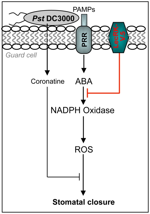
In this study, the L-type lectin receptor kinase LecRK-V.5 was identified as a key player in stomatal immunity. We propose a model in which LecRK-V.5 negatively regulates the signaling pathway leading to PTI-mediated stomatal closure downstream of ABA and upstream of NADPH oxidase to fine tune ROS accumulation in stomatal guard cells (Figure 9). The OST1/SnRK2E/SnRK2.6 protein kinase involved in PAMP-induced stomatal closure [4] was initially identified as a positive regulator upstream of ROS in guard cell ABA signaling [41]. A recent study showed that OST1 interacts with and phosphorylates AtrbohF [42]. Further studies are required to identify protein partners of LecRK-V.5 and implicated in PTI-mediated stomatal closure signaling upstream of ROS production.
Figure 9. Model of LecRK-V.5 role in PAMP-induced stomatal closure.
This model is based on the information provided in this study and references cited in the Discussion. PAMPs are perceived by pattern recognition receptors (PRRs) in the guard cell. PAMPs perception is mechanistically linked to ABA-regulated stomatal closure via ROS production by NADPH oxidase. The virulence factor COR is secreted by Pst DC3000 to interfere with stomatal closure by reverting flg22-inhibition. LecRK-V.5 negatively regulates PAMP-mediated stomatal closure downstream of ABA but upstream of ROS production.
Materials and Methods
Biological materials and growth conditions
Arabidopsis thaliana (L. Heyhn.) ecotypes Columbia (Col-0) and Landsberg erecta (Ler), and derived mutant lines were grown as previously described [43]. The lecrk-V.5-1 (GT12539) Ds transposon insertion line (Ler) is from the Cold Spring Harbor Laboratory (http://genetrap.cshl.org/) and the lecrk-V.5-2 (GK-623G01) T-DNA insertion line (Col-0), abi1-1D and abi2-1D were obtained from the Nottingham Arabidopsis Stock Centre (NASC, http://arabidopsis.info/). Details about PCR analyses performed to screen for homozygous mutants are described under “Gene Expression Studies”. Bacterial strains Pst DC3000, Pst DC3000 COR− (DB29) and Pst DC3000 hrcC mutant (CB200) were provided by B.N. Kunkel (Washington University, St. Louis, USA) [26]. Pst bacteria were cultivated at 28°C, 340 rpm in King's B medium containing rifampicin (Pst DC3000), rifampicin, spectinomycin and kanamycin (DB29) or rifampicin and kanamycin (CB200).
Bacterial infection assays
Bacterial disease assays were conducted as previously described [44]. For surface inoculation, plants were dipped in a bacterial solution of 1×10⁷ cfu.ml−1 containing 0.01% Silwet L-77 (Bioman Scientific Co., Ltd.). Alternatively, three fully expanded leaves per plant were infiltrated on the abaxial surface with Pst DC3000 at a concentration of 1×10⁵ cfu.ml−1 using a needleless 1 ml syringe.
Plasmid constructions and generation of transgenic plants
All cloning experiments were performed using the genomic clone AF001168 kindly provided by Christine Hervé (INRA Toulouse, France). According to [18], 707 pb of the LecRK-V.5 promoter was PCR amplified using the GW-pRK-F (5′-CTGCAACAATTGGGAGGAGGG-3′) and GW-pRK-R (5′-GTTCACGAGACTTTGGTGGGTG-3′) primers. The coding sequence (CDS) of LecRK-V.5 was PCR amplified using the GW-RK-F (5′-ATGTCTCGTGAACTTATTATTCTCTGCC-3′) and GW-RK-R (5′-TCAGCGGCCGTGGGAGACAA-3′) primers. The resulting PCR products were directly cloned by the TOPO cloning reaction into the pCR8/GW/TOPO vector (Invitrogen) following manufacturer's instructions. The promoter and CDS (without stop codon) of LecRK-V.5 was PCR amplified with the GW-attB1-RK-F (5′-GGGGACAAGTTTGTACAAAAAAGCAGGCTCTGCAACAATTGGGAGGAGGG-3′) and GW-attB2-RK-R (5′-GGGGACCACTTTGTACAAGAAAGCTGGGTCGCGGCCGTGGGAGACAAAAG-3′) primers and recombined by the BP reaction into the pDNOR221 vector (Invitrogen). The LecRK-V.5 promoter was sub-cloned by LR reaction in the pMDC163 vector [45] to produce the ProLecRK-V.5::GUS construct. The LecRK-V.5 CDS and promoter plus CDS were then introduced into the gateway pEarleyGATE100, pEarleyGATE201, pEarleyGATE301 and pMDC83 binary vectors [46] to respectively obtain the 35S::LecRK-V.5, 35S::HA-LecRK-V.5, ProLecRK-V.5::LecRK-V.5-HA and 35S::LecRK-V.5-GFP constructs. The fidelity of all constructs was confirmed by sequencing. Arabidopsis plants were transformed using the GV3101 strain of Agrobacterium tumefaciens according to the floral dip protocol [47]. Transgenic insertion lines with single insertion loci were selected on 1/2 MS plates containing 50 µM Glufosinate ammonium (Fluka) and raised to homozygous T3 lines.
Generation of double mutants
The lecrk-V.5-1 abi1-1D and lecrk-V.5-1 abi2-1D double mutants were selected in the F2 progeny of crosses between the two corresponding homozygous parents. Verifications of genotypes involving abi1-1 and abi2-1 crosses were performed as described [31].
Gene expression studies
Semi-quantitative PCR and qRT-PCR were as described [43], with some modifications. Total RNA was extracted and purified using Qiagen RNeasy plant Mini Kit with additional genomic DNA cleanup using Qiagen RNase-Free DNase Set. For cDNA synthesis, RNA was first diluted to 2 µg in a total volume of 22 µL DEPC water and denatured at 65°C for 5 min. Eighteen point five µL of master mix (1× M-MLV buffer, 1 mM dNTP, 5 µM OligoT, 100 U M-MLV reverse transcriptase, [Invitrogen]) was added into each tube and then incubated at 37°C for 1 hr, 70°C for 10 min. cDNA was diluted 5-fold before real-time PCR or semi-quantitative PCR. PCR amplification was done with 2 µL of the first-strand cDNA as template, 1 unit of Taq DNA polymerase (Viogene), 156 µM dNTP and 0.5 µM of primers in a total volume of 20 µL. Primers used for screening of homozygous mutants were GT12539-F (5′-TCGGCTTCAACGTTTACTTC-3′) and GT12539-R (5′-CGATGGAAAGCCTCATTACC-3′) for the lecrk-V.5-1 mutant (P1) and Salk_083045-F (5′-ATGGGTTGGTTAGTTAATGG-3′) and Salk_083045-R (5′-CCTCGCATTCATTTCATTGTC-3′) for the lecrk-V.5-2 mutant (P2). The cycling conditions were 94°C for 5 min for one initial step followed by 94°C for 30 s, 60°C for 30 s and 72°C for 1 min, for 40 cycles. The PCR was terminated with one extra step at 72°C for 10 min. iQ SYBR Green supermix [Bio-RAD] (2 µL of cDNA, 9 µL SYBR Green supermix, 5 µL filtered water, 1 µL of 10 µM forward primer, 1 µL of 10 µM reverse primer, in a total volume of 18 µL per well) was employed for real-time PCR analysis. The cycling conditions were composed of an initial 3 min denaturation step at 95°C, followed by 40 cycles of 95°C for 30 s, 54°C for 35 s, 72°C for 35 s (iQ5 Real-Time PCR Detection System, Bio-RAD). Melting curve was run from 55°C to 95°C with 10-second time interval to ensure the specificity of product. Data were analyzed using Bio-Rad iQ5 software (version 2.0). Elongation factor 1 (EF-1) and ubiquitin (UBQ10) were used as reference genes for normalization of gene expression levels in all samples. The WT without any treatment or mock treatment were considered as controls (expression level = 1) in each experiment. qRT-PCR forward and reverse primers of each gene were as follows: 5′-GAATGGAGTTTCACAGCTACCA-3′ and 5′-GTCGGTTAACTCCAATGAACTC-3′ for LecRK-V.5 expression in complemented lines (CL-1 and CL-2); 5′-TCATGGCATACTTCGTCTCAC-3′ and 5′-CTCATCGACAGGTGTCATCT-3′ for LecRK-V.5 expression in overexpression lines (OE-1 and OE-2); 5′-AAA TGG AGA GAG CAA CAC AAT G-3′ and 5′-ATC GCC CAT TCC AAT GTT AC-3′ for CYP81F2 (At5g57220); 5′-TTC CTG TCC GTA ACC CAA AC-3′ and 5′-CCC TCG TAG TAG GCA TGA GC-3′ for NHL10 (At2g35980); 5′-GCC AAC GGA GAC ATT AGA G-3′ and 5′-CCA TAA CGA CCT GAC TCA TC-3′ for FRK1 (At2g19190); 5′-TGA GCA CGC TCT TCT TGC TTT CA-3′ and 5′-GGT GGT GGC ATC CAT CTT GTT ACA-3′ for EF-1 (At1g07920) and 5′-GGCCTTGTATAATCCCTGATGAAT-3′ and 5′-AAAGAGATAACAGGAACGGAAACA-3′ for UBQ10 (At4g05320).
Subcellular localization in protoplast
For transient expression of the GFP fusion proteins, constructs expressing 35S::LecRK-V.5-GFP and vector alone were co-transfected into Arabidopsis mesophyll protoplasts according to a previously described protocol [48]. Briefly, leaves from 5-week-old plants were digested in an enzyme solution containing 1.5% cellulose R10 (Yakult Pharmaceutical Ind. Co.) and 0.3% macerozyme R10 (Yakult Pharmaceutical Ind. Co.). Transfected protoplasts were incubated overnight under light at room temperature. Confocal laser scanning microscopy with excitation at 488 nm and emission at 500–530 nm (Leica TCS SP5 Confocal, Leica, Wetzlar, Germany) was carried out to visualize subcellular localization of LecRK-V.5-GFP. Autofluorescence was monitored at 488 nm, and transmission images were collected in parallel.
Stomatal experiments
Leaf peels were collected from the abaxial side of fully expanded leaves and floated in stomatal buffer (10 mM MES-KOH, 30 mM KCl, pH 6.15) for 2.5 h under light (100 µmol m−2 s−1) to ensure that most of stomata were opened before treatments [4]. Purified chemical lipopolysaccharide (LPS from P. aeruginosa, Sigma), flg22 peptide (Biomer Technology, CA), elf26 (Biomer Technology, CA) or COR (Sigma) were used at indicated concentrations. flg22 and elf26 were diluted in 10 mM MgSO4. COR and LPS were respectively diluted in milliQ water and in MES buffer containing 0.25 mM MgCl2 and 0.1 mM CaCl2 [5]. ABA, methyl jasmonate (MeJA) and SA used at indicated concentrations were dissolved in 10% ethanol (Sigma). Diphenyleneiodonium chloride (DPI, Sigma) and K252a (Sigma) were dissolved in dimethylsulfoxide (DMSO). Ascorbic acid (ASC) and sodium butyrate (Butyrate, Sigma) were prepared in milliQ water. Mock controls were MES buffer containing 0.1% ethanol for MeJA, ABA and SA, 0.1% DMSO for DPI and K252a, and milliQ water for ASC and Butyrate. Bacterial concentration used was 1×10⁸ cfu.ml−1 in 10 mM MgSO4. Stomatal apertures were measured as described [43].
Monitoring ROS in guard cells
2′,7′-dichlorodihydrofluorescein diacetate (H2DCFDA) fluorescence analysis were performed essentially as described [49]. After 2.5 hrs incubation in stomatal buffer, epidermal peels were transferred to 50 µM H2DCFDA in 10 mM Tris-HCl pH 7.2 for 15 min. Excess H2DCFDA was then removed by washing 3 times in 10 mM Tris-HCl pH 7.2. Then, PAMPs (5 µM flg22, 5 µM elf26 or 100 ng.µL−1 LPS) or 10 µM ABA were added to the incubation buffer for 10 min. H2DCFDA fluorescence was observed with an Olympus BX51 fluorescence microscope with an excitation at 460–480 nm and emission at 495–540 nm. Images were captured with an Olympus DP72 digital camera linked to the Olympus DP2-BSW software. Fluorescence was analyzed using ImageJ 1.42 sofware.
Apoplastic oxidative burst evaluation
ROS released by leaf tissue was assayed as described [23]. Leaf of 5-week-old Arabidopsis plants were cut in 2 mm2 pieces and floated overnight in water. ROS production was triggered with 1 µM flg22 applied together with 100 µM luminol (Sigma) and 1 µg/mL of horseradish peroxidase (Sigma). Luminescence was measured by a Centro LB 962 microplate luminometer (Berthold Technologies) for 20 min after addition of flg22.
Gus staining
Surface-sterilized seeds were sown on 1/2 MS agar plates and cold-treated at 4°C in the dark for 3 days. The plates were then moved to germination conditions 22–24°C day, 17–19°C night temperature under a 15-h-light/9-h-dark photoperiod. Ten-day-old seedlings were transferred to 12 well plates containing liquid 1/2 MS. After overnight incubation, seedlings were treated with 1×10⁸ cfu.ml−1 Pst DC3000 or with 1 µM flg22 for 15 min. β-glucuronidase activity was determined as described [50].
Callose staining
Arabidopsis were syringe infiltrated with 1×10⁸ cfu.ml−1 Pst DC3000 hrcC or 10 mM MgSO4 as a control. Nine leaf discs from 3 different plants were selected for analyses. Harvested leaf samples were cleared overnight by incubation in 95% ethanol at room temperature and then washed three times (2 hrs for each washing) with sterilized water. Cleared leaves were stained with 0.01% aniline blue in 0.15 M phosphate buffer (pH 9.5) for 24 hrs. Callose deposits were visualized under ultraviolet illumination using an Olympus DP72 digital camera linked to the Olympus DP2-BSW software. Callose deposits were counted using the “analyze particles” function of ImageJ 1.42 sofware.
Growth conditions for PTI marker gene expression study
Surface-sterilized seeds were sown on 1/2 MS agar plates and cold-treated at 4°C in the dark for 3 days. The plates were then moved to germination conditions 22–24°C day, 17–19°C night temperature under a 15-h-light/9-h-dark photoperiod. Ten-day-old seedlings were transferred to 24 well plates. Three seedlings per well were soaked in 200 µL liquid 1/2 MS containing 1% sucrose. After incubation overnight, seedlings were treated with 1×10⁸ cfu.ml−1 Pst DC3000 hrcC mutant (CB200) for 1 hr and 3 hrs.
Accession numbers
Sequence data from this article can be found in the Arabidopsis Genome Initiative under accession number(s): LecRK-V.5 (At3g59700), FRK1 (At2g19190), NHL10 (At2g35980), CYP81F2 (At5g57220), EF-1 (At1g07920), UBQ10 (At4g05320), ABI1 (At4g26080) and ABI2 (At5g57050).
Supporting Information
LecRK-V.5 expression levels in transgenic lines. Relative expression levels in WT (Ler) and two complemented lines (CL-1 and CL-2) and WT (Col-0) and two overexpression lines (OE-1 and OE-2). Transcript levels were determined by qRT-PCR and normalized to both EF-1 and UBQ10. Expression levels were compared to WT controls with a defined expression value of 1. Bars indicate SD (n = 6). Experiments were repeated 3 times with similar results.
(TIF)
Susceptibility of lecrk-V.5 mutants to Pst DC3000 infiltration-inoculation. Bacterial growth (colony forming units (cfu) per cm leaf area) was determined in Ler, Col-0 and lecrk-V.5 mutants infiltrated with 1×10⁵ cfu.ml−1 Pst DC3000 (Pst). Data represent average ±SD. Means were not significantly different between WT and mutants when evaluated by a t-test (P<0.01, n = 9). dpi, day post inoculation. Experiments were repeated 3 times with similar results.
(TIF)
Stomatal aperture in lecrk-V.5-2 mutant and overexpression lines after bacterial inoculation. (A) Stomatal aperture of WT Col-0 and lecrk-V.5-2 after a 1 hr and 3 hr incubation time with MES buffer (Control) or 1×10⁸ cfu.ml−1 Pst DC3000 (Pst). (B) Stomatal aperture in WT Col-0 and overexpression lines (OE-1 and OE-2) after 1, 1.5 and 3 hrs incubation in MES buffer (Control) or 1×10⁸ cfu.ml−1 Pst DC3000 (Pst). Results are shown as mean of ≥60 stomata ± SE. Asterisks indicates significant differences between WT and mutant/OE based on a t test (P<0.001). All experiments were repeated at least three times with similar results. hpi, hour post inoculation.
(TIF)
Altered PAMP-induced stomatal closure in lines overexpressing LecRK-V.5 . The stomatal response of lines overexpressing LecRK-V.5 (OE-1 and OE-2) to different concentrations of flg22 (A), elf26 (B) and LPS (C). Results are shown as mean of ≥60 stomata ± SE. Asterisks indicate significant differences between WT Col-0 and OE lines based on a t test (P<0.001). All experiments were repeated at least three times with similar results.
(TIF)
ROS production upon PAMPs treatments. ROS detected by H2DCFDA fluorescence in guard cells of WT Ler and lecrk-V.5-1 mutant after treatments with MES buffer (Control), 5 µM flg22, 5 µM elf26 or 100 ng.µL−1 LPS. Results are shown as mean ± SE. Asterisks indicate significant differences to WT control based on a t test analysis (n≥60; P<0.001). Experiment was repeated at least three times with similar results.
(TIF)
Lines overexpressing LecRK-V.5 demonstrate a WT stomatal response to MeJA and SA. Effect of MeJA (A) and SA (B) on stomatal aperture in WT Col-0 and overexpression lines OE-1 and OE-2. Results are shown as mean of ≥60 stomata ± SE. No significant differences between Col-0 and OE lines were observed based on a t test (P<0.001). All experiments were repeated at least three times with similar results.
(TIF)
Acknowledgments
We are grateful to C. Hervé (INRA Toulouse, France) for providing the genomic clone AF001168. We thank the Cold Spring Harbor Laboratory and NASC for providing seeds and B.N. Kunkel for bacteria. We also acknowledge J. Weber, J. Leung and members of Zimmerli's laboratory for critical comments. We thank the Technology Commons (TechComm), College of Life Science, National Taiwan University for providing qRT-PCR equipment. We also thank Y.C. Chuang from TechComm for help with confocal microscopy.
Footnotes
The authors have declared that no competing interests exist.
This work was supported by the National Science Council of Taiwan grants 96-2628-B-002-112-MY3 and 99-2628-B-002-053-MY3 (to L.Z.) and the Frontier and Innovative Research grant of the National Taiwan University code number 99R70436 (to L.Z.). The funders had no role in study design, data collection and analysis, decision to publish, or preparation of the manuscript.
References
- 1.Boller T, Felix G. A renaissance of elicitors: perception of microbe-associated molecular patterns and danger signals by pattern-recognition receptors. Annu Rev Plant Biol. 2009;60:379–406. doi: 10.1146/annurev.arplant.57.032905.105346. [DOI] [PubMed] [Google Scholar]
- 2.Nicaise V, Roux M, Zipfel C. Recent advances in PAMP-Triggered Immunity against bacteria: pattern recognition receptors watch over and raise the alarm. Plant Physiol. 2009;150:1638–1647. doi: 10.1104/pp.109.139709. [DOI] [PMC free article] [PubMed] [Google Scholar]
- 3.Acharya BR, Assmann SM. Hormone interactions in stomatal function. Plant Mol Biol. 2009;69:451–462. doi: 10.1007/s11103-008-9427-0. [DOI] [PubMed] [Google Scholar]
- 4.Melotto M, Underwood W, Koczan J, Nomura K, He S-Y. Plant stomata function in innate immunity against bacterial invasion. Cell. 2006;126:969–980. doi: 10.1016/j.cell.2006.06.054. [DOI] [PubMed] [Google Scholar]
- 5.Melotto M, Underwood W, He S-Y. Role of stomata in plant innate immunity and foliar bacterial diseases. Annu Rev Phytopathol. 2008;46:101–122. doi: 10.1146/annurev.phyto.121107.104959. [DOI] [PMC free article] [PubMed] [Google Scholar]
- 6.Zhang W, He S-Y, Assmann SM. The plant innate immunity response in stomatal guard cells invokes G-protein-dependent ion channel regulation. Plant J. 2008;56:984–996. doi: 10.1111/j.1365-313X.2008.03657.x. [DOI] [PMC free article] [PubMed] [Google Scholar]
- 7.Zeng W, Melotto M, He S-Y. Plant stomata: a checkpoint of host immunity and pathogen virulence. Curr Opin Biotechnol. 2010;21:599–603. doi: 10.1016/j.copbio.2010.05.006. [DOI] [PMC free article] [PubMed] [Google Scholar]
- 8.Klüsener B, Young JJ, Murata Y, Allen GJ, Mori IC, et al. Convergence of calcium signaling pathways of pathogenic elicitors and abscisic acid in Arabidopsis guard cells. Plant Physiol. 2002;130:2152–2163. doi: 10.1104/pp.012187. [DOI] [PMC free article] [PubMed] [Google Scholar]
- 9.Desikan R, Horák J, Chaban C, Mira-Rodado V, Witthöft J, et al. The histidine kinase AHK5 integrates endogenous and environmental signals in Arabidopsis guard cells. PLoS One. 2008;3:e2491. doi: 10.1371/journal.pone.0002491. [DOI] [PMC free article] [PubMed] [Google Scholar]
- 10.Kwak JM, Mori IC, Pei ZM, Leonhardt N, Torres MA, et al. NADPH oxidase AtrbohD and AtrbohF genes function in ROS-dependent ABA signaling in Arabidopsis. EMBO J. 2003;22:2623–2633. doi: 10.1093/emboj/cdg277. [DOI] [PMC free article] [PubMed] [Google Scholar]
- 11.Zhang J, Shao F, Li Y, Cui H, Chen L, et al. A Pseudomonas syringae effector inactivates MAPKs to suppress PAMP-induced immunity in plants. Cell Host Microbe. 2007;1:175–185. doi: 10.1016/j.chom.2007.03.006. [DOI] [PubMed] [Google Scholar]
- 12.Nühse TS, Bottrill AR, Jones AM, Peck SC. Quantitative phosphoproteomic analysis of plasma membrane proteins reveals regulatory mechanisms of plant innate immune responses. Plant J. 2007;51:931–940. doi: 10.1111/j.1365-313X.2007.03192.x. [DOI] [PMC free article] [PubMed] [Google Scholar]
- 13.Mersmann S, Bourdais G, Rietz S, Robatzek S. Ethylene signaling regulates accumulation of the FLS2 receptor and is required for the oxidative burst contributing to plant immunity. Plant Physiol. 2010;154:391–400. doi: 10.1104/pp.110.154567. [DOI] [PMC free article] [PubMed] [Google Scholar]
- 14.Hervé C, Dabos P, Galaud JP, Rougé P, Lescure B. Characterization of an Arabidopsis thaliana gene that defines a new class of putative plant receptor kinases with an extracellular lectin-like domain. J Mol Biol. 1996;258:778–788. doi: 10.1006/jmbi.1996.0286. [DOI] [PubMed] [Google Scholar]
- 15.Barre A, Hervé C, Lescure B, Rougé P. Lectin receptor kinases in plants. Crit Rev Plant Science. 2002;21:379–399. [Google Scholar]
- 16.Bouwmeester K, Govers F. Arabidopsis L-type lectin receptor kinases: phylogeny, classification, and expression profiles. J Exp Bot. 2009;60:4383–4396. doi: 10.1093/jxb/erp277. [DOI] [PubMed] [Google Scholar]
- 17.Hervé C, Serres J, Dabos P, Canut H, Barre A, et al. Characterization of the Arabidopsis lecRK-a genes: members of a superfamily encoding putative receptors with an extracellular domain homologous to legume lectins. Plant Mol Biol. 1999;39:671–682. doi: 10.1023/a:1006136701595. [DOI] [PubMed] [Google Scholar]
- 18.Riou C, Hervé C, Pacquit V, Dabos P, Lescure B. Expression of an Arabidopsis lectin kinase receptor gene, lecRK-a1, is induced during senescence, wounding and in response to oligogalacturonic acids. Plant Physiol Bioch. 2002;40:431–438. [Google Scholar]
- 19.Chen X, Shang J, Chen D, Lei C, Zou Y, et al. A B-lectin receptor kinase gene conferring rice blast resistance. Plant J. 2006;46:794–804. doi: 10.1111/j.1365-313X.2006.02739.x. [DOI] [PubMed] [Google Scholar]
- 20.Kanzaki H, Saitoh H, Takahashi Y, Berberich T, Ito A, et al. NbLRK1, a lectin-like receptor kinase protein of Nicotiana benthamiana, interacts with Phytophthora infestans INF1 elicitin and mediates INF1-induced cell death. Planta. 2008;228:977–987. doi: 10.1007/s00425-008-0797-y. [DOI] [PubMed] [Google Scholar]
- 21.Bouwmeester K, de Sain M, Weide R, Gouget A, Klamer S, et al. The lectin receptor kinase LecRK-I.9 is a novel phytophthora resistance component and a potential host target for a RXLR effector. PLoS Pathog. 2011;3:e1001327. doi: 10.1371/journal.ppat.1001327. [DOI] [PMC free article] [PubMed] [Google Scholar]
- 22.Zeng W, He S-Y. A prominent role of the flagellin receptor FLAGELLIN-SENSING2 in mediating stomatal response to Pseudomonas syringae pv tomato DC3000 in Arabidopsis. Plant Physiol. 2010;153:1188–1198. doi: 10.1104/pp.110.157016. [DOI] [PMC free article] [PubMed] [Google Scholar]
- 23.Chinchilla D, Zipfel C, Robaatzek S, Kemmerling B, Nürnberger T, et al. A flagellin-induced complex of the receptor FLS2 and BAK1 initiates plant defence. Nature. 2007;448:497–500. doi: 10.1038/nature05999. [DOI] [PubMed] [Google Scholar]
- 24.Gómez-Gómez L, Felix G, Boller T. A single locus determines sensitivity to bacterial flagellin in Arabidopsis thaliana. Plant J. 1999;18:277–284. doi: 10.1046/j.1365-313x.1999.00451.x. [DOI] [PubMed] [Google Scholar]
- 25.Boudsocq M, Willmann MR, McCormack M, Lee H, Shan L, et al. Differential innate immune signalling via Ca2+ sensor protein kinases. Nature. 2010;464:418–422. doi: 10.1038/nature08794. [DOI] [PMC free article] [PubMed] [Google Scholar]
- 26.Brooks DM, Hernández-Guzmán G, Kloek AP, Alarcón-Chaidez F, Sreedharan A, et al. Identification and characterization of a well-defined series of coronatine biosynthetic mutants of Pseudomonas syringae pv. tomato DC3000. Mol Plant Microbe Interact. 2004;17:162–174. doi: 10.1094/MPMI.2004.17.2.162. [DOI] [PubMed] [Google Scholar]
- 27.Lee S, Choi H, Suh S, Doo IS, Oh KY, et al. Oligogalacturonic acid and chitosan reduce stomatal aperture by inducing the evolution of reactive oxygen species from guard cells of tomato and Commelina communis. Plant Physiol. 1999;121:147–152. doi: 10.1104/pp.121.1.147. [DOI] [PMC free article] [PubMed] [Google Scholar]
- 28.Pei Z-M, Murata Y, Benning G, Thomine S, Klüsener B, et al. Calcium channels activated by hydrogen peroxide mediate abscisic acid signalling in guard cells. Nature. 2000;406:731–734. doi: 10.1038/35021067. [DOI] [PubMed] [Google Scholar]
- 29.Suhita D, Raghavendra AS, Kwak JM, Vavasseur A. Cytoplasmic alkalization precedes reactive oxygen species production during methyl jasmonate- and abscisic acid-induced stomatal closure. Plant Physiol. 2004;134:1536–1545. doi: 10.1104/pp.103.032250. [DOI] [PMC free article] [PubMed] [Google Scholar]
- 30.Meyer K, Leube MP, Grill E. A protein phosphatase 2C involved in ABA signal transduction in Arabidopsis thaliana. Science. 1994;264:1452–1455. doi: 10.1126/science.8197457. [DOI] [PubMed] [Google Scholar]
- 31.Leung J, Merlot S, Giraudat J. The Arabidopsis ABSCISIC ACID–INSENSITIVE 2 (ABI2) and ABI1 genes encode redundant protein phosphatases 2C involved in abscisic acid signal transduction. Plant Cell. 1997;9:759–771. doi: 10.1105/tpc.9.5.759. [DOI] [PMC free article] [PubMed] [Google Scholar]
- 32.Mori IC, Pinontoan R, Kawano T, Muto S. Involvement of superoxide generation in salicylic acid-induced stomatal closure in Vicia faba. Plant Cell Physiol. 2001;42:1383–1388. doi: 10.1093/pcp/pce176. [DOI] [PubMed] [Google Scholar]
- 33.Gudesblat GE, Torres PS, Vojnov AA. Xanthomonas campestris overcomes Arabidopsis stomatal innate immunity through a DSF cell-to-cell signal-regulated virulence factor. Plant Physiol. 2009;149:1017–1027. doi: 10.1104/pp.108.126870. [DOI] [PMC free article] [PubMed] [Google Scholar]
- 34.Schellenberg B, Ramel C, Dudler R. Pseudomonas syringae virulence factor syringolin A counteracts stomatal immunity by proteasome inhibition. Mol Plant Microbe Interact. 2010;23:1287–1293. doi: 10.1094/MPMI-04-10-0094. [DOI] [PubMed] [Google Scholar]
- 35.Liu J, Elmore JM, Fuglsang AT, Palmgren MG, Staskawicz BJ, et al. RIN4 functions with plasma membrane H+-ATPases to regulate stomatal apertures during pathogen attack. PLoS Biol. 2009;7:e1000139. doi: 10.1371/journal.pbio.1000139. [DOI] [PMC free article] [PubMed] [Google Scholar]
- 36.Zeng W, Brutus A, Kremer JM, Withers JC, Gao X, et al. A Genetic Screen Reveals Arabidopsis Stomatal and/or Apoplastic Defenses against Pseudomonas syringae pv. tomato DC3000. PLoS Pathog. 2011;7:e1002291. doi: 10.1371/journal.ppat.1002291. [DOI] [PMC free article] [PubMed] [Google Scholar]
- 37.Hamilton DW, Hills A, Kohler B, Blatt MR. Ca2+ channels at the plasma membrane of stomatal guard cells are activated by hyperpolarization and abscisic acid. Proc Natl Acad Sci U SA. 2000;97:4967–4972. doi: 10.1073/pnas.080068897. [DOI] [PMC free article] [PubMed] [Google Scholar]
- 38.Zhang X, Zhang L, Dong F, Gao J, Galbraith DW, et al. Hydrogen peroxide is involved in abscisic acid-induced stomatal closure in Vicia faba. Plant Physiol. 2001;126:1438–1448. doi: 10.1104/pp.126.4.1438. [DOI] [PMC free article] [PubMed] [Google Scholar]
- 39.Jannat R, Uraji M, Morofuji M, Hossain MA, Islam MM, et al. The roles of CATALASE2 in abscisic acid signaling in Arabidopsis guard cells. Biosci Biotechnol Biochem. 2011;75:2034–2036. doi: 10.1271/bbb.110344. [DOI] [PubMed] [Google Scholar]
- 40.Jannat R, Uraji M, Morofuji M, Islam MM, Bloom RE, et al. Roles of intracellular hydrogen peroxide accumulation in abscisic acid signaling in Arabidopsis guard cells. J Plant Physiol. 2011;168:1919–1926. doi: 10.1016/j.jplph.2011.05.006. [DOI] [PMC free article] [PubMed] [Google Scholar]
- 41.Mustilli A-C, Merlot S, Vavasseur A, Fenzi F, Giraudat J. Arabidopsis OST1 protein kinase mediates the regulation of stomatal aperture by abscisic acid and acts upstream of reactive oxygen species production. Plant Cell. 2002;14:3089–3099. doi: 10.1105/tpc.007906. [DOI] [PMC free article] [PubMed] [Google Scholar]
- 42.Sirichandra C, Gu D, Hu HC, Davanture M, Lee S, et al. Phosphorylation of the Arabidopsis AtrbohF NADPH oxidase by OST1 protein kinase. FEBS Lett. 2009;583:2982–2986. doi: 10.1016/j.febslet.2009.08.033. [DOI] [PubMed] [Google Scholar]
- 43.Tsai CH, Singh P, Chen CW, Thomas J, Weber J, et al. Priming for enhanced defence responses by specific inhibition of the Arabidopsis response to coronatine. Plant J. 2011;65:469–479. doi: 10.1111/j.1365-313X.2010.04436.x. [DOI] [PubMed] [Google Scholar]
- 44.Zimmerli L, Jakab C, Metraux JP, Mauch-Mani B. Potentiation of pathogen-specific defense mechanisms in Arabidopsis by beta-aminobutyric acid. Proc Natl Acad Sci U S A. 2000;97:12920–12925. doi: 10.1073/pnas.230416897. [DOI] [PMC free article] [PubMed] [Google Scholar]
- 45.Curtis MD, Grossniklaus U. A gateway cloning vector set for high-throughput functional analysis of genes in planta. Plant Physiol. 2003;133:462–469. doi: 10.1104/pp.103.027979. [DOI] [PMC free article] [PubMed] [Google Scholar]
- 46.Earley KW, Haag JR, Pontes O, Opper K, Juehne T, et al. Gateway-compatible vectors for plant functional genomics and proteomics. Plant J. 2006;45:616–629. doi: 10.1111/j.1365-313X.2005.02617.x. [DOI] [PubMed] [Google Scholar]
- 47.Clough SJ, Bent AF. Floral dip: a simplified method for Agrobacterium-mediated transformation of Arabidopsis thaliana. Plant J. 1998;16:735–743. doi: 10.1046/j.1365-313x.1998.00343.x. [DOI] [PubMed] [Google Scholar]
- 48.Yoo SD, Cho YH, Sheen J. Arabidopsis mesophyll protoplasts: a versatile cell system for transient gene expression analysis. Nat Protocols. 2007;2:1565–1572. doi: 10.1038/nprot.2007.199. [DOI] [PubMed] [Google Scholar]
- 49.Murata Y, Pei ZM, Mori IC, Schroeder J. Abscisic acid activation of plasma membrane Ca(2+) channels in guard cells requires cytosolic NAD(P)H and is differentially disrupted upstream and downstream of reactive oxygen species production in abi1-1 and abi2-1 protein phosphatase 2C mutants. Plant Cell. 2001;13:2513–2523. doi: 10.1105/tpc.010210. [DOI] [PMC free article] [PubMed] [Google Scholar]
- 50.Zimmerli L, Stein M, Lipka V, Schulze-Lefert P, Somerville S. Host and non-host pathogens elicit different jasmonate/ethylene responses in Arabidopsis. Plant J. 2004;40:633–646. doi: 10.1111/j.1365-313X.2004.02236.x. [DOI] [PubMed] [Google Scholar]
Associated Data
This section collects any data citations, data availability statements, or supplementary materials included in this article.
Supplementary Materials
LecRK-V.5 expression levels in transgenic lines. Relative expression levels in WT (Ler) and two complemented lines (CL-1 and CL-2) and WT (Col-0) and two overexpression lines (OE-1 and OE-2). Transcript levels were determined by qRT-PCR and normalized to both EF-1 and UBQ10. Expression levels were compared to WT controls with a defined expression value of 1. Bars indicate SD (n = 6). Experiments were repeated 3 times with similar results.
(TIF)
Susceptibility of lecrk-V.5 mutants to Pst DC3000 infiltration-inoculation. Bacterial growth (colony forming units (cfu) per cm leaf area) was determined in Ler, Col-0 and lecrk-V.5 mutants infiltrated with 1×10⁵ cfu.ml−1 Pst DC3000 (Pst). Data represent average ±SD. Means were not significantly different between WT and mutants when evaluated by a t-test (P<0.01, n = 9). dpi, day post inoculation. Experiments were repeated 3 times with similar results.
(TIF)
Stomatal aperture in lecrk-V.5-2 mutant and overexpression lines after bacterial inoculation. (A) Stomatal aperture of WT Col-0 and lecrk-V.5-2 after a 1 hr and 3 hr incubation time with MES buffer (Control) or 1×10⁸ cfu.ml−1 Pst DC3000 (Pst). (B) Stomatal aperture in WT Col-0 and overexpression lines (OE-1 and OE-2) after 1, 1.5 and 3 hrs incubation in MES buffer (Control) or 1×10⁸ cfu.ml−1 Pst DC3000 (Pst). Results are shown as mean of ≥60 stomata ± SE. Asterisks indicates significant differences between WT and mutant/OE based on a t test (P<0.001). All experiments were repeated at least three times with similar results. hpi, hour post inoculation.
(TIF)
Altered PAMP-induced stomatal closure in lines overexpressing LecRK-V.5 . The stomatal response of lines overexpressing LecRK-V.5 (OE-1 and OE-2) to different concentrations of flg22 (A), elf26 (B) and LPS (C). Results are shown as mean of ≥60 stomata ± SE. Asterisks indicate significant differences between WT Col-0 and OE lines based on a t test (P<0.001). All experiments were repeated at least three times with similar results.
(TIF)
ROS production upon PAMPs treatments. ROS detected by H2DCFDA fluorescence in guard cells of WT Ler and lecrk-V.5-1 mutant after treatments with MES buffer (Control), 5 µM flg22, 5 µM elf26 or 100 ng.µL−1 LPS. Results are shown as mean ± SE. Asterisks indicate significant differences to WT control based on a t test analysis (n≥60; P<0.001). Experiment was repeated at least three times with similar results.
(TIF)
Lines overexpressing LecRK-V.5 demonstrate a WT stomatal response to MeJA and SA. Effect of MeJA (A) and SA (B) on stomatal aperture in WT Col-0 and overexpression lines OE-1 and OE-2. Results are shown as mean of ≥60 stomata ± SE. No significant differences between Col-0 and OE lines were observed based on a t test (P<0.001). All experiments were repeated at least three times with similar results.
(TIF)


