Abstract
Supraorbital neuralgia is a rare disorder accounting for 4% of incidence with hallmark of localized pain in or above the eyebrow, clinically characterized by the following triad: (1) forehead pain in the area supplied by the supraorbital nerve, (2) tenderness on either the supraorbital notch and (3) absolute, but transitory relief of symptoms upon supraorbital nerve blockade. The pain presents with a chronic or intermittent pattern. The persistence of protracted unilateral forehead/occular pain, tenderness over the nerve and repeated blockade effect strongly suggest the diagnosis. Surgical treatment can be used when the medical treatment fails or in patients who do not tolerate the pharmacological treatment.
Keywords: Incidence, management, pain, supraorbital neuralgia
INTRODUCTION
Supraorbital neuralgia is a form of localized headache in or above the eyebrow with possible extension in the entire skin region of nerve V1.[1] Supraorbital neuralgia is defined by the International Classification of headache disorders[2] by the following diagnostic criteria: paroxysmal or constant pain in the region of the supraorbital notch and the medial aspect of the forehead of the area supplied by the supraorbital nerve.[2] The most important hallmark is the same pain experienced by pressure on the supraorbital notch.[1] In general, trigeminal neuralgia (TN) is unilateral affecting the maxillary (35%), mandibular (30%), both (20%), ophthalmic and maxillary (10%) and ophthalmic (4%) branches and all branches of the trigeminal nerve (1%).[3]
A case of TN involving supraorbital neuralgia and infraorbital neuralgia as the incidence of involvement of ophthalmic division and maxillary division is rare.
CASE REPORT
A 35-year-old man with a history of severe (score of 10 in the verbal numerical scale), shock like and throbbing pain in the right V1–V2 region, lasting for 5–10 s that increased on talking, chewing, smiling, with strong breeze and cold water while washing his face since last 6–7 years. He visited a dental clinic for the same, for which he has undergone extraction of the upper right posterior teeth (16). The pain did not subside even he visited various clinics in various places where he was medicated with tablet carbamazepine 200 mg eight hourly. He reported to Department of Oral and Maxillofacial Surgery, Modern Dental College and Research Centre, Indore. After taking a detailed history, the trigger zones were noted which were nasolabial fold, ala of nose, cheek region, supraorbital rim on right side of the face. Diagnostic block was given in the infraorbital and supraorbital regions on different occasions for which pain was relieved for several hours. This confirmed the infraorbital and supraorbital neuralgia. To rule out the possible etiology of the neuralgia, MRI of cranium was advised and there was no intracranial involvement of the nerve was found. The treatment of peripheral neurectomy was planned as he was on already medication since last 6 years. Blood investigations were within normal limits.
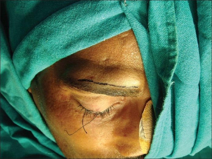
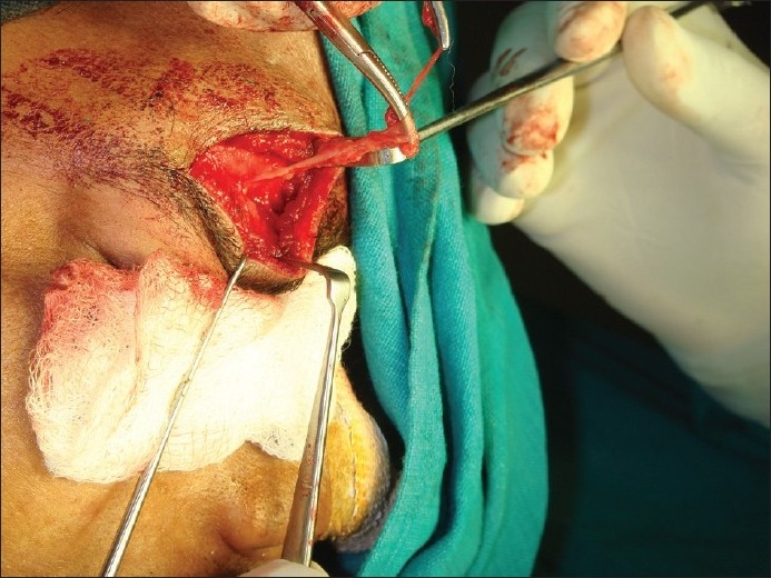
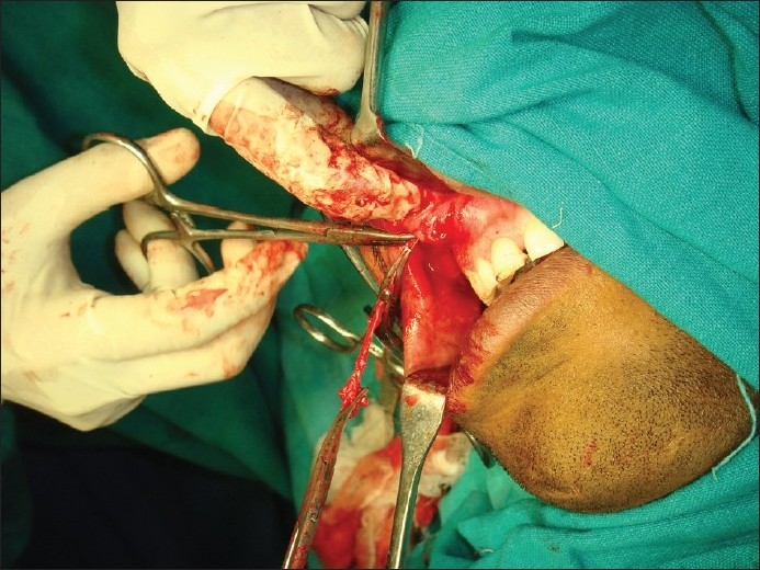
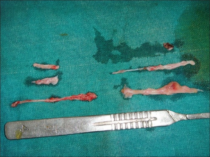
The patient was taken under GA with endotracheal intubation and the supraorbital nerve was exposed via upper eyebrow incision [Figure 1]. The nerve was identified and avulsed by twisting the nerve on the artery forcep [Figure 2]. Double layer closure was done with 3–0 vicryl and 3–0 ethilon. Then, the infraorbital nerve was exposed through intraoral approach by giving upper vestibular incision from 13 to 16 region [Figure 3]. The infraorbital nerve was avulsed in the same way [Figure 4]. The double layer closure is done. Postoperative recovery was uneventful. Patient has been relieved of pain since last 1 year.
Figure 1.

Upper eyebrow incision
Figure 2.

Supraorbital nerve held with artery forceps
Figure 3.

Infraorbital nerve held with artery forceps
Figure 4.

Avulsed nerve
DISCUSSION
The term neuralgia is used to describe unexplained peripheral nerve pain and the head and neck are the most common sites of such neuralgias. TN is by far the most frequently diagnosed form of neuralgia with mean incidence of 4 per 100,000 populations and mean age of 50 years at the time of examination. There is a female predominance ranging from 1:2 to 2:3.[4,5] TN is usually unilateral affecting the maxillary (35%), mandibular (30%), both (20%), ophthalmic and maxillary (10%) and ophthalmic (4%) branches and all branches of the trigeminal nerve (1%).[3]
TN has long been recognized in the medical literature; infact, it was described as early as the first century AD in the writings of Aretaeus. It was later discussed by Johannes Bausch in 1672. Nicholas Andre in 1756 used the term tic doulourex (painful sensation) to describe the disorder. Fothergill provided a vivid description of TN in 1773, hence also called as Fothergill's disease.[6]
Although several hypotheses have been put forward, the cause of TN has not been fully explained in the literature. For most patients, the cause is unknown as was in our case. The most common reported cause of TN is compression of the nerve root. Mechanical compression of the TN can occur as the nerve leaves the pons and traverses the subarchonoid space toward Meckel's cave. Most commonly, the nerve compressed by a major artery, usually the superior cerebellar artery. When pain is felt in second and third divisions, the usual finding is the compression of the rostral and anterior portions of the nerve by superior cerebellar artery, if the pain is felt in ophthalmic division, the usual finding is compression of the nerve by the anterior inferior cerebellar artery. However, in our case MRI showed no compression of the nerve along its course. Other causes can be multiple sclerosis, vascular factors—transient ischemia, autoimmune hypersensitivity, secondary to dental diseases such as maxillary sinusitis, third molar infections, trauma and congenital abnormalities in bony canal. Lesions with trigeminal ganglion and their rootlets such as central brain lesions, pressure from adjacent cranial structures, tumors of the posterior cranial fossa and vascular compression of nerve root fibers have been reported to be the other causes.[6]
TN characterized by paroxysms of severe, lancinating, electric-like bouts of pain restricted to the distribution of the trigeminal nerve. The pain predominantly occurs unilaterally, most commonly on the right side and involves the mandibular and/or maxillary branch or rarely the ophthalmic branch. Pain attacks occur spontaneously and also triggered by a nonpainful sensory stimulus to the skin, intraoral mucosa surrounding the teeth and tongue. Anatomical trigger zones often can be identified. Each attack usually lasts only seconds to minutes, but they may be repeated at short intervals; consequently, individual attacks can overlap each other and may be described as lingering, painful sensation. Attacks can occur at any time and are triggered not only by sensory stimuli to the face, but also by movement of the face. Pain during the night that interrupts sleep is rare.[4,5]
No medical test exists that can be used to diagnose clearly all cases of TN. The diagnosis of TN rests on the clinician's ability to recognize a distinctive series of signs and symptoms that define this disorder. Sweet's criteria have been commonly used worldwide for the diagnosis of TN. The criteria emphasize five major clinical features that in essence, define the diagnosis of TN. They are:
The pain is paroxysmal.
The pain may be provoked by light touch to the face (trigger zone).
The pain is confined to trigeminal distribution.
The pain is unilateral.
The clinical sensory examination is normal.[5]
Diagnostic criteria are defined by the International Association for the Study of Pain (IASP) and by the International Classification of Headache Disorders/International Headache Society (ICHD/IHS) and include:
Paroxysmal episodes lasting from a fraction of a 1 sec. to 2 min., affecting one or more divisions of the trigeminal nerve.
-
The pain has at least one of the following characteristics:
-
a)severe, sudden, superficial, or stabbing; and
-
b)initiated by trigger factors or trigger points.
-
a)
Episodes are similar among patients.
Patients do not have clinically evident neurologic changes.
The most common disorders involved in the differential diagnosis include bursts of headaches, dental pain, giant cell arteritis, glossopharyngeal nerve neuralgia, intracranial tumor, migraine, multiple sclerosis, otitis media, paroxysmal hemicrania, postherpetic neuralgia, sinusitis, SUNCT headache (short-lasting, unilateral, neuralgiform pain with conjunctival injection and tearing), temporomandibular joint syndrome and TN.[3]
Initially, administration of anticonvulsant drugs was the treatment of choice. Now a variety of other effective treatments are available both pharmacologic and surgical. There are several factors that can affect a treatment plan, including the patient's age, life expectancy, associated medical and psychiatric conditions, compliance with medical therapy and tolerability of adverse effects of drugs. Invasive procedures (peripheral neurectomy, radiofrequency thermocoagulation at gasserian ganglion, retrogasserian rhizotomy, medullary tractotomy, midbrain tractotomy and intracranial nerve decompression) are usually reserved for those patients who are unable to obtain relief with medical management or experience unacceptable adverse effects from the use of pharmacologic treatments.[6]
Supraorbital neuralgia is a rare disorder clinically characterized by the following triad:
forehead pain in the area supplied by the supraorbital nerve, without side shift;
tenderness on either the supraorbital notch or traject of the nerve; and
absolute, but transitory relief of symptoms upon supraorbital nerve blockade.
The pain presents with a chronic or intermittent pattern. In addition, there may be signs and symptoms of sensory dysfunction (hypoesthesia, paresthesia) and typical “neuralgic features” (lightning pain and exteroceptive precipitating mechanisms).[7] The hallmark of supraorbital neuralgia is localized pain in or above the eyebrow (sometimes extending into the parietotemporal scalp region). The pain is intermittent; however, longer periods with different severity occur. It can be triggered by pressure on the supraorbital notch.[8] Although etiology and pathogenesis of supraorbital neuralgia are largely unknown, entrapment of the supraorbital nerve at its outlet,[9] trauma to the face,[10] such as when the head hits the windshield or after a punch to the face are thought to be the causative factor and neuralgic pain. The headache might not present for many years until the scar cicatrix tightens enough around the nerve to finally cause entrapment.[11] Supratrochlear nerve in this region can be injured by poor-fitting eyeglasses,[12] presenting as a more midline forehead pain.[10] Entrapments of first division of the trigeminal nerve can cause unilateral or bilateral throbbing headaches, often just before menses or triggered by bright lights that cause squinting. Supraorbital neuralgia can be mistaken for frontal sinusitis.[11] The proposed treatment is an injection of 2 ml of 2% lidocaine, 1:60,000 adrenalin in the supraorbital canal, which relieves and mostly obviates the pain in 80% of the patients over a considerable time of span.[1] Generally, there will be lack of, or only minor benefit from drug treatment, including carbamazepine and indomethacin. There will clearly be tenderness over the supraorbital nerve, especially at its outlet and in some subjects occasionally, a slight local loss of sensation.[13] The persistence of protracted unilateral forehead/ocular pain, tenderness over the nerve and repeated blockade effect strongly suggests the diagnosis[13] and patient's willingness for surgical treatment.
Complications reported are local hemorrhage, transient diplopia, transient anesthesia of supraorbital skin, mostly lasting only for few days.[8]
CONCLUSION
Correct diagnosis of supraorbital neuralgia is critical in choosing therapy. For persistent or recurrent pain, acupuncture, injection of phenol/glycerol or botulism toxin, neurolysis and root section of the trigeminal nerve are methods that have been successfully employed to treat this condition.[12] Consensus on the best treatment for TN, clinical or surgical, does not exist. However, pain relief, recurrence, morbidity and mortality should be considered.[3]
Footnotes
Source of Support: Nil.
Conflict of Interest: None declared.
REFERENCE
- 1.Bastiaensen LA. Supra-orbital neuralgia and its treatment. Neuroophthalmology. 1996;16:225–7. [Google Scholar]
- 2.Evans RW, Pareja J. Supra-orbital neuralgia. Headache. 2009;49:278–81. doi: 10.1111/j.1526-4610.2008.001328.x. [DOI] [PubMed] [Google Scholar]
- 3.Oliveira CM, Baaklini LG, Issy AM, Sakata RK. Bilateral Trigeminal Neuralgia.Case Report. Rev Bras Anestesiol. 2009;59:476–80. doi: 10.1590/s0034-70942009000400010. [DOI] [PubMed] [Google Scholar]
- 4.Bagheri S, Farhidvash F, Perciaccante V. Diagnosis and treatment of patients with trigeminal neuralgia. J Am Dent Assoc. 2004;135:1713–7. doi: 10.14219/jada.archive.2004.0124. [DOI] [PubMed] [Google Scholar]
- 5.Serivani S, Mathews E, Maciewicz R. Trigeminal neuralgia. Oral Surg Oral Med Oral Pathol Oral Radiol Endod. 2005;100:527–38. doi: 10.1016/j.tripleo.2005.06.004. [DOI] [PubMed] [Google Scholar]
- 6.Siddiqui M, Siddiqui S, Ranasinghe J, Furgang F. Pain management: Trigeminal neuralgia. Hosp Physician. 2003;39(1):64–70. [Google Scholar]
- 7.Pareja JA, Caminero AB. Supraorbital neuralgia. Curr Pain Headache Rep. 2006;10:302–5. doi: 10.1007/s11916-006-0036-9. [DOI] [PubMed] [Google Scholar]
- 8.Bastiaensen LA. Supraorbital neuralgia and its treatment. J Neuroophthalmology. 1997;17:221. [Google Scholar]
- 9.Caminerol AB, Pareja JA. Supraorbital Neuralgia: A Clinical Study. Cephalalgia. 2001;21:216–23. doi: 10.1046/j.1468-2982.2001.00190.x. [DOI] [PubMed] [Google Scholar]
- 10.Ducic, Ivica, Larson, Ethan E. Posttraumatic headache: Surgical management of supraorbital neuralgia. Plast Reconstr Surg. 2008;121:1943–8. doi: 10.1097/PRS.0b013e3181707063. [DOI] [PubMed] [Google Scholar]
- 11.Trescot AM. Headache Management in an Interventional Pain Practice. Pain Physician. 2000;3:197–200. [PubMed] [Google Scholar]
- 12.O’brien JC., Jr Swimmer's headache, or supraorbital neuralgia. Proc (Bayl Univ Med Cent) 2004;17:418–9. doi: 10.1080/08998280.2004.11928006. [DOI] [PMC free article] [PubMed] [Google Scholar]
- 13.Sjaastad O, Stolt-Nielsen A, Pareja JA, Fredriksen TA, Vincent M. Supraorbital neuralgia.On the clinical manifestations and a possible therapeutic approach. Headache. 1999;39:204–12. doi: 10.1046/j.1526-4610.1999.3903204.x. [DOI] [PubMed] [Google Scholar]


