Abstract
Normal rhythms originate in the sino-atrial node, a specialized cardiac tissue consisting of only a few thousands of pacemaker cells. Malfunction of pacemaker cells due to diseases or aging leads to rhythm generation disorders (for example, bradycardias and sick-sinus syndrome (SSS)), which often necessitate the implantation of electronic pacemakers. Although effective, electronic devices are associated with such shortcomings as limited battery life, permanent implantation of leads, lead dislodging, the lack of autonomic responses and so on. Here, various gene- and cell-based approaches, with a particular emphasis placed on the use of pluripotent stem cells and the hyperpolarization-activated cyclic nucleotide-gated-encoded pacemaker gene family, that have been pursued in the past decade to reconstruct bio-artificial pacemakers as alternatives will be discussed in relation to the basic biological insights and translational regenerative potential.
Keywords: human embryonic stem cells, pluripotent, bio-artificial, pacemaker, cardiac differentiation, electrophysiology
The heart beats with a regular rhythm to pump blood throughout the body. These mechanical actions require the highly coordinated efforts of different types of cardiomyocytes (CMs) such as atrial, ventricular and pacemaker cells. Chamber-specific CM types differ substantially in their electrical properties, which in turn govern cardiac excitability. Normal heart (sinus) rhythms originate in the sino-atrial node (SAN), a specialized cardiac tissue consisting of only a few thousands of nodal or so-called pacemaker cells.1,2 The SAN spontaneously and rhythmically generates action potentials (APs) by the process of pacemaking. These rhythmic APs propagate through the atria to the atrioventricular node, and after a slow pause, subsequently to the ventricles via a unique conduction system for coordinated chamber contractions and therefore blood pumping. As such, the SAN is responsible for initiating and controlling the heart rate.
CLASSICAL VIEW OF CARDIAC PACEMAKING: SAN IS A COMPLEX STRUCTURE THAT FUNCTIONS WITH A COMPLEX MECHANISM
Structurally, the native SAN is a complex, three-dimensional tissue containing a heterogeneous population of pacemaker cells that display a range of phenotypic properties and anatomic boundary effects.1,3 For instance, there are gradual gradient changes in the AP profile,4,5 ionic current densities and gap-junction expression from central (dominant or the leading pacemaker site) to peripheral (subsidiary) SAN cells.1 These differences and anatomic arrangements (for example, interdigitation) have been thought to ensure that the leading center cells are protected from any over-hyperpolarizing effects from the surrounding mass of atrial CMs and that the depolarization wave front is propagated in the proper directions. In addition to pacemaker cells, the SAN also contains atrial CMs, fibroblasts and adipocytes. The collagen content of the SAN region is relatively high.6 During aging, the SAN undergoes structural remodeling along with an increased collagen content,7 along with other functional changes. For a more thorough review of the structure–function relationships of SAN, please refer to other excellent reviews of the topic.1,3
At the molecular and cellular levels, the process of pacemaking involves the complex interplay of a range of ionic channels and pumps, which in turn give pacemaker AP a unique signature waveform. For instance, the upstroke velocity is much lower than in the ventricle and uniquely display a re-depolarization phase at the end of each AP that results in repetitive firing. Among the key players, the hyperpolarization-activated current (If, f for ‘funny’) or the so-called pacemaker current8 is robustly present in pacemaker CMs but absent in healthy adult ventricular cells. In contrast, the inward-rectifier current (IK1) responsible for stabilizing the resting membrane potential is robustly expressed in ventricular CMs but absent or lowly expressed in the SAN. Additional components such as the l-type (ICa,L) and T-type (ICa,T) Ca currents, the transient outward current (Ito), the rapid (IKr) and slow (IKs) components of the delayed rectifier current (IK), as well as the Na+-Ca2+ exchanger, also significantly contribute to shape the pacemaker AP.
At the multicellular level, cell-to-cell electrical coupling within the SAN is poor because of the low density of connexin-encoded gap junctions. A poor electric connection in the center of the node, together with a progressively improved coupling when approaching the border with atrial muscle, is thought to favor electrical signal propagation from the SAN center while protecting from hyperpolarization by the atrial muscle mass.
ELECTRONIC PACEMAKERS FOR HEART RHYTHM DISORDERS
Malfunction of cardiac pacemaker cells due to diseases or aging leads to a range of rhythm generation disorders (for example, bradycardias and SSS). Traditional treatments require pharmacological intervention and/or implantation of electronic pacemakers. Although effective, such therapy is also associated with significant risks (for example infection, hemorrhage, lung collapse and death) and other disadvantages such as limited battery life, permanent implantation of catheters, lack of autonomic neurohumoral responses and so on. As for pediatric patients with conditions such as symptomatic bradycardias, bradycardia-dependent ventricular arrhythmias and other postoperative arrhythmias that require electronic pacemakers for life-sustaining rhythms, several considerations can further complicate their management.7,9–12 For instance, somatic growth may result in lead tension and thereby increase the risk of lead dislodging and fracturing that are fairly common in active young patients. The placement of an electronic system could also be hindered by congenital heart defects and structural abnormalities.
Structure–function relationships of hyperpolarization-activated cyclic nucleotide-gated (HCN) channels, a key player of pacemaker activity
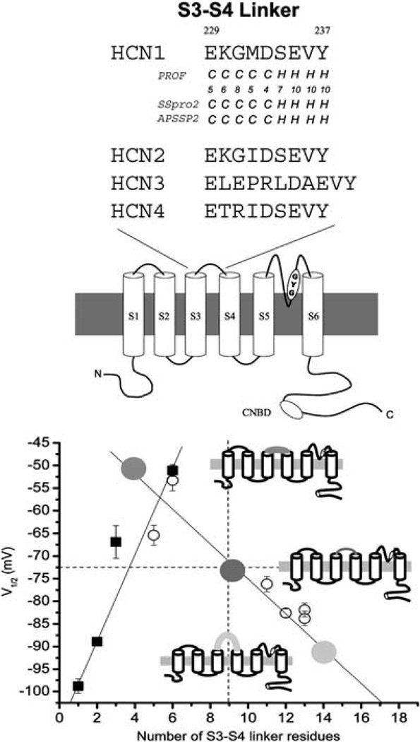
As mentioned above, the diastolic depolarizing current If, encoded by the HCN or the so-called ‘pacemaker channel’ gene family,13–17 is a key player in cardiac pacing. Structurally, HCN channels share the same basic design motif of classical depolarization-activated voltage-gated K+ (Kv) channels whose fundamental building blocks are monomers consisting of six transmembrane segments (S1–6)(Figure 1a). Functionally, two distinct features set HCN channels apart: (1) they nonselectively permeate Na+ and K+ (with a ratio of 1:4 vs 1:100 of K+ channels), despite the fact that their pore also contains the signature sequence GYG and (2) HCN channels are activated by hyperpolarization rather than depolarization, although their S4 voltage sensor also carries a ribbon of regularly spaced positively-charged amino-acid residues similar to their depolarization-activated counterparts. HCN channels also contain in their C-termini a cyclic nucleotide-binding domain homologous to those of cyclic nucleotide-gated channels.18,19 Upon cAMP-binding, cyclic nucleotide-binding domain undergoes conformational changes to accelerate HCN current kinetics and positively shift steady-state activation, thereby promoting channel activity and subsequently the rhythmic firing rate that If modulates.20
Figure 1.
(a) Schematic representation of a monomeric subunit of HCN channels. The approximate locations of the TVGYG motif and the cyclic nucleotide-binding domain are highlighted. Sequence comparison (middle and right) of the S5–S6, the P-loops and the S4 of various HCN and K+ channels. Adapted from Siu et al.8 (b) Summary of the effects of the S3–S4 linker length on HCN1 steady-state activation (V1/2), a measure of the energetic stability for opening and closing. A strong correlation was observed between the V1/2 of the channels and their linker length. Schematics representing three HCN1 channels with shorter, wild-type (WT) and longer S3–S4 linkers are given for illustration. Adapted from Tsang et al.42
To date, four isoforms, namely HCN1–4, have been identified. These isoforms exhibit different patterns of gene expression and tissue distribution,14,16,17,21,22 and co-assemble to form hetero-tetrameric complexes (except between HCN2 and HCN3) that underlie the native If.23–26 HCN1 is the most abundant isoform in the brain and is substantially expressed in the SAN (but not in the ventricles or atria).27,28 Although HCN3 is present in the central nervous system but absent in the heart, HCN2 and HCN4 are found in both. When heterologously expressed, HCN1–4 channels have distinct cAMP sensitivities and gating properties. Of the two predominant isoforms found in the SAN, time-dependent HCN1 currents are 40 times faster than those of HCN416.29–32 If modulates heart rate in response to neural inputs by regulating the rates of cellular depolarization to AP threshold (that is, the slope of phase 4 depolarization), thereby determining the oscillation frequency. Mutations in the human HCN gene lead to familial sinus node dysfunction.33,34 Although If in the human atria has been suggested to contribute to atrial ectopy,35 upregulation of HCN2 and HCN4 in the ventricles in disease states, such as heart failure, hypertrophy and hypertension, may predispose certain arrhythmias.36,37
Because the four HCN isoforms co-assemble to form heteromultimeric channels, the molecular identity of endogenous If is complex. Also, given the poorly defined subunit interactions of HCN, it is therefore virtually impossible to reproduce the permutations of endogenous If via genetic expression of a single HCN isoform. To bioengineer a single recombinant HCN construct that mimics the native channel, our laboratory first undertook a series of structure–function studies of HCN channels24,38–47 to better understand their basic biology. In some of the initial studies, we discovered that HCN gating (that is, opening and closing) is highly dependent on the length of the linker between the third and fourth transmembrane segments (that is, S3–S4 linker, see Figure 1b)44 and several charged amino acids therein,42,43,45 further highlighting several evolutionary similarities and differences between HCN and Kv channels.10,48 Long and short linkers generally shift steady-state activation in the negative and positive directions, making them energetically more difficult and easier to open, respectively. As such, not only does engineering the S3–S4 linker length provide a flexible approach to mimic the native heteromultimeric If but also a means to customize HCN activity for ‘programming’ bio-artificial pacing,49 as further elaborated in the next section.
Engineering biological alternatives to electronic pacemakers
Gene-based bio-artificial SAN (bio-SAN)
Unlike rhythmically firing pacemaker cells, adult atrial and ventricular muscle cells are normally electrically silent unless they get stimulated by signals transmitted from neighboring cells that originate from the SAN. This quiescent nature of cardiac muscle CMs is due to the absence of If and the intense expression of IK1 (a.k.a. the inward-rectifier K+ current), encoded by the Kir2 gene family, which stabilizes a negative resting membrane potential (RMPB~ −80 mV). Given the limitations of electronic pacemakers, several gene- and cell-based approaches have been explored to confer upon normally quiescent cardiac muscle cells the ability to intrinsically fire APs similar to genuine nodal pacemaker cells as potential biological alternatives or supplements to electronic devices. In addition to the translational potential, these studies also shed mechanistic insights into cardiac automaticity. Miake et al.50 demonstrated that genetic suppression of IK1 in normally silent ventricular myocytes by >80% can cause spontaneous firing activity in a binary ‘on-and-off’ fashion. The induced frequency is only a third of the normal physiological range and as such not suitable for acting as a reliable biological pacemaker. Alternatively, we49,51–54 and others55 have chosen to employ a genuine pacemaker gene product, the HCN channels, for the induction of automaticity. As mentioned, native If is heteromultimeric and difficult to reproduce with a single gene product. Indeed, forced expression of wild-type HCN149 or HCN256 channels alone is insufficient to induce pacemaking activity in adult ventricular CMs. As such, efforts of converting a recombinant Kv1.4 channel into a hyperpolarization-activated channel by specific amino-acid substitutions have been pursued to give rise to spontaneous APs.57
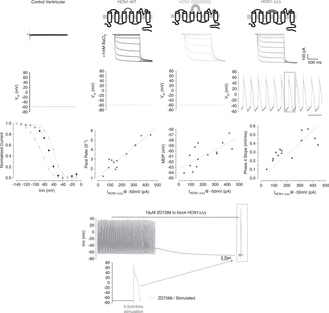
By taking an in silico or mathematical approach, our laboratory first developed a computational model for converting normally quiescent adult ventricular or atrial muscle CMs into AP-firing pacemaker.51 Using such a conceptual strategy as our guide, we went on to experimentally validate by gene-transferring various engineered HCN (S3–S4) constructs to adult guinea pig left ventricle (LV) CMs.49 Electrophsyiological recordings at the single-cell level show that control ventricular CMs are always silent (Figure 2) but excitable to generate typical ventricular APs upon stimulation. Interestingly, adenovirus (Ad)-mediated forced expression of HCN1-ΔΔΔ (whose S3–S4 linker has been shortened to favor opening)42–44 exclusively induces physiological pacemaking activities that resemble those of native SAN cells. Interestingly, the induced pacing rate, maximum diastolic potential (1D) and phase 4 slope are all positively correlated to HCN1-ΔΔΔ current amplitude. In contrast, neither wild-type nor QQ293QQQ (with a lengthened linker) HCN channels that activate more negatively exert any effect on the ventricular phenotype.49 Inhibition by the HCN-specific blocker ZD7288 ceases bio-artificial pacing. Interestingly, the silenced cells can generate normal ventricular APs upon stimulations. These findings suggest that HCN-converted bio-SAN cells display a dual identity that is not seen in native cells; they retain their ventricular identity as a muscle cell when not functioning as a bio-artificial pacemaker.
Figure 2.
(a) Top: representative tracings of HCN-encoded pacemaker current from Ad-CMV-GFP-, Ad-CMV-WT-HCN1-, Ad-CMV-HCN1-ΔΔΔ- and Ad-CMV-HCN1-QQ253QQ-transduced ventricular CMs. Bottom: automaticity could be recorded only from the Ad-CMV-HCN1-ΔΔΔ-transduced group. Steady-state activation curves indicate that HCN1-ΔΔΔ channels open most easily. Correlation of HCN current to induced pacing rate, MDP and phase 4 slope of HCN1-ΔΔΔ-mediated biopacemaking. (b) Effects of the If blocker ZD7288 on WT or engineered HCN1-transduced LV CMs. Spontaneously firing APs recorded from an HCN1-ΔΔΔ-transduced LV CM are abolished after addition of ZD7288. Upon silencing, an electrical stimulus elicits a normal ventricular AP. Adapted from Xue et al.49
To date, we have similarly successfully employed HCN1-ΔΔΔ to convert left atrial and right atrial (RA), as well as ventricular (V) CMs, and various species (guinea pigs, pigs, neonatal rat, mouse and human), indicating the versatility of this approach. Interestingly, the same HCN1-ΔΔΔ always induces AP-firing with rates similar to the heart rates of the corresponding species, implicating that although If is key to rhythm generation (by primarily modulating the phase 4 slope), other ionic components are responsible for modulating APD or cycle length. These results are consistent with our finding that engineering the ratio of two ionic components, If and IK1, alone is sufficient to confer the pacemaking phenotype on even inexcitable cell types (see later).
Engineering If and IK1 ratio is an effective strategy for inducing and fine-tuning bio-artificial pacing—cardiac pacemaking has a simple mechanistic basis
Our recent computational and experimental results indicate that induction of automaticity in RA CMs occurs only within a finite range of If/IK1 ratio53 (see58 for editorial comment). In other words, engineering the If/IK1 ratio is a more effective strategy for fine-tuning biopacing than If or IK1 alone. We have determined the optical range of If for inducing automaticity in RA CMs; excessive If can silence bio-artificial pacemakers,53 and the strategies of IK1 suppression and If overexpression are therefore not necessarily synergistic.54 Unique interactions exist between If and IK1, and their co-expression promotes bioengineered cardiac automaticity. Intriguingly, engineering the If/IK1 ratio even suffices to confer on even inexcitable cells such as undifferentiated human embryonic stem cell (hESC) pacemaking activity (Fu JD and Li RA, unpublished data), suggesting that pacemaking or the process of rhythm generation has a simple mechanistic basis and that all the other ionic components present are for modulation and species-specific signatures.
A SIMPLE injection to construct an in vivo bio-SAN suffices to mimic the complex native SAN
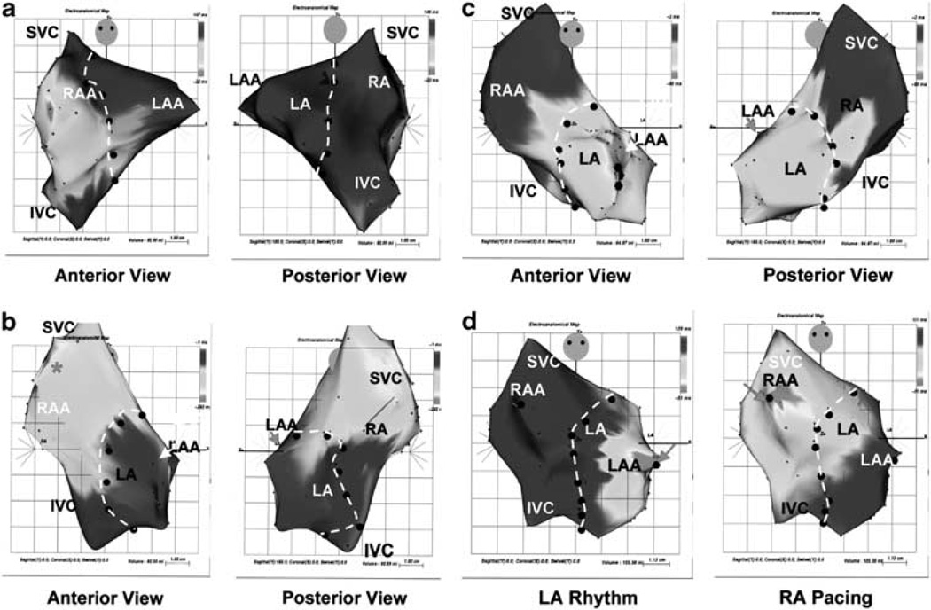
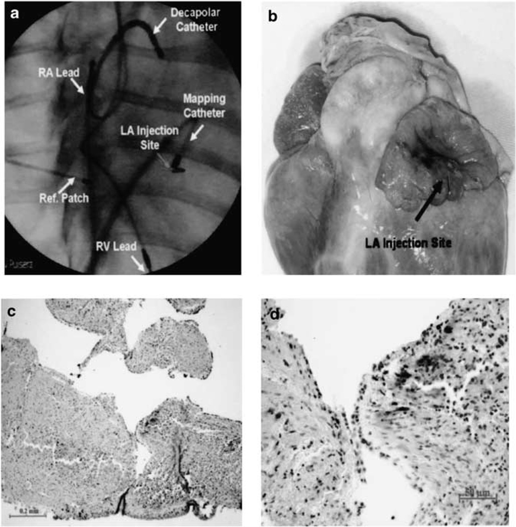
Although HCN1- expression can confer on adult atrial and ventricular CMs, the ability to spontaneously fire APs at physiological rates at the single-cell level,49 the massive surrounding atrial or ventricular tissue can serve as an electrotonic sink that prevents signal propagation away from bio-SAN due to cell-to-cell coupling. In some unpublished experiments, heart block was indeed observed (RA Li and FG Akar, unpublished data). To test this, we developed a large animal (swine) model of SSS such that clinically relevant procedures and assessments can be applied.52 Importantly, swine heart has anatomical and physiological properties that are more comparable to those of human (for example, heart rates of both species are ~80 vs 4–500 bpm of rodents). To create an SSS model, we radiofrequency-ablated the native SAN and implanted a dual-chamber electronic pacemaker for life-sustaining rhythms. Interestingly, focal injection of Ad-CMV-GFP-IRES-HCN1-ΔΔΔ in the left atrium of SSS swines reproducibly induces a stable, catecholamine-responsive in vivo bio-SAN that exhibits a physiological heart rate (Figures 3 and 4), substantially reducing device-supported pacing (from 80 to 15%) in 2 weeks. In this experiment, left atrial (rather than RA where the native SAN resides) was strategically chosen to create a reverse pattern. In other words, we have successfully translocated the SAN from the right to the left. Furthermore, our construction of a bio-SAN requires the focal transduction of only a relatively small number of cells within the heart, consistent with the small size of the native SAN, but suggest that a complex three-dimensional structure is not needed for bio-SAN to functionally pace the entire heart.
Figure 3.
Left: anterior (left) and posterior (right) views of electroanatomical mapping of (a) control and (b) a SSS swine heart that has been implanted an electronic pacemaker whose wired lead generates heart beat in the RA. (c) SSS animal after Ad-CGI-HCN1-ΔΔΔ injection demonstrate the atrial activation at the injection site in the left atrial (LA). (d) Anterior view after Ad-CGI-HCN1-ΔΔΔ injection during spontaneous LA rhythm (left) and device-supported RA pacing (right). Note that the earliest endocardial activation shifted from the left injection site to the right pacing site during device-supported RA overdrive pacing. Adapted from Tse et al.52
Figure 4.
(a) Fluoroscopic image showing pacing leads at the RA and RV apex, the decapolar catherter at the roof of RA against the intra-atrial septum and the mapping catheter in the LA during electroanatomic mapping. The site of injection was marked by a surgical clip (red arrow). Reffered patch indicates reference patch for anatomical mapping. (b) Gross pathology showing the injection site at the LA apendage (arrow). (c and d) Sectioned heart demonstrating the expression of GFP with GFP staining (brown) ath low (left) and high (right) magnifications.
Cell-based bio-artificial pacemakers
As an alternative to the gene-based approaches for creating bio-SAN, several cell-based approaches have also been pursued. Human mesenchymal stem cells pretransfected with HCN2 channels have been employed as a delivery vehicle for introducing If into neighboring CMs via gap-junction-mediated coupling.59,60 However, questions related to the stability of such coupling between non-cardiac cells and the myocardium, time-dependent loss of the HCN activity introduced by transfection, HCN-transduced human mesenchymal stem cells are not electrically active per se, the heterogeneous nature of human mesenchymal stem cells and the possibility of differentiating into cardiac or even non-cardiac cells with electrical properties, different from those of pre-transplanted cells, have been raised61 and discussed.62,63 Along the same line, ventricular myocytes have been converted into pacemaker-like heterokaryons via chemically-induced fusion with fibroblasts transduced to express HCN1 channels.64 When injected into the guinea pig LV, electrocardiography confirmed ectopic rhythms. Previous studies have indicated that similar heterokaryons can remain stable for several months.65–67 In yet another proof-of-principle experiments, autologous SAN pacemaker cells were harvested, then transplanted back into the same animal in a dog model of sinus disorders for rhythm regeneration.68
Human ESCs, isolated from the inner cell mass of blastocyst, can self-renew while maintaining their pluripotency to differentiate into all cell types,69 including CMs.70,71 Therefore, in principle, hESCs can serve as an unlimited ex vivo source of CMs for cell-based heart therapies.72,73 We have differentiated hESCs into electrically active CMs, followed by transplantation into the guinea pig LVs to generate an in vivo epicardial pacing origin or cell-based pacemaker as confirmed by high-resolution optimal mapping.74 Independently, Gepstein and colleagues75 similarly transplanted spontaneously beating hESC-derived clusters into swines to induce ectopic pacing. Both the laboratories considered the entire transplant of beating clusters as single electrically active entities. When dissociated into single cells, we have now learned from patch-clamp recordings that the clusters are highly heterogenous containing a mixture of ventricular-, atrial- as well as pacemaker-like derivatives.76 Furthermore, it is uncertain as to how long and to what extent electrically active hESC-derived CMs will continue to retain their ‘nodal-like’ phenotype in vivo. In addition, the graft size, transplantation site, purity of nodal cells in the graft, time course for maturation and so on are other contributing factors that also need to be considered.
FUTURE DIRECTIONS AND LIMITATIONS
Taken collectively, a number of options are now available with proof-of-concept experiments performed. Although much has been learned in the past few years, these previous studies were performed by different groups using somewhat different strategies, protocols, cell types and animal models, making detailed comparison for fine tuning difficult. For instance, cellular AP recordings were mostly done with ventricular25,49,50,56,77,78 rather atrial CMs,52,53 although the latter will be the likely therapeutic recipient; similarly, some in vivo gene transfer or transplantation experiments were done in the LV50,52,57,64,74 or left atrial rather than the RA where the native SAN resides. Although surface electrocardiography hints at ectopic ventricular beats in some studies, but no further mapping was done to confirm the pacing origin.50,57,64 As such, detailed conduction properties from bio-SAN and susceptibility to reentrant arrhythmias after implantation are not known. In some studies, HCN constructs were confirmed in HEK cells79 (or cultured neonatal ventricular CMs80) followed by in vivo transduction in the left atrium, but the recipient atrial cells were never isolated and recorded to show spontaneous APs.81 Furthermore, almost all in vivo studies reported data not more than 2 weeks52,60,64,74,75,80,81 (except a recent report on human mesenchymal stem cell transplantation with data up to 6 weeks59). Clearly, the field can now advance more rapidly by taking a unified experimental approach; significant proof-of-concept data also suggest that experiments for testing the long-term functional efficacy and safety of bio-SAN are now appropriate. Knowledge of bradycardic drug-binding site and the cAMP-binding domain may also be applied to engineer the responses of bio-SAN to biological and physiological stimuli. It is imperative to know whether bio-SAN displays over time the crucial features that a surrogate pacemaker is expected to function. For instance, are there time-dependent changes? If so, are they only secondary to altered responses of bio-SAN to neurohumoral inputs? Are there changes in the way the recipient heart receives and propagates bio-SAN signals? It is also unclear whether the introduction of a bio-SAN per se confers any short- and/or long-term arrhythmogenic risks. By identifying if how, where, when and why bio-SAN fails under physiological as well as stressful conditions (for example, native SAN automaticity is known to be susceptible and vulnerable to metabolic inhibition such as hypoxia82), we will be able to develop the next-generation prototypes with improved functional efficacy and durability.
Adeno-associated virus-mediated somatic gene transfer
The optimal vector for developing gene (HCN)-based bio-SAN should enable permanent transgene expression without undesirable side effects such as immunological responses. In our previous studies,49,52 adenoviral vectors were employed for initial proof-of-concept experiments because of their high titers, ease of production, and high in vivo transduction efficiency at the injection site. However, Ad-mediated transgene expression is only transient due to the lack of integration. Previous in vivo adenoviral gene-transfer studies83–85 have reported an expression profile that peaks at ~1 week, plateaus, then declines for a period of ~3–5 weeks. By week 10, no transgene expression could be detected. Furthermore, Ad vectors evoke intense immune and inflammatory responses. As such, Ad is not suitable for investigating the long-term efficacy and safety of HCN-based bio-SAN. In contrast, recombinant adeno-associated viral (rAAV) vectors, derived from replication-deficient, nonpathogenic parvovirus with a single-stranded DNA genome, appears to be uniquely suitable. First, previous studies have demonstrated that the heart can be efficiently transduced with rAAV.86–91 Second, rAAV virions are generated without wild-type Ad coinfection, eliminating adenoviral immune response. Third, rAAV can be maintained in the human host as epichromosomal material or by integration into the host genome, establishing long-term gene expression. Fourth, high primary titers can be readily generated for high-efficiency gene delivery (107–1013 viral particles or vp ml−1), unlike other delivery vehicles such as lentiviruses that are also capable of mediating persistent genetic modification. This is an important consideration because our experience indicates that only a relatively limited volume (~500 µl) can be focally injected into the atrium (unlike the thick LV wall). Fifth, rAAV has an infectivity of a broad range of dividing and nondividing mammalian cells, including striated skeletal and cardiac muscle in immunocompetent hosts92,93 for long-term transgene expression. Sixth, rAAV vectors are stable under various physical and environmental conditions. To date, at least 12 serotypes (AAV1-12) have been identified, each with different tissue tropisms. Indeed, AAV presents an option for studying long-term in vivo genetic modifications and has been used in clinical trials for heart failure,94,95 cystic fibrosis,96,97 hemophilia98,99 and Parkinson’s disease.100 The use of rAAV and various viral and nonviral vectors for gene therapies of cardiovascular diseases, as well as their pros and cons, have been recently extensively reviewed by Hajjar and colleagues.90
CONCLUSION
Numerous proof-of-concept data are now available to convincingly demonstrate that bio-SAN-displayed functions similar or superior to electronic pacemaker can be constructed. The challenge now is to test their long-term efficacy and safety, and whether bio-SAN displays over time the crucial features that a surrogate pacemaker is expected to function. It is also imperative to identify where and when (that is, under what conditions) bio-SAN fails so as to develop the next-generation improved prototypes. It remains to be seen whether and when bio-SAN will ever become a clinical routine as have electronic devices. This process depends on numerous scientific as well as nonscientific factors. However, it is certain that invaluable knowledge and experience will be gained during the quest. Having entered the era of regenerative medicine, there has been an exponential increase of viable options that were previously not available. Although many are still in the experimental stage or clinical trials, it is reasonable to expect that better treatments or even cures for the most debilitating and currently incurable diseases will become available in the foreseeable future.
ACKNOWLEDGEMENTS
This work was supported by grants from the NIH—R01 HL72857, the CC Wong Foundation Stem Cell Fund and the Research Grant Council (T13-706/11 and 103544).
Footnotes
CONFLICT OF INTEREST
The author declares no conflict of interest.
References
- 1.Boyett MR, Honjo H, Kodama I. The sinoatrial node, a heterogeneous pacemaker structure. Cardiovasc Res. 2000;47:658–687. doi: 10.1016/s0008-6363(00)00135-8. [DOI] [PubMed] [Google Scholar]
- 2.Dobrzynski H, Boyett MR, Anderson RH. New insights into pacemaker activity: promoting understanding of sick sinus syndrome. Circulation. 2007;115:1921–1932. doi: 10.1161/CIRCULATIONAHA.106.616011. [DOI] [PubMed] [Google Scholar]
- 3.Boyett MR, Dobrzynski H, Lancaster MK, Jones SA, Honjo H, Kodama I. Sophisticated architecture is required for the sinoatrial node to perform its normal pacemaker function. J Cardiovasc Electrophysiol. 2003;14:104–106. doi: 10.1046/j.1540-8167.2003.02307.x. [DOI] [PubMed] [Google Scholar]
- 4.Boyett MR, Honjo H, Yamamoto M, Nikmaram MR, Niwa R, Kodama I. Downward gradient in action potential duration along conduction path in and around the sinoatrial node. Am J Physiol. 1999;276:H686–H698. doi: 10.1152/ajpheart.1999.276.2.H686. [DOI] [PubMed] [Google Scholar]
- 5.Boyett MR, Honjo H, Yamamoto M, Nikmaram MR, Niwa R, Kodama I. Regional differences in effects of 4-aminopyridine within the sinoatrial node. Am J Physiol. 1998;275:H1158–H1168. doi: 10.1152/ajpheart.1998.275.4.H1158. [DOI] [PubMed] [Google Scholar]
- 6.Opthof T, de Jonge B, Jongsma HJ, Bouman LN. Functional morphology of the mammalian sinuatrial node. Eur Heart J. 1987;8:1249–1259. doi: 10.1093/oxfordjournals.eurheartj.a062200. [DOI] [PubMed] [Google Scholar]
- 7.Berul CI, Cecchin F. Indications and techniques of pediatric cardiac pacing. Expert Rev Cardiovasc Ther. 2003;1:165–176. doi: 10.1586/14779072.1.2.165. [DOI] [PubMed] [Google Scholar]
- 8.Siu CW, Lieu DK, Li RA. HCN-encoded pacemaker channels: from physiology and biophysics to bioengineering. J Membr Biol. 2006;214:115–122. doi: 10.1007/s00232-006-0881-9. [DOI] [PubMed] [Google Scholar]
- 9.Dubin AM, Berul CI. Electrophysiological interventions for treatment of congestive heart failure in pediatrics and congenital heart disease. Expert Rev Cardiovasc Ther. 2007;5:111–118. doi: 10.1586/14779072.5.1.111. [DOI] [PubMed] [Google Scholar]
- 10.Silka MJ, Bar-Cohen Y. Pacemakers and implantable cardioverter-defibrillators in pediatric patients. Heart Rhythm. 2006;3:1360–1366. doi: 10.1016/j.hrthm.2006.02.009. [DOI] [PubMed] [Google Scholar]
- 11.Walsh EP, Cecchin F. Recent advances in pacemaker and implantable defibrillator therapy for young patients. Curr Opin Cardiol. 2004;19:91–96. doi: 10.1097/00001573-200403000-00004. [DOI] [PubMed] [Google Scholar]
- 12.SlizJr NB, Johns JA. Cardiac pacing in infants and children. Cardiol Rev. 2000;8:223–239. doi: 10.1097/00045415-200008040-00008. [DOI] [PubMed] [Google Scholar]
- 13.Gauss R, Seifert R, Kaupp UB. Molecular identification of a hyperpolarization-activated channel in sea urchin sperm. Nature. 1998;393:583–587. doi: 10.1038/31248. [DOI] [PubMed] [Google Scholar]
- 14.Ludwig A, Zong X, Jeglitsch M, Hofmann F, Biel M. A family of hyperpolarization-activated mammalian cation channels. Nature. 1998;393:587–591. doi: 10.1038/31255. [DOI] [PubMed] [Google Scholar]
- 15.Santoro B, Grant SG, Bartsch D, Kandel ER. Interactive cloning with the SH3 domain of N-src identifies a new brain specific ion channel protein, with homology to eag and cyclic nucleotide-gated channels. Proc Natl Acad Sci USA. 1997;94:14815–14820. doi: 10.1073/pnas.94.26.14815. [DOI] [PMC free article] [PubMed] [Google Scholar]
- 16.Santoro B, Liu DT, Yao H, Bartsch D, Kandel ER, Siegelbaum SA. Identification of a gene encoding a hyperpolarization-activated pacemaker channel of brain. Cell. 1998;93:717–729. doi: 10.1016/s0092-8674(00)81434-8. [DOI] [PubMed] [Google Scholar]
- 17.Santoro B, Tibbs GR. The HCN gene family: molecular basis of the hyperpolarization-activated pacemaker channels. Ann NY Acad Sci. 1999;868:741–764. doi: 10.1111/j.1749-6632.1999.tb11353.x. [DOI] [PubMed] [Google Scholar]
- 18.Biel M, Zong X, Ludwig A, Sautter A, Hofmann F. Structure and function of cyclic nucleotide-gated channels. Rev Physiol Biochem Pharmacol. 1999;135:151–171. doi: 10.1007/BFb0033672. [DOI] [PubMed] [Google Scholar]
- 19.Zagotta WN, Siegelbaum SA. Structure and function of cyclic nucleotide-gated channels. Annu Rev Neurosci. 1996;19:235–263. doi: 10.1146/annurev.ne.19.030196.001315. [DOI] [PubMed] [Google Scholar]
- 20.Wainger BJ, DeGennaro M, Santoro B, Siegelbaum SA, Tibbs GR. Molecular mechanism of cAMP modulation of HCN pacemaker channels. Nature. 2001;411:805–810. doi: 10.1038/35081088. [DOI] [PubMed] [Google Scholar]
- 21.Moosmang S, Stieber J, Zong X, Biel M, Hofmann F, Ludwig A. Cellular expression and functional characterization of four hyperpolarization-activated pacemaker channels in cardiac and neuronal tissues. Eur J Biochem. 2001;268:1646–1652. doi: 10.1046/j.1432-1327.2001.02036.x. [DOI] [PubMed] [Google Scholar]
- 22.Santoro B, Chen S, Luthi A, Pavlidis P, Shumyatsky GP, Tibbs GR. Molecular and functional heterogeneity of hyperpolarization-activated pacemaker channels in the mouse CNS. J Neurosci. 2000;20:5264–5275. doi: 10.1523/JNEUROSCI.20-14-05264.2000. [DOI] [PMC free article] [PubMed] [Google Scholar]
- 23.Ulens C, Tytgat J. Functional heteromerization of HCN1 and HCN2 pacemaker channels. J Biol Chem. 2001;276:6069–6072. doi: 10.1074/jbc.C000738200. [DOI] [PubMed] [Google Scholar]
- 24.Xue T, Marban E, Li RA. Dominant-negative suppression of HCN1- and HCN2-encoded pacemaker currents by an engineered HCN1 construct: insights into structure-function relationships and multimerization. Circ Res. 2002;90:1267–1273. doi: 10.1161/01.res.0000024390.97889.c6. [DOI] [PubMed] [Google Scholar]
- 25.Er F, Larbig R, Ludwig A, Biel M, Hofmann F, Beuckelmann DJ. Dominant-negative suppression of HCN channels markedly reduces the native pacemaker current I(f) and undermines spontaneous beating of neonatal cardiomyocytes. Circulation. 2003;107:485–489. doi: 10.1161/01.cir.0000045672.32920.cb. [DOI] [PubMed] [Google Scholar]
- 26.Chen J, Mitcheson JS, Tristani-Firouzi M, Lin M, Sanguinetti MC. The S4–S5 linker couples voltage sensing and activation of pacemaker channels. Proc Natl Acad Sci USA. 2001;98:11277–11282. doi: 10.1073/pnas.201250598. [DOI] [PMC free article] [PubMed] [Google Scholar]
- 27.Shi W, Wymore R, Yu H, Wu J, Wymore RT, Pan Z. Distribution and prevalence of hyperpolarization-activated cation channel (HCN) mRNA expression in cardiac tissues. Circ Res. 1999;85:e1–e6. doi: 10.1161/01.res.85.1.e1. [DOI] [PubMed] [Google Scholar]
- 28.Moroni A, Gorza L, Beltrame M, Gravante B, Vaccari T, Bianchi ME. Hyperpolarization-activated cyclic nucleotide-gated channel 1 is a molecular determinant of the cardiac pacemaker current I(f) J Biol Chem. 2001;276:29233–29241. doi: 10.1074/jbc.M100830200. [DOI] [PubMed] [Google Scholar]
- 29.Altomare C, Bucchi A, Camatini E, Baruscotti M, Viscomi C, Moroni A. Integrated allosteric model of voltage gating of HCN channels. J Gen Physiol. 2001;117:519–532. doi: 10.1085/jgp.117.6.519. [DOI] [PMC free article] [PubMed] [Google Scholar]
- 30.Ishii TM, Takano M, Ohmori H. Determinants of activation kinetics in mammalian hyperpolarization-activated cation channels. J Physiol. 2001;537(Part 1):93–100. doi: 10.1111/j.1469-7793.2001.0093k.x. [DOI] [PMC free article] [PubMed] [Google Scholar]
- 31.Ludwig A, Zong X, Hofmann F, Biel M. Structure and function of cardiac pacemaker channels. Cell Physiol Biochem. 1999;9:179–186. doi: 10.1159/000016315. [DOI] [PubMed] [Google Scholar]
- 32.Seifert R, Scholten A, Gauss R, Mincheva A, Lichter P, Kaupp UB. Molecular characterization of a slowly gating human hyperpolarization-activated channel predominantly expressed in thalamus, heart, and testis. Proc Natl Acad Sci USA. 1999;96:9391–9396. doi: 10.1073/pnas.96.16.9391. [DOI] [PMC free article] [PubMed] [Google Scholar]
- 33.Schulze-Bahr E, Neu A, Friederich P, Kaupp UB, Breithardt G, Pongs O. Pacemaker channel dysfunction in a patient with sinus node disease. J Clin Invest. 2003;111:1537–1545. doi: 10.1172/JCI16387. [DOI] [PMC free article] [PubMed] [Google Scholar]
- 34.Milanesi R, Baruscotti M, Gnecchi-Ruscone T, DiFrancesco D. Familial sinus bradycardia associated with a mutation in the cardiac pacemaker channel. N Engl J Med. 2006;354:151–157. doi: 10.1056/NEJMoa052475. [DOI] [PubMed] [Google Scholar]
- 35.Zorn-Pauly K, Schaffer P, Pelzmann B, Lang P, Machler H, Rigler B. If in left human atrium: a potential contributor to atrial ectopy. Cardiovasc Res. 2004;64:250–259. doi: 10.1016/j.cardiores.2004.07.001. [DOI] [PubMed] [Google Scholar]
- 36.Cerbai E, Pino R, Porciatti F, Sani G, Toscano M, Maccherini M. Characterization of the hyperpolarization-activated current, I(f), in ventricular myocytes from human failing heart. Circulation. 1997;95:568–571. doi: 10.1161/01.cir.95.3.568. [DOI] [PubMed] [Google Scholar]
- 37.Cerbai E, Sartiani L, DePaoli P, Pino R, Maccherini M, Bizzarri F. The properties of the pacemaker current I(F)in human ventricular myocytes are modulated by cardiac disease. J Mol Cell Cardiol. 2001;33:441–448. doi: 10.1006/jmcc.2000.1316. [DOI] [PubMed] [Google Scholar]
- 38.Siu CW, Azene EM, Au KW, Lau CP, Tse HF, Li RA. State-dependent accessibility of the P-S6 linker of pacemaker (HCN) channels supports a dynamic pore-to-gate coupling model. J Membr Biol. 2009;230:35–47. doi: 10.1007/s00232-009-9184-2. [DOI] [PMC free article] [PubMed] [Google Scholar]
- 39.Chan YC, Wang K, Au KW, Lau CP, Tse HF, Li RA. Probing the bradycardic drug binding receptor of HCN-encoded pacemaker channels. Pflugers Arch. 2009;459:25–38. doi: 10.1007/s00424-009-0719-2. [DOI] [PMC free article] [PubMed] [Google Scholar]
- 40.Au KW, Siu CW, Lau CP, Tse HF, Li RA. Structural and functional determinants in the S5-P region of HCN-encoded pacemaker channels revealed by cysteine-scanning substitutions. Am J Physiol Cell Physiol. 2008;294:C136–C144. doi: 10.1152/ajpcell.00340.2007. [DOI] [PubMed] [Google Scholar]
- 41.Azene EM, Sang D, Tsang SY, Li RA. Pore-to-gate coupling of HCN channels revealed by a pore variant that contributes to gating but not permeation. Biochem Biophys Res Commun. 2005;327:1131–1142. doi: 10.1016/j.bbrc.2004.12.127. [DOI] [PubMed] [Google Scholar]
- 42.Tsang SY, Lesso H, Li RA. Dissecting the structural and functional roles of the S3–S4 linker of pacemaker (hyperpolarization-activated cyclic nucleotide-modulated) channels by systematic length alterations. J Biol Chem. 2004;279:43752–43759. doi: 10.1074/jbc.M408747200. [DOI] [PubMed] [Google Scholar]
- 43.Tsang SY, Lesso H, Li RA. Critical intra-linker interactions of HCN1-encoded pacemaker channels revealed by interchange of S3–S4 determinants. Biochem Biophys Res Commun. 2004;322:652–658. doi: 10.1016/j.bbrc.2004.07.167. [DOI] [PubMed] [Google Scholar]
- 44.Lesso H, Li RA. Helical secondary structure of the external S3–S4 linker of pacemaker (HCN) channels revealed by site-dependent perturbations of activation phenotype. J Biol Chem. 2003;278:22290–22297. doi: 10.1074/jbc.M302466200. [DOI] [PubMed] [Google Scholar]
- 45.Henrikson CA, Xue T, Dong P, Sang D, Marban E, Li RA. Identification of a surface charged residue in the S3–S4 linker of the pacemaker (HCN) channel that influences activation gating. J Biol Chem. 2003;278:13647–13654. doi: 10.1074/jbc.M211025200. [DOI] [PubMed] [Google Scholar]
- 46.Azene EM, Xue T, Li RA. Molecular basis of the effect of potassium on heterologously expressed pacemaker (HCN) channels. J Physiol. 2003;547(Part 2):349–356. doi: 10.1113/jphysiol.2003.039768. [DOI] [PMC free article] [PubMed] [Google Scholar]
- 47.Xue T, Li RA. An external determinant in the S5-P linker of the pacemaker (HCN) channel identified by sulfhydryl modification. J Biol Chem. 2002;277:46233–46242. doi: 10.1074/jbc.M204915200. [DOI] [PubMed] [Google Scholar]
- 48.Prole DL, Yellen G. Reversal of HCN channel voltage dependence via bridging of the S4–S5 linker and Post-S6. J Gen Physiol. 2006;128:273–282. doi: 10.1085/jgp.200609590. [DOI] [PMC free article] [PubMed] [Google Scholar]
- 49.Xue T, Siu CW, Lieu DK, Lau CP, Tse HF, Li RA. Mechanistic role of I(f) revealed by induction of ventricular automaticity by somatic gene transfer of gating-engineered pacemaker (HCN) channels. Circulation. 2007;115:1839–1850. doi: 10.1161/CIRCULATIONAHA.106.659391. [DOI] [PMC free article] [PubMed] [Google Scholar]
- 50.Miake J, Marban E, Nuss HB. Biological pacemaker created by gene transfer. Nature. 2002;419:132–133. doi: 10.1038/419132b. [DOI] [PubMed] [Google Scholar]
- 51.Azene EM, Xue T, Marban E, Tomaselli GF, Li RA. Non-equilibrium behavior of HCN channels: insights into the role of HCN channels in native and engineered pacemakers. Cardiovasc Res. 2005;67:263–273. doi: 10.1016/j.cardiores.2005.03.006. [DOI] [PubMed] [Google Scholar]
- 52.Tse HF, Xue T, Lau CP, Siu CW, Wang K, Zhang QY. Bioartificial sinus node constructed via in vivo gene transfer of an engineered pacemaker HCN Channel reduces the dependence on electronic pacemaker in a sick-sinus syndrome model. Circulation. 2006;114:1000–1011. doi: 10.1161/CIRCULATIONAHA.106.615385. [DOI] [PubMed] [Google Scholar]
- 53.Lieu DK, Chan YC, Lau CP, Tse HF, Siu CW, Li RA. Overexpression of HCN-encoded pacemaker current silences bioartificial pacemakers. Heart Rhythm. 2008;5:1310–1317. doi: 10.1016/j.hrthm.2008.05.010. [DOI] [PubMed] [Google Scholar]
- 54.Chan YC, Siu CW, Lau YM, Lau CP, Li RA, Tse HF. Synergistic effects of inward rectifier (I) and pacemaker (I) currents on the induction of bioengineered cardiac automaticity. J Cardiovasc Electrophysiol. 2009;20:1048–1054. doi: 10.1111/j.1540-8167.2009.01475.x. [DOI] [PMC free article] [PubMed] [Google Scholar]
- 55.Robinson RB, Brink PR, Cohen IS, Rosen MR. I(f) and the biological pacemaker. Pharmacol Res. 2006;53:407–415. doi: 10.1016/j.phrs.2006.03.007. [DOI] [PubMed] [Google Scholar]
- 56.Qu J, Barbuti A, Protas L, Santoro B, Cohen IS, Robinson RB. HCN2 overexpression in newborn and adult ventricular myocytes: distinct effects on gating and excitability. Circ Res. 2001;89:E8–E14. doi: 10.1161/hh1301.094395. [DOI] [PubMed] [Google Scholar]
- 57.Kashiwakura Y, Cho HC, Barth AS, Azene E, Marban E. Gene transfer of a synthetic pacemaker channel into the heart: a novel strategy for biological pacing. Circulation. 2006;114:1682–1686. doi: 10.1161/CIRCULATIONAHA.106.634865. [DOI] [PubMed] [Google Scholar]
- 58.Nattel S. Inward rectifier-funny current balance and spontaneous automaticity: cautionary notes for biologic pacemaker development. Heart Rhythm. 2008;5:1318–1319. doi: 10.1016/j.hrthm.2008.06.014. [DOI] [PubMed] [Google Scholar]
- 59.Plotnikov AN, Shlapakova I, Szabolcs MJ, Danilo P, Jr, Lorell BH, Potapova IA. Xenografted adult human mesenchymal stem cells provide a platform for sustained biological pacemaker function in canine heart. Circulation. 2007;116:706–713. doi: 10.1161/CIRCULATIONAHA.107.703231. [DOI] [PubMed] [Google Scholar]
- 60.Plotnikov AN, Sosunov EA, Qu J, Shlapakova IN, Anyukhovsky EP, Liu L. Biological pacemaker implanted in canine left bundle branch provides ventricular escape rhythms that have physiologically acceptable rates. Circulation. 2004;109:506–512. doi: 10.1161/01.CIR.0000114527.10764.CC. [DOI] [PubMed] [Google Scholar]
- 61.Boheler KR. Functional markers and the ‘homogeneity’ of human mesenchymal stem cells. J Physiol. 2004;554:592. doi: 10.1113/jphysiol.2003.057224. [DOI] [PMC free article] [PubMed] [Google Scholar]
- 62.Robinson RB, Rosen MR, Brink PR, Cohen IS. Letter regarding the article by Xue ‘Functional integration of electrically active cardiac derivatives from genetically engineered human embryonic stem cells with quiescent recipient ventricular cardiomyocytes’. Circulation. 2005;112:e82. doi: 10.1161/CIRCULATIONAHA.104.534214. [DOI] [PubMed] [Google Scholar]
- 63.Xue T, Li RA. Circulation. 2005;112:e82–e83. [Google Scholar]
- 64.Cho HC, Kashiwakura Y, Marban E. Creation of a biological pacemaker by cell fusion. Circ Res. 2007;100:1112–1115. doi: 10.1161/01.RES.0000265845.04439.78. [DOI] [PubMed] [Google Scholar]
- 65.Alvarez-Dolado M, Pardal R, Garcia-Verdugo JM, Fike JR, Lee HO, Pfeffer K. Fusion of bone-marrow-derived cells with Purkinje neurons, cardiomyocytes and hepatocytes. Nature. 2003;425:968–973. doi: 10.1038/nature02069. [DOI] [PubMed] [Google Scholar]
- 66.Gussoni E, Bennett RR, Muskiewicz KR, Meyerrose T, Nolta JA, Gilgoff I. Long-term persistence of donor nuclei in a Duchenne muscular dystrophy patient receiving bone marrow transplantation. J Clin Invest. 2002;110:807–814. doi: 10.1172/JCI16098. [DOI] [PMC free article] [PubMed] [Google Scholar]
- 67.Weimann JM, Johansson CB, Trejo A, Blau HM. Stable reprogrammed heterokaryons form spontaneously in Purkinje neurons after bone marrow transplant. Nat Cell Biol. 2003;5:959–966. doi: 10.1038/ncb1053. [DOI] [PubMed] [Google Scholar]
- 68.Zhang H, Lau DH, Shlapakova IN, Zhao X, Danilo P, Robinson RB. Implantation of sinoatrial node cells into canine right ventricle: biological pacing appears limited by the substrate. Cell Transplant. 2011 doi: 10.3727/096368911X565038. [DOI] [PMC free article] [PubMed] [Google Scholar]
- 69.Thomson JA, Itskovitz-Eldor J, Shapiro SS, Waknitz MA, Swiergiel JJ, Marshall VS. Embryonic stem cell lines derived from human blastocysts. Science. 1998;282:1145–1147. doi: 10.1126/science.282.5391.1145. [DOI] [PubMed] [Google Scholar]
- 70.Kehat I, Kenyagin-Karsenti D, Snir M, Segev H, Amit M, Gepstein A. Human embryonic stem cells can differentiate into myocytes with structural and functional properties of cardiomyocytes. J Clin Invest. 2001;108:407–414. doi: 10.1172/JCI12131. [DOI] [PMC free article] [PubMed] [Google Scholar]
- 71.Xu C, Police S, Rao N, Carpenter MK. Characterization and enrichment of cardiomyocytes derived from human embryonic stem cells. Circ Res. 2002;91:501–508. doi: 10.1161/01.res.0000035254.80718.91. [DOI] [PubMed] [Google Scholar]
- 72.Poon E, Kong CW, Li RA. Human pluripotent stem cell-based approaches for myocardial repair: from the electrophysiological perspective. Mol Pharm. 2011;8:1495–1504. doi: 10.1021/mp2002363. [DOI] [PMC free article] [PubMed] [Google Scholar]
- 73.Kong CW, Akar FG, Li RA. Translational potential of human embryonic and induced pluripotent stem cells for myocardial repair: insights from experimental models. Thromb Haemost. 2010;104:30–38. doi: 10.1160/TH10-03-0189. [DOI] [PubMed] [Google Scholar]
- 74.Xue T, Cho HC, Akar FG, Tsang SY, Jones SP, Marban E. Functional integration of electrically active cardiac derivatives from genetically engineered human embryonic stem cells with quiescent recipient ventricular cardiomyocytes: insights into the development of cell-based pacemakers. Circulation. 2005;111:11–20. doi: 10.1161/01.CIR.0000151313.18547.A2. [DOI] [PubMed] [Google Scholar]
- 75.Kehat I, Khimovich L, Caspi O, Gepstein A, Shofti R, Arbel G. Electromechanical integration of cardiomyocytes derived from human embryonic stem cells. Nat Biotechnol. 2004;22:1282–1289. doi: 10.1038/nbt1014. [DOI] [PubMed] [Google Scholar]
- 76.Moore JC, Fu J, Chan YC, Lin D, Tran H, Tse HF. Distinct cardiogenic preferences of two human embryonic stem cell (hESC) lines are imprinted in their proteomes in the pluripotent state. Biochem Biophys Res Commun. 2008;372:553–558. doi: 10.1016/j.bbrc.2008.05.076. [DOI] [PMC free article] [PubMed] [Google Scholar]
- 77.Miake J, Marban E, Nuss HB. Functional role of inward rectifier current in heart probed by Kir2.1 overexpression and dominant-negative suppression. J Clin Invest. 2003;111:1529–1536. doi: 10.1172/JCI17959. [DOI] [PMC free article] [PubMed] [Google Scholar]
- 78.Potapova IA, Gaudette GR, Brink PR, Robinson RB, Rosen MR, Cohen IS. Mesenchymal stem cells support migration, extracellular matrix invasion, proliferation, and survival of endothelial cells in vitro. Stem Cells. 2007;25:1761–1768. doi: 10.1634/stemcells.2007-0022. [DOI] [PubMed] [Google Scholar]
- 79.Plotnikov AN, Bucchi A, Shlapakova I, Danilo P, Jr, Brink PR, Robinson RB. HCN212-channel biological pacemakers manifesting ventricular tachyarrhythmias are responsive to treatment with I(f) blockade. Heart Rhythm. 2008;5:282–288. doi: 10.1016/j.hrthm.2007.09.028. [DOI] [PMC free article] [PubMed] [Google Scholar]
- 80.Bucchi A, Plotnikov AN, Shlapakova I, Danilo P, Jr, Kryukova Y, Qu J. Wild-type and mutant HCN channels in a tandem biological-electronic cardiac pacemaker. Circulation. 2006;114:992–999. doi: 10.1161/CIRCULATIONAHA.106.617613. [DOI] [PubMed] [Google Scholar]
- 81.Qu J, Plotnikov AN, Danilo P, Jr, Shlapakova I, Cohen IS, Robinson RB. Expression and function of a biological pacemaker in canine heart. Circulation. 2003;107:1106–1109. doi: 10.1161/01.cir.0000059939.97249.2c. [DOI] [PubMed] [Google Scholar]
- 82.Nishi K, Yoshikawa Y, Sugahara K, Morioka T. Changes in electrical activity and ultrastructure of sinoatrial nodal cells of the rabbit’s heart exposed to hypoxic solution. Circ Res. 1980;46:201–213. doi: 10.1161/01.res.46.2.201. [DOI] [PubMed] [Google Scholar]
- 83.Kass-Eisler A, Falck-Pedersen E, Alvira M, Rivera J, Buttrick PM, Wittenberg BA. Quantitative determination of adenovirus-mediated gene delivery to rat cardiac myocytes in vitro and in vivo. Proc Natl Acad Sci USA. 1993;90:11498–11502. doi: 10.1073/pnas.90.24.11498. [DOI] [PMC free article] [PubMed] [Google Scholar]
- 84.Muhlhauser J, Jones M, Yamada I, Cirielli C, Lemarchand P, Gloe TR. Safety and efficacy of in vivo gene transfer into the porcine heart with replication-deficient, recombinant adenovirus vectors. Gene Ther. 1996;3:145–153. [PubMed] [Google Scholar]
- 85.French BA, Mazur W, Geske RS, Bolli R. Direct in vivo gene transfer into porcine myocardium using replication-deficient adenoviral vectors. Circulation. 1994;90:2414–2424. doi: 10.1161/01.cir.90.5.2414. [DOI] [PubMed] [Google Scholar]
- 86.Dandapat A, Hu CP, Li D, Liu Y, Chen H, Hermonat PL. Overexpression of TGFbeta1 by adeno-associated virus type-2 vector protects myocardium from ischemia-reperfusion injury. Gene Ther. 2008;15:415–423. doi: 10.1038/sj.gt.3303071. [DOI] [PubMed] [Google Scholar]
- 87.Muller OJ, Leuchs B, Pleger ST, Grimm D, Franz WM, Katus HA. Improved cardiac gene transfer by transcriptional and transductional targeting of adeno-associated viral vectors. Cardiovasc Res. 2006;70:70–78. doi: 10.1016/j.cardiores.2005.12.017. [DOI] [PubMed] [Google Scholar]
- 88.Su H, Joho S, Huang Y, Barcena A, Arakawa-Hoyt J, Grossman W. Adeno-associated viral vector delivers cardiac-specific and hypoxia-inducible VEGF expression in ischemic mouse hearts. Proc Natl Acad Sci USA. 2004;101:16280–16285. doi: 10.1073/pnas.0407449101. [DOI] [PMC free article] [PubMed] [Google Scholar]
- 89.Djurovic S, Iversen N, Jeansson S, Hoover F, Christensen G. Comparison of nonviral transfection and adeno-associated viral transduction on cardiomyocytes. Mol Biotechnol. 2004;28:21–32. doi: 10.1385/MB:28:1:21. [DOI] [PubMed] [Google Scholar]
- 90.Ly H, Kawase Y, Yoneyama R, Hajjar RJ. Gene therapy in the treatment of heart failure. Physiology (Bethesda) 2007;22:81–96. doi: 10.1152/physiol.00037.2006. [DOI] [PubMed] [Google Scholar]
- 91.Maeda Y, Ikeda U, Shimpo M, Ueno S, Ogasawara Y, Urabe M. Efficient gene transfer into cardiac myocytes using adeno-associated virus (AAV) vectors. J Mol Cell Cardiol. 1998;30:1341–1348. doi: 10.1006/jmcc.1998.0697. [DOI] [PubMed] [Google Scholar]
- 92.Xiao X, McCown TJ, Li J, Breese GR, Morrow AL, Samulski RJ. Adeno-associated virus (AAV) vector antisense gene transfer in vivo decreases GABA(A) alpha1 containing receptors and increases inferior collicular seizure sensitivity. Brain Res. 1997;756:76–83. doi: 10.1016/s0006-8993(97)00120-0. [DOI] [PubMed] [Google Scholar]
- 93.Yang CC, Xiao X, Zhu X, Ansardi DC, Epstein ND, Frey MR. Cellular recombination pathways and viral terminal repeat hairpin structures are sufficient for adeno-associated virus integration in vivo and in vitro. J Virol. 1997;71:9231–9247. doi: 10.1128/jvi.71.12.9231-9247.1997. [DOI] [PMC free article] [PubMed] [Google Scholar]
- 94.Giacca M, Baker AH. Heartening results: the CUPID gene therapy trial for heart failure. Mol Ther. 2011;19:1181–1182. doi: 10.1038/mt.2011.123. [DOI] [PMC free article] [PubMed] [Google Scholar]
- 95.Tilemann L, Ishikawa K, Weber T, Hajjar RJ. Gene therapy for heart failure. Circ Res. 2012;110:777–793. doi: 10.1161/CIRCRESAHA.111.252981. [DOI] [PMC free article] [PubMed] [Google Scholar]
- 96.Croteau GA, Martin DB, Camp J, Yost M, Conrad C, Zeitlin PL. Evaluation of exposure and health care worker response to nebulized administration of tgAAVCF to patients with cystic fibrosis. Ann Occup Hyg. 2004;48:673–681. doi: 10.1093/annhyg/meh066. [DOI] [PubMed] [Google Scholar]
- 97.Wagner JA, Nepomuceno IB, Messner AH, Moran ML, Batson EP, Dimiceli S. A phase II, double-blind, randomized, placebo-controlled clinical trial of tgAAVCF using maxillary sinus delivery in patients with cystic fibrosis with antrostomies. Hum Gene Ther. 2002;13:1349–1359. doi: 10.1089/104303402760128577. [DOI] [PubMed] [Google Scholar]
- 98.Chuah MK, Collen D, VandenDriessche T. Clinical gene transfer studies for hemophilia A. Semin Thromb Hemost. 2004;30:249–256. doi: 10.1055/s-2004-825638. [DOI] [PubMed] [Google Scholar]
- 99.High KA. Clinical gene transfer studies for hemophilia B. Semin Thromb Hemost. 2004;30:257–267. doi: 10.1055/s-2004-825639. [DOI] [PubMed] [Google Scholar]
- 100.Kaplitt MG, Feigin A, Tang C, Fitzsimons HL, Mattis P, Lawlor PA. Safety and tolerability of gene therapy with an adeno-associated virus (AAV) borne GAD gene for Parkinson’s disease: an open label, phase I trial. Lancet. 2007;369:2097–2105. doi: 10.1016/S0140-6736(07)60982-9. [DOI] [PubMed] [Google Scholar]


