Abstract
Exercise confers numerous health benefits, many of which are thought to stem from exercise-induced mitochondrial biogenesis (EIMB) in skeletal muscle. The transcriptional coactivator PGC-1α, a potent regulator of metabolism in numerous tissues, is widely believed to be required for EIMB. We show here that this is not the case. Mice engineered to lack PGC-1α specifically in skeletal muscle (Myo-PGC-1αKO mice) retained intact EIMB. The exercise capacity of these mice was comparable to littermate controls. Induction of metabolic genes after 2 weeks of in-cage voluntary wheel running was intact. Electron microscopy revealed no gross abnormalities in mitochondria, and the mitochondrial biogenic response to endurance exercise was as robust in Myo-PGC-1αKO mice as in wildtype mice. The induction of enzymatic activity of the electron transport chain by exercise was likewise unperturbed in Myo-PGC-1αKO mice. These data demonstrate that PGC-1α is dispensable for exercise-induced mitochondrial biogenesis in skeletal muscle, in sharp contrast to the prevalent assumption in the field.
Introduction
Endurance exercise is a powerful inducer of mitochondrial biogenesis in skeletal muscle [1], [2], [3]. This process is thought to underlie numerous of the benefits seen with endurance exercise. Increases in mitochondrial function have been associated with a reduction in diabetes and obesity outcomes, delayed effects of aging, and improved exercise capacity [3], [4], [5], [6]. Many of these benefits are likely due to increased expression of electron transport chain (ETC) enzymes and oxidative phosphorylation (OXPHOS).
Mitochondrial biogenesis occurs via the coordinated expression of 100s of genes on both the nuclear and mitochondrial genomes [7], [8]. The mitochondrial genome encodes 13 essential subunits of ETC complexes I, III, IV and V, while the nuclear genome encodes the remaining subunits and all subunits of complex II. Members of the nuclear respiratory factor (NRF) and estrogen-related receptor (ERR) families of transcription factors regulate the expression of the nuclear encoded genes [7], [8], [9], while Transcription factor A, mitochondrial (Tfam) regulates the expression of mitochondrial encoded genes [10].
Peroxisome proliferator-activated receptor gamma coactivator-1α (PGC-1α) interacts with a variety of transcription factors to activate broad genetic programs in various tissues. In skeletal muscle, as in other tissues, PGC-1α potently induces mitochondrial biogenesis and oxidative capacity [11], [12], [13], [14], [15], [16]. To do so, PGC-1α coactivates ERRs and NRFs, and induces the expression of Tfam, thus coordinating the expression of genes from both mitochondrial and nuclear genomes. Animals transgenically expressing PGC-1α in skeletal muscle contain highly oxidative myofibers, rich in mitochondria and supporting capillary network [11], [13], [17]. The animals are able to run longer and further in endurance-based training [18], [19]. These animals are also protected from denervation atrophy, and muscle dystrophy [20], [21]. Many of these changes closely mirror the changes seen with endurance exercise. Whole body deletion and muscle-specific deletion of PGC-1α result in mild decrease in exercise performance [22], [23], [24]. PGC-1β, a homolog of PGC-1α, exhibits many of these same properties [18], [25], [26], [27], [28].
PGC-1α mRNA and protein are highly responsive to a variety of environmental signals and intracellular signaling cascades, including cAMP, AMPK, Sirt1, and others [29], [30], [31], [32], [33]. PGC-1α is thus generally felt to be a critical node for signaling to mitochondrial biology [34], [35]. Exercise potently induces the expression of PGC-1α mRNA in skeletal muscle in both humans and rodents [36], [37], [38], [39], [40], [41]. This occurs in large part via the robust activation of an otherwise dormant alternative promoter [35], [42], [43], in part via β2 adrenergic stimulation. In contrast, the expression of PGC-1β is not induced by exercise. These observations, in conjunction with the observation that PGC-1α over-expression in skeletal muscle recapitulates many of the hallmarks of exercise-induced changes, has led to the widespread assumption by us and others that PGC-1α is required for exercise-induced mitochondrial biogenesis (EIMB) [3], [17], [36], [44], [45], [46], [47]. We show here, however, that this is not the case. Using mice genetically engineered to lack PGC-1α in skeletal myocytes, we show that muscle PGC-1α is entirely dispensable for EIMB.
Materials and Methods
Ethics Statement
All animal experiments were performed according to procedures approved by the Beth Israel Deaconess Medical Center Institutional Animal Care and Use Committee (Protocol Number 058–2008).
Animal Experiments
10 to 12-week old female PGC-1α muscle-specific knockout mice (Myo-PGC-1αKO) as previously described [35] and female control littermates were used for all experiments.
Voluntary Wheel Runs
Animals were subjected to either overnight or 2-week in-cage voluntary running wheel endurance exercise. Exercise performance was measured using electronic monitoring system (VitalView). All mice were housed individually. All study groups received identical chow, were sacrificed and the same time of day, and were controlled for gender.
Real-Time PCR
Total RNA was isolated from mouse tissue using TRIzol (Invitrogen) method respectively. Samples were reverse transcribed (Applied Biosystems) and quantitative real-time PCR performed on the cDNAs in the presence of fluorescent dye (SYBR green, BioRad). Expression levels were determined using the comparative cycle threshold (2−ΔΔCt) method [48].
Transmission Electron Micrographs
Muscles were dissected, trimmed (1 mm × 2 mm) and placed into fixative. Samples were processed for transmission electron micrographs (TEM) in the BIDMC Electron Micrograph Core using standard procedures. Quantification of EMs was performed computationally, using NIH Image software. Random fields were chosen and the mitochondria area were measured per total area. All quantifications were performed blindly.
Electron Complex Activity
Muscles were dissected out, and trimmed (1 mm X 2 mm) fragments and snap frozen on liquid nitrogen. Electron transport activity was determined as previously described [49], [50].
Statistical Analysis
The data are presented as means ± standard error of the mean (SEM). Statistical analysis was performed with Student’s t test for all in vitro experiments and ANOVAs for all in vivo experiments. P values of less than 0.05 were considered statistically significant.
Additional method details on western blotting can be found in Methods S1.
Results
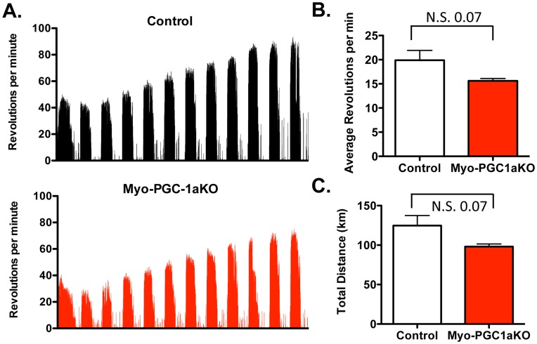
Mice lacking PGC-1α throughout the body have numerous systemic effects, including hypermetabolism, hyperactivity, and a reluctance to exercise [35], [51], rendering them a poor model to study EIMB in skeletal muscle. Mice bearing myocyte-specific deletion of PGC-1α (Myo-PGC-1αKO mice) were thus used. The mice were generated by Cre/Lox recombination and transgenic expression of Cre with a myogenin/MEF2 promoter/enhancer construct, as previously described [35], [52], [53]. 12-week old female Myo-PGC-1αKO mice and littermate control mice were allowed to exercise in individually housed cages with hanging voluntary running wheels for 12 days. The wheel revolutions per minute were assessed over the time course using an in-cage monitoring system (Figure 1A). As shown in Figure 1, both Myo-PGC-1αKO and control mice voluntarily ran nightly, and rested daily, running approximately 10 hours/day for an approximate calculated total distance of 100 km over the 2-week period. Mice of both genotypes initially ran at 20–40 revolutions/minute, and increased their running performance over the subsequent 9 days to a plateau of approximately 60–80 revolutions/minute. Overall, the Myo-PGC-1αKO mice revealed a mild, non-statistically significant, reduction in exercise performance, as assessed by both the average revolutions per min (Figure 1B) and total distance run (Figure 1C). Therefore the Myo-PGC-1αKO mice provide a good system for assessing the effects of deleting PGC-1α in skeletal muscle without any of the negative effects of germline deletion of PGC-1α.
Figure 1. Mild decrease voluntary-wheel performance in Myo-PGC-1α animals.
A.) 10 to 12 week old Myo-PGC-1αKO mice and control littermates were individually housed in voluntary running wheel cages with electronic monitoring system for 2 weeks. Tracing of wheel activity, in revolutions per minute is shown. B.) Average number of revolutions per minute C.)Total distance ran in kilometers (km). Error bars indicate s.e.m.; n >6 per group in all panels. * - P<0.05 compared to control.
Induction of OXPHOS Genes in Myo-PGC-1α Mice
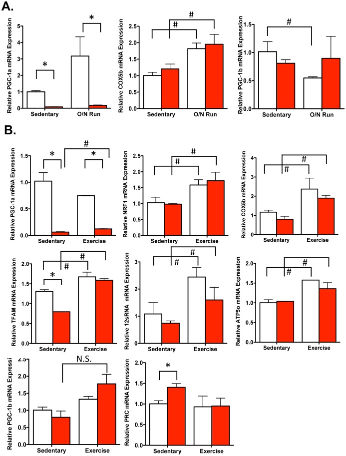
We next sought to determine whether exercise induced-genes where affected in Myo-PGC-1αKO mice after an overnight bout of voluntary wheel running. Myo-PGC-1αKO mice and control littermates were placed in individually housed cages either with wheels, or without as sedentary controls. The Myo-PGC-1αKO mice and the control mice ran overnight to a similar extent (data not shown). The next morning, quadriceps (QUADs) were harvested, RNA was isolated using the Trizol method, and the relative levels of mRNA expression of various genes were assessed by quantitative real-time PCR (qPCR). PGC-1α mRNA was induced in response to exercise ∼3-fold (Figure 2A), as has been reported [35]. Importantly, the expression of PGC-1α was almost completely absent in the Myo-PGC-1αKO animals (Figure 2A), consistent with efficient deletion of PGC-1α in the myocytes compartment, and relatively low PGC-1α expression in non-myocytic cells in skeletal muscle. Cytochrome c oxidase subunit Vb (COX5b), known to be induced by exercise, and a known target of PGC-1α, was induced in response to exercise, as expected (Figure 2A). Surprisingly, however, COX5b was similarly induced by exercise in the absence of PGC-1α (Figure 2A). No significant compensatory changes in PGC-1β expression were seen in the absence of PGC-1α (Figure 2A). We next sought to extend these findings to the response to a longer bout of exercise. Myo-PGC-1α KO and control littermates were subjected to a 12-day wheel run, again using littermate controls, and sedentary controls. Prolonged exercise induced in quadriceps of control mice the expression of COX5b, ATP synthase subunit 5o (ATP5o) as well as the nuclear respiratory factor 1 (NRF1), the mitochondria encoded 12 s RNA, and transcription factor A (Tfam) (Figure 2B), as expected. Again, surprisingly, all of these genes were induced as efficiently in the absence of muscle PGC-1α (Figure 2B). The soleus, a predominantly type I muscle, did not reveal any significant exercise-induced response (Figure S1). Taken together, these results show that PGC-1α is dispensable for the exercise-mediated induction of genes encoding key components of oxidative function and mitochondrial biogenesis.
Figure 2. Induction of OXPHOS genes in Myo-PGC-1α animals.
A.) After overnight bout of voluntary running wheel, RNA was prepared from quadriceps muscles of the Myo-PGC-1αKO (red bar) and littermate controls (white bar), and the expression of the indicated genes measured by quantitative RT-PCR. B.) After 2 week bout of voluntary running wheel, RNA was prepared from quadriceps muscles of the Myo-PGC-1αKO (red bar) and littermate controls (white bar), and the expression of the indicated genes measured by quantitative RT-PCR. Error bars indicate s.e.m.; n >3 per group in all panels. * - P<0.05 compared to control; # - P<0.05 compared to sedentary.
Normal Exercise-Induced Mitochondrial Biogenesis in Myo-PGC-1α KO mice
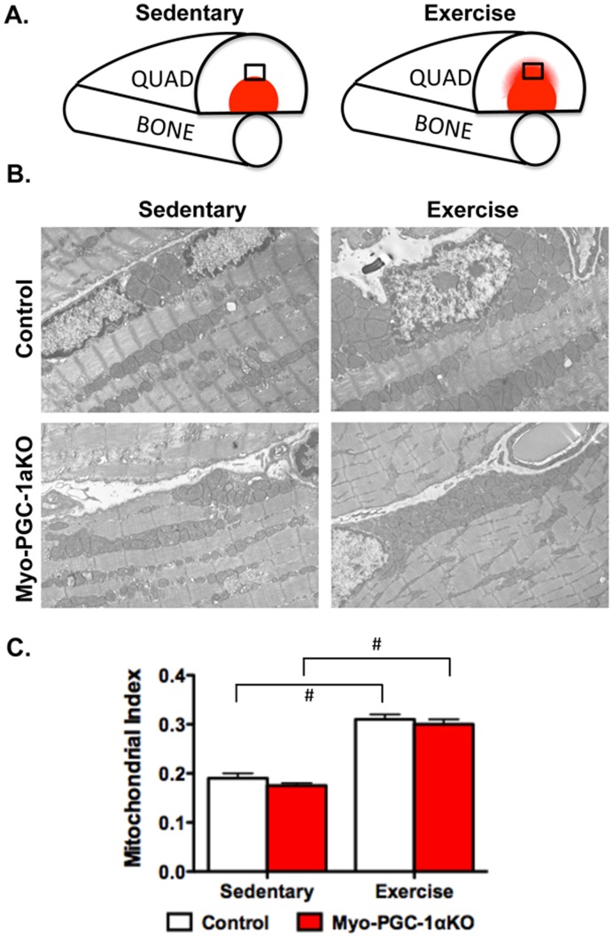
We next sought to evaluate mitochondrial biogenesis in response to exercise in the absence of PGC-1α. Endurance exercise recruits different muscles, and even different portions of muscles, differently [54], [55]. In the quadriceps muscle, for example, endurance exercise primarily recruits the middle portion of the muscle (Figure 3A). The deep portion in close proximity to the bone is likely used for locomotion and postural maintenance even in sedentary animals, while the superficial portion of the muscle is more likely to be recruited for low-endurance functions such as strength tasks. We thus focused our attention on the middle portion of the quadriceps (Figure 3A). Myo-PGC-1αKO and control mice were again allowed to run for 12 days on voluntary wheels, versus sedentary controls. Electron micrographs of the recruited portion of the quadriceps muscle were then imaged, and the mitochondrial density in the muscle was quantified using morphometric analyses. As shown in Figure 3B, 12 days of endurance exercise led to a marked 60% induction of mitochondrial density in this part of the muscle of wildtype run mice. This exercise-induced increase in mitochondrial density was entirely normal in the Myo-PGC-1αKO mice (Figure 3B). PGC-1α is thus dispensable for mitochondrial biogenesis in skeletal muscle in response to exercise.
Figure 3. Exercise induced mitochondrial biogenesis in Myo-PGC-1α animals.
A.) Schematic of mid-portion of muscle recruited in response to exercise. Shading represents oxidative portion of muscle. Black square represents region of interest (ROI) used for studies B.) Transmission electron micrographs (TEM) of transverse sections of recruited ROI of quadriceps before and after 2-week voluntary wheel run C.) Quantification of mitochondrial density from TEMsof the Myo-PGC-1αKO (red bar) and littermate controls (white bar).n>4 fields from 4 animals per group. Error bars indicate s.e.m.; n >3 per group in all panels. * - P<0.05 compared to control.
Preserved Exercise-Induced Electron Transport Complex Activity in Myo-PGC-1αKO mice
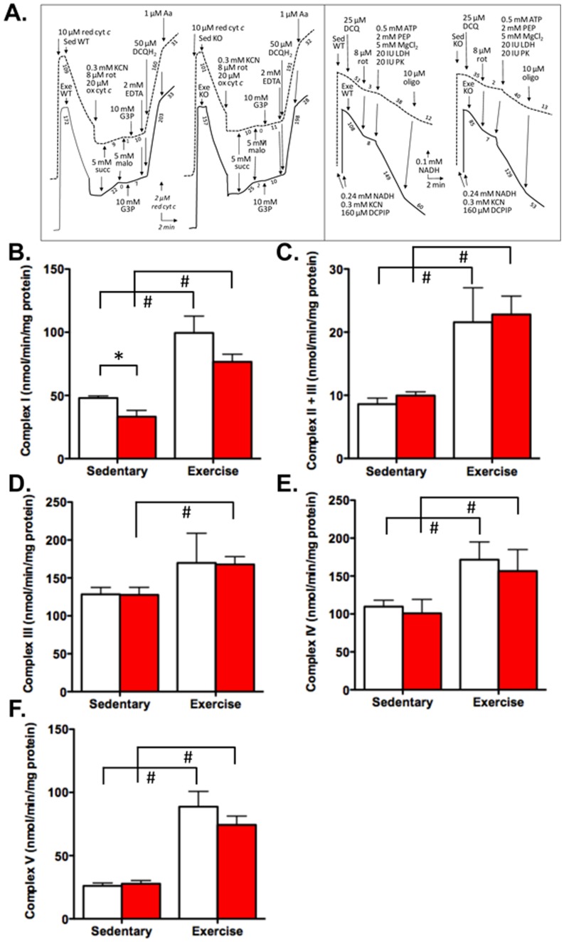
We next sought to directly measure mitochondrial oxidative capacity in response to exercise. Myo-PGC-1αKO and control mice were again allowed to run for 12 days, after which the recruited portions of the quadriceps muscle were harvested. The activity of all 4 complexes of the electron transport chain, as well as the ATPase (Complex V) were then measured directly, using a state-of-the-art enzymatic spectrophotometric assay (Figure 4A, B, C, D, E, F) [50]. As shown in Figures 4B, C, D, E, F, exercise led to strong induction of the functional capacity of each of the 5 complexes, ranging from 30% increase (ComplexIII) (Figure 4D) to a tripling of activity (Complex V) (Figure 4F). Strikingly, this strong induction of respiratory capacity was almost entirely preserved in the absence of PGC-1α. Complex I activity after exercise was reduced approximately 25% in the Myo-PGC-1αKO (Figure 4B), but the activity was also reduced in the sedentary animals, so that the induction of activity induced by exercise was similar in both genotypes. Complex V activity was reduced approximately 15% in the exercised Myo-PGC-1αKO mice, but not in the sedentary animals (Figure 4F). The induction of the activity of the 3 other complexes by exercise was entirely preserved in the Myo-PGC-1αKO mice. PGC-1α in skeletal muscle cells is thus not required for exercise-induced adaptations of mitochondrial capacity.
Figure 4. Intact Electron Complex Activity in Myo-PGC-1α in response to exercise.
A.) Enzymatic traces of complex activities B.) Rotenone sensitive NADH dehydrogenase activity (Complex I). C.) Succinate-cytochrome creductase activity (Complex II+III). D.) Glycerol-3-phosphate dehydrogenase + Complex III, rate dependent on glycerol-3-phosphate dehydrogenase (Complex III). E.) Cytochrome oxidase activity (Complex IV).F.) ATPase activity (Complex V). Myo-PGC-1αKO (red bar) and littermate controls (white bar). Error bars indicate s.e.m.; n >3 per group in all panels. * - P<0.05 compared to control; # - P<0.05 compared to sedentary.
Discussion
Mitochondrial biogenesis is strongly induced in skeletal muscle by exercise [1], [2], [3]. PGC-1α potently induces mitochondrial biogenesis in skeletal muscle [11], [12], [13], and PGC-1α expression in skeletal muscle is strongly induced by exercise. Gain-of-function observations in mouse models have led to the widely accepted belief that PGC-1α mediates exercise-induced mitochondrial biogenesis [11], [17], [19]. Formal testing of this hypothesis had not been done, however. We show here, using a loss-of-function approach, that, surprisingly, PGC-1α is in fact dispensable for exercise-induced mitochondrial adaptations in skeletal muscle. This surprising finding underscores the importance of loss-of-function studies, and highlights the still incomplete understanding of the mechanisms by which exercise strongly activates mitochondrial biogenesis in skeletal muscle. The finding also cautions against concluding that elevations of PGC-1α expression concomitant with increases in mitochondrial biogenesis necessarily indicate a causal relationship. Interestingly, Uguccioni et al. showed in a muscle cell culture model that siPGC-1α failed to block the induction of many oxidative genes and function by forced contractile activity, consistent with our findings here [56].
What other mechanisms could activate exercise-induced mitochondrial biogenesis in the absence of PGC-1α? Pogozelski et al. have recently shown that p38γmitogen-activated protein kinase (MAPK) (but not p38α or β) is required for EIMB, indicating that the MAPK pathway is critical [57]. Similarly, AMP-activated protein kinase (AMPK) has been identified as important in regulating mitochondrial content in response to exercise [58]. However, we do not observe any significant changes in either total or phospho-AMPK in our Myo-PGC-1α mice (Figure S2). PGC-1α protein and message are known to be affected by both p38 and AMPK [33], [59], but our data indicate that other targets must thus also exist in this context. PGC-1β is a homolog of PGC-1α (40 and 48% identity in the conserved N-terminal activation domain and RNA binding domain, respectively) [25] that shares many, though not all, of the roles of PGC-1α. In skeletal muscle, PGC-1β can induce mitochondrial biogenesis as robustly as PGC-1α [18]. Deletion of both PGC-1α and β in skeletal muscle reduces mitochondrial function markedly more than deletion of either alone [24]. Unlike PGC-1α, however, expression of PGC-1β is not induced by exercise or adrenergic stimulation, and may in fact be decreased [40], [60]. For this reason, PGC-1β has not generally been thought to play a role in EIMB. We also did not observe compensatory increases in PGC-1β expression in the Myo-PGC-1α mice. Thus, if PGC-1β is compensating for the absence of PGC-1α, then exercise must be modulating PGC-1β post-transcriptionally, for example via adrenergic and/or p38-mediated or AMPK-mediated phosphorylation. The contribution of PGC-1β to EIMB will need to be addressed with muscle-specific deletion of PGC-1β, alone or concurrent with deletion of PGC-1α. Lastly, the hypoxia inducible factors 1 and 2 (HIFs) have also been shown to be induced in response to exercise [61]. However, HIFs likely suppress mitochondrial function and OXPHOS [62], [63]and thus are unlikely candidates to mediate EIMB.
The current study does not evaluate every form of exercise, but rather focuses on voluntary endurance exercise. This choice was guided by the extensive amount of exercise that mice perform voluntarily, as well as the strong mitochondrial biogenic response to the stimulus. The study cannot rule out, however, that mitochondrial adaptations to other forms of exercise, such as controlled endurance exercise or strength training, might be mediated by PGC-1α. Nevertheless, voluntary running in in-cage wheels robustly induces PGC-1α in skeletal muscle [35], and despite this induction, reveals no dependency on PGC-1α for the induction of mitochondrial biogenesis (Figure 3C).
It is possible, though unlikely, that PGC-1α was insufficiently deleted from skeletal muscle in our study. The myogenin/MEF2-CRE driver has been used widely, and indeed the mRNA expression of PGC-1α was dramatically reduced in all muscles tested, and especially so in the muscles used for subsequent mitochondrial experiments. Moreover, the same muscle-specific PGC-1α knockout mice have been used by us and others to highlight other phenotypes, including the existence of a muscle/beta cell crosstalk, and the requirement for PGC-1α for exercise-induced angiogenesis [35], [53]. Finally, it is highly unlikely that the few myonuclei that have retained a non-deleted PGC-1α locus could induce, at a distance, target genes throughout the myofiber. Therefore it is unlikely that any of the exercise-induced effects observed in this study are due to incomplete deletion of PGC-1α. It is also possible that ontogenic deletion of PGC-1α led to compensatory changes during development, although, as noted above, no changes were observed in the expression of the most likely candidates, PGC-1β and PGC-1-related coactivator (PRC).
Recently, a naturally occurring alternatively spliced form of PGC-1α that is truncated at exon 7 (NT-PGC-1α) has been shown to have some of the biological activity of full length PGC-1α [64], [65], [66]. The role of NT-PGC-1α in skeletal muscle is not known. Importantly, however the Myo-PGC-1α KO animals used in this study lack exons 3 thru 5 of PGC-1α, which also affects NT-PGC-1α. Therefore NT-PGC-1α is unlikely to play a role in mediating the exercise-induced responses observed on the Myo-PGC-1α KO mice.
It is interesting that, while PGC-1α is not required for EIMB (this study), or for exercise-induced changes in fiber types [67], it does appear to be critical for exercise-induced angiogenesis [35], underscoring the complexity of the muscle response to exercise, and the role of PGC-1α.Therefore, while dispensable for some aspects of exercise-induced changes, PGC-1α clearly still has a pivotal role in skeletal muscle adaptions in response to exercise.
In summary, the current study demonstrates that PGC-1α is dispensable within skeletal muscle for exercise-induced mitochondrial adaptations, in sharp contrast to the prevalent assumption in the field, and indicating that other important pathway(s) clearly exist. Exercise is a powerful intervention for the treatment of many diseases [68], [69], [70], [71], [72], [73], and exercise-induced changes on mitochondria are likely important for many of the benefits of exercise [3], [4], [5], [6]. Identifying and understanding the PGC-1α-independent pathways that mediate EIMB will thus be of great interest.
Supporting Information
Expression of OXPHOS genes in soleus of Myo-PGC-1α animals. After 2-week bout of voluntary running wheel, RNA was prepared from soleus muscles of the Myo-PGC-1αKO (red bar) and littermate controls (white bar), and the expression of the indicated genes measured by quantitative RT-PCR. Error bars indicate s.e.m.; n >3 per group in all panels. * - P<0.05 compared to control; # - P<0.05 compared to sedentary.
(TIF)
Phospho and Total AMPK levels in quadriceps of Myo-PGC-1α animals. After 2-week bout of voluntary running wheel, protein was prepared from quadriceps muscles of the Myo-PGC-1αKO and littermate controls, levels of P-AMPK and total AMPK were assessed by western blotting analysis.
(TIF)
Additional details on method for western blotting analysis.
(DOCX)
Footnotes
Competing Interests: The authors have declared that no competing interests exist.
Funding: GCR has received support from NIH (5T32HL007374-31) and UNCF-Merck Science Initiative (post-doctoral fellowship) (umsi.uncf.org), and ZA is supported by the Ellison Foundation (www.ellisonfoundation.org) and the NHLBI (5R01HL094499-02). The funders had no role in study design, data collection and analysis, decision to publish, or preparation of the manuscript.
References
- 1.Gollnick PD, King DW. Effect of exercise and training on mitochondria of rat skeletal muscle. The American journal of physiology. 1969;216:1502–1509. doi: 10.1152/ajplegacy.1969.216.6.1502. [DOI] [PubMed] [Google Scholar]
- 2.Freyssenet D, Berthon P, Denis C. Mitochondrial biogenesis in skeletal muscle in response to endurance exercises. Arch Physiol Biochem. 1996;104:129–141. doi: 10.1076/apab.104.2.129.12878. [DOI] [PubMed] [Google Scholar]
- 3.Little JP, Safdar A, Benton CR, Wright DC. Skeletal muscle and beyond: the role of exercise as a mediator of systemic mitochondrial biogenesis. Applied physiology, nutrition, and metabolism = Physiologie appliquee, nutrition et metabolisme. 2011;36:598–607. doi: 10.1139/h11-076. [DOI] [PubMed] [Google Scholar]
- 4.Ostergard T, Andersen JL, Nyholm B, Lund S, Nair KS, et al. Impact of exercise training on insulin sensitivity, physical fitness, and muscle oxidative capacity in first-degree relatives of type 2 diabetic patients. American journal of physiology Endocrinology and metabolism. 2006;290:E998–1005. doi: 10.1152/ajpendo.00012.2005. [DOI] [PubMed] [Google Scholar]
- 5.Safdar A, Bourgeois JM, Ogborn DI, Little JP, Hettinga BP, et al. Endurance exercise rescues progeroid aging and induces systemic mitochondrial rejuvenation in mtDNA mutator mice. Proceedings of the National Academy of Sciences of the United States of America. 2011;108:4135–4140. doi: 10.1073/pnas.1019581108. [DOI] [PMC free article] [PubMed] [Google Scholar] [Retracted]
- 6.Steiner JL, Murphy EA, McClellan JL, Carmichael MD, Davis JM. Exercise Training Increases Mitochondrial Biogenesis in the Brain. Journal of applied physiology. 2011. [DOI] [PubMed]
- 7.Kelly DP, Scarpulla RC. Transcriptional regulatory circuits controlling mitochondrial biogenesis and function. Genes Dev. 2004;18:357–368. doi: 10.1101/gad.1177604. [DOI] [PubMed] [Google Scholar]
- 8.Scarpulla RC. Nuclear activators and coactivators in mammalian mitochondrial biogenesis. Biochim Biophys Acta. 2002;1576:1–14. doi: 10.1016/s0167-4781(02)00343-3. [DOI] [PubMed] [Google Scholar]
- 9.Gleyzer N, Vercauteren K, Scarpulla RC. Control of mitochondrial transcription specificity factors (TFB1M and TFB2M) by nuclear respiratory factors (NRF-1 and NRF-2) and PGC-1 family coactivators. Mol Cell Biol. 2005;25:1354–1366. doi: 10.1128/MCB.25.4.1354-1366.2005. [DOI] [PMC free article] [PubMed] [Google Scholar]
- 10.Virbasius JV, Scarpulla RC. Activation of the human mitochondrial transcription factor A gene by nuclear respiratory factors: a potential regulatory link between nuclear and mitochondrial gene expression in organelle biogenesis. Proc Natl Acad Sci U S A. 1994;91:1309–1313. doi: 10.1073/pnas.91.4.1309. [DOI] [PMC free article] [PubMed] [Google Scholar]
- 11.Lin J, Wu H, Tarr PT, Zhang CY, Wu Z, et al. Transcriptional co-activator PGC-1 alpha drives the formation of slow-twitch muscle fibres. Nature. 2002;418:797–801. doi: 10.1038/nature00904. [DOI] [PubMed] [Google Scholar]
- 12.Wende AR, Schaeffer PJ, Parker GJ, Zechner C, Han DH, et al. A role for the transcriptional coactivator PGC-1alpha in muscle refueling. J Biol Chem. 2007;282:36642–36651. doi: 10.1074/jbc.M707006200. [DOI] [PubMed] [Google Scholar]
- 13.St-Pierre J, Lin J, Krauss S, Tarr PT, Yang R, et al. Bioenergetic analysis of peroxisome proliferator-activated receptor gamma coactivators 1alpha and 1beta (PGC-1alpha and PGC-1beta) in muscle cells. J Biol Chem. 2003;278:26597–26603. doi: 10.1074/jbc.M301850200. [DOI] [PubMed] [Google Scholar]
- 14.Wu Z, Puigserver P, Andersson U, Zhang C, Adelmant G, et al. Mechanisms controlling mitochondrial biogenesis and respiration through the thermogenic coactivator PGC-1. Cell. 1999;98:115–124. doi: 10.1016/S0092-8674(00)80611-X. [DOI] [PubMed] [Google Scholar]
- 15.Mootha VK, Handschin C, Arlow D, Xie X, St Pierre J, et al. Erralpha and Gabpa/b specify PGC-1alpha-dependent oxidative phosphorylation gene expression that is altered in diabetic muscle. Proc Natl Acad Sci U S A. 2004;101:6570–6575. doi: 10.1073/pnas.0401401101. [DOI] [PMC free article] [PubMed] [Google Scholar]
- 16.Wende AR, Huss JM, Schaeffer PJ, Giguere V, Kelly DP. PGC-1alpha coactivates PDK4 gene expression via the orphan nuclear receptor ERRalpha: a mechanism for transcriptional control of muscle glucose metabolism. Mol Cell Biol. 2005;25:10684–10694. doi: 10.1128/MCB.25.24.10684-10694.2005. [DOI] [PMC free article] [PubMed] [Google Scholar]
- 17.Arany Z, Foo SY, Ma Y, Ruas JL, Bommi-Reddy A, et al. HIF-independent regulation of VEGF and angiogenesis by the transcriptional coactivator PGC-1alpha. Nature. 2008;451:1008–1012. doi: 10.1038/nature06613. [DOI] [PubMed] [Google Scholar]
- 18.Arany Z, Lebrasseur N, Morris C, Smith E, Yang W, et al. The transcriptional coactivator PGC-1beta drives the formation of oxidative type IIX fibers in skeletal muscle. Cell Metab. 2007;5:35–46. doi: 10.1016/j.cmet.2006.12.003. [DOI] [PubMed] [Google Scholar]
- 19.Calvo JA, Daniels TG, Wang X, Paul A, Lin J, et al. Muscle-specific expression of PPARγ coactivator-1α improves exercise performance and increases peak oxygen uptake. J Appl Physiol. 2008;104:1304–1312. doi: 10.1152/japplphysiol.01231.2007. [DOI] [PubMed] [Google Scholar]
- 20.Sandri M, Lin J, Handschin C, Yang W, Arany ZP, et al. PGC-1alpha protects skeletal muscle from atrophy by suppressing FoxO3 action and atrophy-specific gene transcription. Proc Natl Acad Sci U S A. 2006;103:16260–16265. doi: 10.1073/pnas.0607795103. [DOI] [PMC free article] [PubMed] [Google Scholar]
- 21.Handschin C, Kobayashi YM, Chin S, Seale P, Campbell KP, et al. PGC-1alpha regulates the neuromuscular junction program and ameliorates Duchenne muscular dystrophy. Genes Dev. 2007;21:770–783. doi: 10.1101/gad.1525107. [DOI] [PMC free article] [PubMed] [Google Scholar]
- 22.Leick L, Wojtaszewski JF, Johansen ST, Kiilerich K, Comes G, et al. PGC-1alpha is not mandatory for exercise- and training-induced adaptive gene responses in mouse skeletal muscle. Am J Physiol Endocrinol Metab. 2008;294:E463–474. doi: 10.1152/ajpendo.00666.2007. [DOI] [PubMed] [Google Scholar]
- 23.Handschin C, Chin S, Li P, Liu F, Maratos-Flier E, et al. Skeletal muscle fiber-type switching, exercise intolerance, and myopathy in PGC-1alpha muscle-specific knock-out animals. J Biol Chem. 2007;282:30014–30021. doi: 10.1074/jbc.M704817200. [DOI] [PubMed] [Google Scholar]
- 24.Zechner C, Lai L, Zechner JF, Geng T, Yan Z, et al. Total skeletal muscle PGC-1 deficiency uncouples mitochondrial derangements from fiber type determination and insulin sensitivity. Cell Metab. 2010;12:633–642. doi: 10.1016/j.cmet.2010.11.008. [DOI] [PMC free article] [PubMed] [Google Scholar]
- 25.Lin J, Puigserver P, Donovan J, Tarr P, Spiegelman BM. Peroxisome proliferator-activated receptor gamma coactivator 1beta (PGC-1beta), a novel PGC-1-related transcription coactivator associated with host cell factor. J Biol Chem. 2002;277:1645–1648. doi: 10.1074/jbc.C100631200. [DOI] [PubMed] [Google Scholar]
- 26.Kamei Y, Ohizumi H, Fujitani Y, Nemoto T, Tanaka T, et al. PPARgamma coactivator 1beta/ERR ligand 1 is an ERR protein ligand, whose expression induces a high-energy expenditure and antagonizes obesity. Proc Natl Acad Sci U S A. 2003;100:12378–12383. doi: 10.1073/pnas.2135217100. [DOI] [PMC free article] [PubMed] [Google Scholar]
- 27.Lelliott CJ, Medina-Gomez G, Petrovic N, Kis A, Feldmann HM, et al. Ablation of PGC-1beta results in defective mitochondrial activity, thermogenesis, hepatic function, and cardiac performance. PLoS Biol. 2006;4:e369. doi: 10.1371/journal.pbio.0040369. [DOI] [PMC free article] [PubMed] [Google Scholar]
- 28.Rowe GC, Jang C, Patten IS, Arany Z. PGC-1beta regulates angiogenesis in skeletal muscle. American journal of physiology Endocrinology and metabolism. 2011;301:E155–163. doi: 10.1152/ajpendo.00681.2010. [DOI] [PMC free article] [PubMed] [Google Scholar]
- 29.Puigserver P, Rhee J, Lin J, Wu Z, Yoon JC, et al. Cytokine stimulation of energy expenditure through p38 MAP kinase activation of PPARgamma coactivator-1. Mol Cell. 2001;8:971–982. doi: 10.1016/s1097-2765(01)00390-2. [DOI] [PubMed] [Google Scholar]
- 30.Teyssier C, Ma H, Emter R, Kralli A, Stallcup MR. Activation of nuclear receptor coactivator PGC-1alpha by arginine methylation. Genes Dev. 2005;19:1466–1473. doi: 10.1101/gad.1295005. [DOI] [PMC free article] [PubMed] [Google Scholar]
- 31.Rodgers JT, Lerin C, Haas W, Gygi SP, Spiegelman BM, et al. Nutrient control of glucose homeostasis through a complex of PGC-1alpha and SIRT1. Nature. 2005;434:113–118. doi: 10.1038/nature03354. [DOI] [PubMed] [Google Scholar]
- 32.Lerin C, Rodgers JT, Kalume DE, Kim SH, Pandey A, et al. GCN5 acetyltransferase complex controls glucose metabolism through transcriptional repression of PGC-1alpha. Cell Metab. 2006;3:429–438. doi: 10.1016/j.cmet.2006.04.013. [DOI] [PubMed] [Google Scholar]
- 33.Jager S, Handschin C, St-Pierre J, Spiegelman BM. AMP-activated protein kinase (AMPK) action in skeletal muscle via direct phosphorylation of PGC-1alpha. Proc Natl Acad Sci U S A. 2007;104:12017–12022. doi: 10.1073/pnas.0705070104. [DOI] [PMC free article] [PubMed] [Google Scholar]
- 34.Lin J, Yang R, Tarr PT, Wu PH, Handschin C, et al. Hyperlipidemic effects of dietary saturated fats mediated through PGC-1beta coactivation of SREBP. Cell. 2005;120:261–273. doi: 10.1016/j.cell.2004.11.043. [DOI] [PubMed] [Google Scholar]
- 35.Chinsomboon J, Ruas J, Gupta RK, Thom R, Shoag J, et al. The transcriptional coactivator PGC-1α mediates exercise-induced angiogenesis in skeletal muscle. Proc Natl Acad Sci U S A. 2009;106:21401–21406. doi: 10.1073/pnas.0909131106. [DOI] [PMC free article] [PubMed] [Google Scholar]
- 36.Baar K, Wende AR, Jones TE, Marison M, Nolte LA, et al. Adaptations of skeletal muscle to exercise: rapid increase in the transcriptional coactivator PGC-1. Faseb J. 2002;16:1879–1886. doi: 10.1096/fj.02-0367com. [DOI] [PubMed] [Google Scholar]
- 37.Terada S, Goto M, Kato M, Kawanaka K, Shimokawa T, et al. Effects of low-intensity prolonged exercise on PGC-1 mRNA expression in rat epitrochlearis muscle. Biochem Biophys Res Commun. 2002;296:350–354. doi: 10.1016/s0006-291x(02)00881-1. [DOI] [PubMed] [Google Scholar]
- 38.Pilegaard H, Saltin B, Neufer PD. Exercise induces transient transcriptional activation of the PGC-1alpha gene in human skeletal muscle. J Physiol. 2003;546:851–858. doi: 10.1113/jphysiol.2002.034850. [DOI] [PMC free article] [PubMed] [Google Scholar]
- 39.Akimoto T, Pohnert SC, Li P, Zhang M, Gumbs C, et al. Exercise stimulates Pgc-1alpha transcription in skeletal muscle through activation of the p38 MAPK pathway. J Biol Chem. 2005;280:19587–19593. doi: 10.1074/jbc.M408862200. [DOI] [PubMed] [Google Scholar]
- 40.Mathai AS, Bonen A, Benton CR, Robinson DL, Graham TE. Rapid exercise-induced changes in PGC-1alpha mRNA and protein in human skeletal muscle. J Appl Physiol. 2008;105:1098–1105. doi: 10.1152/japplphysiol.00847.2007. [DOI] [PubMed] [Google Scholar]
- 41.Handschin C, Spiegelman BM. The role of exercise and PGC1alpha in inflammation and chronic disease. Nature. 2008;454:463–469. doi: 10.1038/nature07206. [DOI] [PMC free article] [PubMed] [Google Scholar]
- 42.Miura S, Kawanaka K, Kai Y, Tamura M, Goto M, et al. An increase in murine skeletal muscle peroxisome proliferator-activated receptor-gamma coactivator-1alpha (PGC-1alpha) mRNA in response to exercise is mediated by beta-adrenergic receptor activation. Endocrinology. 2007;148:3441–3448. doi: 10.1210/en.2006-1646. [DOI] [PubMed] [Google Scholar]
- 43.Miura S, Kai Y, Kamei Y, Ezaki O. Isoform-Specific Increases in Murine Skeletal Muscle Peroxisome Proliferator-Activated Receptor-γ Coactivator-1α (PGC-1α) mRNA in Response to β2-Adrenergic Receptor Activation and Exercise. Endocrinology. 2008. [DOI] [PubMed]
- 44.Baar K. Involvement of PPAR gamma co-activator-1, nuclear respiratory factors 1 and 2, and PPAR alpha in the adaptive response to endurance exercise. Proc Nutr Soc. 2004;63:269–273. doi: 10.1079/PNS2004334. [DOI] [PubMed] [Google Scholar]
- 45.Lin J, Handschin C, Spiegelman BM. Metabolic control through the PGC-1 family of transcription coactivators. Cell Metab. 2005;1:361–370. doi: 10.1016/j.cmet.2005.05.004. [DOI] [PubMed] [Google Scholar]
- 46.Yan Z, Li P, Akimoto T. Transcriptional control of the Pgc-1alpha gene in skeletal muscle in vivo. Exerc Sport Sci Rev. 2007;35:97–101. doi: 10.1097/JES.0b013e3180a03169. [DOI] [PubMed] [Google Scholar]
- 47.Ljubicic V, Joseph AM, Saleem A, Uguccioni G, Collu-Marchese M, et al. Transcriptional and post-transcriptional regulation of mitochondrial biogenesis in skeletal muscle: effects of exercise and aging. Biochimica et biophysica acta. 2010;1800:223–234. doi: 10.1016/j.bbagen.2009.07.031. [DOI] [PubMed] [Google Scholar]
- 48.Arany ZP. High-throughput quantitative real-time PCR. Curr Protoc Hum Genet Chapter 11: Unit 11 10. 2008. [DOI] [PubMed]
- 49.Rustin P, Chretien D, Bourgeron T, Gerard B, Rotig A, et al. Biochemical and molecular investigations in respiratory chain deficiencies. Clinica chimica acta; international journal of clinical chemistry. 1994;228:35–51. doi: 10.1016/0009-8981(94)90055-8. [DOI] [PubMed] [Google Scholar]
- 50.Benit P, Goncalves S, Philippe Dassa E, Briere JJ, Martin G, et al. Three spectrophotometric assays for the measurement of the five respiratory chain complexes in minuscule biological samples. Clin Chim Acta. 2006;374:81–86. doi: 10.1016/j.cca.2006.05.034. [DOI] [PubMed] [Google Scholar]
- 51.Lin J, Wu PH, Tarr PT, Lindenberg KS, St-Pierre J, et al. Defects in adaptive energy metabolism with CNS-linked hyperactivity in PGC-1alpha null mice. Cell. 2004;119:121–135. doi: 10.1016/j.cell.2004.09.013. [DOI] [PubMed] [Google Scholar]
- 52.Li S, Czubryt MP, McAnally J, Bassel-Duby R, Richardson JA, et al. Requirement for serum response factor for skeletal muscle growth and maturation revealed by tissue-specific gene deletion in mice. Proc Natl Acad Sci U S A. 2005;102:1082–1087. doi: 10.1073/pnas.0409103102. [DOI] [PMC free article] [PubMed] [Google Scholar]
- 53.Handschin C, Choi CS, Chin S, Kim S, Kawamori D, et al. Abnormal glucose homeostasis in skeletal muscle-specific PGC-1alpha knockout mice reveals skeletal muscle-pancreatic beta cell crosstalk. J Clin Invest. 2007;117:3463–3474. doi: 10.1172/JCI31785. [DOI] [PMC free article] [PubMed] [Google Scholar]
- 54.Holloszy JO, Coyle EF. Adaptations of skeletal muscle to endurance exercise and their metabolic consequences. Journal of applied physiology: respiratory, environmental and exercise physiology. 1984;56:831–838. doi: 10.1152/jappl.1984.56.4.831. [DOI] [PubMed] [Google Scholar]
- 55.Altenburg TM, Degens H, van Mechelen W, Sargeant AJ, de Haan A. Recruitment of single muscle fibers during submaximal cycling exercise. Journal of applied physiology. 2007;103:1752–1756. doi: 10.1152/japplphysiol.00496.2007. [DOI] [PubMed] [Google Scholar]
- 56.Uguccioni G, Hood DA. The importance of PGC-1alpha in contractile activity-induced mitochondrial adaptations. American journal of physiology Endocrinology and metabolism. 2011;300:E361–371. doi: 10.1152/ajpendo.00292.2010. [DOI] [PubMed] [Google Scholar]
- 57.Pogozelski AR, Geng T, Li P, Yin X, Lira VA, et al. p38gamma mitogen-activated protein kinase is a key regulator in skeletal muscle metabolic adaptation in mice. PLoS One. 2009;4:e7934. doi: 10.1371/journal.pone.0007934. [DOI] [PMC free article] [PubMed] [Google Scholar]
- 58.O’Neill HM, Maarbjerg SJ, Crane JD, Jeppesen J, Jorgensen SB, et al. AMP-activated protein kinase (AMPK) beta1beta2 muscle null mice reveal an essential role for AMPK in maintaining mitochondrial content and glucose uptake during exercise. Proceedings of the National Academy of Sciences of the United States of America. 2011;108:16092–16097. doi: 10.1073/pnas.1105062108. [DOI] [PMC free article] [PubMed] [Google Scholar]
- 59.Knutti D, Kressler D, Kralli A. Regulation of the transcriptional coactivator PGC-1 via MAPK-sensitive interaction with a repressor. Proc Natl Acad Sci U S A. 2001;98:9713–9718. doi: 10.1073/pnas.171184698. [DOI] [PMC free article] [PubMed] [Google Scholar]
- 60.Mortensen OH, Plomgaard P, Fischer CP, Hansen AK, Pilegaard H, et al. PGC-1beta is downregulated by training in human skeletal muscle: no effect of training twice every second day vs. once daily on expression of the PGC-1 family. Journal of applied physiology. 2007;103:1536–1542. doi: 10.1152/japplphysiol.00575.2007. [DOI] [PubMed] [Google Scholar]
- 61.Lundby C, Gassmann M, Pilegaard H. Regular endurance training reduces the exercise induced HIF-1alpha and HIF-2alpha mRNA expression in human skeletal muscle in normoxic conditions. European journal of applied physiology. 2006;96:363–369. doi: 10.1007/s00421-005-0085-5. [DOI] [PubMed] [Google Scholar]
- 62.Semenza GL. Oxygen-dependent regulation of mitochondrial respiration by hypoxia-inducible factor 1. The Biochemical journal. 2007;405:1–9. doi: 10.1042/BJ20070389. [DOI] [PubMed] [Google Scholar]
- 63.Zhang H, Bosch-Marce M, Shimoda LA, Tan YS, Baek JH, et al. Mitochondrial autophagy is an HIF-1-dependent adaptive metabolic response to hypoxia. The Journal of biological chemistry. 2008;283:10892–10903. doi: 10.1074/jbc.M800102200. [DOI] [PMC free article] [PubMed] [Google Scholar] [Retracted]
- 64.Zhang Y, Huypens P, Adamson AW, Chang JS, Henagan TM, et al. Alternative mRNA splicing produces a novel biologically active short isoform of PGC-1alpha. J Biol Chem. 2009;284:32813–32826. doi: 10.1074/jbc.M109.037556. [DOI] [PMC free article] [PubMed] [Google Scholar]
- 65.Chang JS, Huypens P, Zhang Y, Black C, Kralli A, et al. Regulation of NT-PGC-1alpha subcellular localization and function by protein kinase A-dependent modulation of nuclear export by CRM1. J Biol Chem. 2010;285:18039–18050. doi: 10.1074/jbc.M109.083121. [DOI] [PMC free article] [PubMed] [Google Scholar]
- 66.Chang JS, Fernand V, Zhang Y, Shin J, Jun HJ, et al. NT-PGC-1alpha Protein Is Sufficient to Link beta3-Adrenergic Receptor Activation to Transcriptional and Physiological Components of Adaptive Thermogenesis. The Journal of biological chemistry. 2012;287:9100–9111. doi: 10.1074/jbc.M111.320200. [DOI] [PMC free article] [PubMed] [Google Scholar]
- 67.Yan Z, Okutsu M, Akhtar YN, Lira VA. Regulation of exercise-induced fiber type transformation, mitochondrial biogenesis, and angiogenesis in skeletal muscle. J Appl Physiol. 2011;110:264–274. doi: 10.1152/japplphysiol.00993.2010. [DOI] [PMC free article] [PubMed] [Google Scholar]
- 68.Taivassalo T, Shoubridge EA, Chen J, Kennaway NG, DiMauro S, et al. Aerobic conditioning in patients with mitochondrial myopathies: physiological, biochemical, and genetic effects. Ann Neurol. 2001;50:133–141. doi: 10.1002/ana.1050. [DOI] [PubMed] [Google Scholar]
- 69.Taivassalo T, Haller RG. Exercise and training in mitochondrial myopathies. Med Sci Sports Exerc. 2005;37:2094–2101. doi: 10.1249/01.mss.0000177446.97671.2a. [DOI] [PubMed] [Google Scholar]
- 70.Jeppesen TD, Schwartz M, Olsen DB, Wibrand F, Krag T, et al. Aerobic training is safe and improves exercise capacity in patients with mitochondrial myopathy. Brain. 2006;129:3402–3412. doi: 10.1093/brain/awl149. [DOI] [PubMed] [Google Scholar]
- 71.Wenz T, Diaz F, Hernandez D, Moraes CT. Endurance exercise is protective for mice with mitochondrial myopathy. J Appl Physiol. 2009;106:1712–1719. doi: 10.1152/japplphysiol.91571.2008. [DOI] [PMC free article] [PubMed] [Google Scholar] [Retracted]
- 72.Kerr DS. Treatment of mitochondrial electron transport chain disorders: a review of clinical trials over the past decade. Mol Genet Metab. 2010;99:246–255. doi: 10.1016/j.ymgme.2009.11.005. [DOI] [PubMed] [Google Scholar]
- 73.Lira VA, Benton CR, Yan Z, Bonen A. PGC-1alpha regulation by exercise training and its influences on muscle function and insulin sensitivity. Am J Physiol Endocrinol Metab. 2010;299:E145–161. doi: 10.1152/ajpendo.00755.2009. [DOI] [PMC free article] [PubMed] [Google Scholar]
Associated Data
This section collects any data citations, data availability statements, or supplementary materials included in this article.
Supplementary Materials
Expression of OXPHOS genes in soleus of Myo-PGC-1α animals. After 2-week bout of voluntary running wheel, RNA was prepared from soleus muscles of the Myo-PGC-1αKO (red bar) and littermate controls (white bar), and the expression of the indicated genes measured by quantitative RT-PCR. Error bars indicate s.e.m.; n >3 per group in all panels. * - P<0.05 compared to control; # - P<0.05 compared to sedentary.
(TIF)
Phospho and Total AMPK levels in quadriceps of Myo-PGC-1α animals. After 2-week bout of voluntary running wheel, protein was prepared from quadriceps muscles of the Myo-PGC-1αKO and littermate controls, levels of P-AMPK and total AMPK were assessed by western blotting analysis.
(TIF)
Additional details on method for western blotting analysis.
(DOCX)


