Abstract
Expression of oncogenic K-RAS in primary cells elicits oncogene-induced cellular senescence (OIS), a form of growth arrest that potently opposes tumourigenesis. This effect has been largely attributed to transcriptional mechanisms that depend on the p53 tumour suppressor protein. The PML tumour suppressor was initially identified as a component of the PML-RARα oncoprotein of acute promyelocytic leukaemia (APL). PML, a critical OIS mediator, is upregulated by oncogenic K-RAS in vivo and in vitro. We demonstrate here that oncogenic K-RAS induces PML protein upregulation by activating the RAS/MEK1/mTOR/eIF4E pathway even in the absence of p53. Under these circumstances, PML mRNA is selectively associated to polysomes. Importantly, we find that the PML 5′ untranslated mRNA region plays a key role in mediating PML protein upregulation and that its presence is essential for an efficient OIS response. These findings demonstrate that upregulation of PML translation plays a central role in oncogenic K-RAS-induced OIS. Thus, selective translation initiation plays a critical role in tumour suppression with important therapeutic implications for the treatment of solid tumours and APL.
Keywords: mTOR, oncogene-induced cellular senescence, oncogenic K-RAS, PML, protein translation
INTRODUCTION
When bound to GTP, the RAS family of small guanosine triphosphatases activates several signalling pathways that transduce critical cell proliferation and survival signals. Three members of the family, H-RAS, K-RAS and N-RAS, acquire transforming potential as a result of a single amino-acid substitution most commonly at position 12 (oncogenic RAS; Malumbres & Barbacid, 2003).
About 20% of all human tumours harbour oncogenic K-RAS mutations. These mutants transform most immortalized cell lines and initiate tumourigenesis in mouse cancer models (Downward, 2003). However, it has become apparent that oncogenic K-RAS signalling in primary cells also triggers oncogene-induced cellular senescence (OIS), a potent growth-suppressive response that opposes cancer progression (Campisi & d'Adda di Fagagna, 2007). Oncogenic K-RAS controls the RAF/MEK1/ERK1/2 and the PI3K/AKT signalling pathways. Both of these have been implicated in OIS induction (Chen et al, 2005; Lin et al, 1998).
Oncogenic K-RAS may lead to increased translation efficiency of existing mRNAs through several downstream effectors such as ERK1/2 and AKT. These protein kinases regulate initiation of translation by phosphorylating and inactivating TSC2, a tumour suppressor that in a complex with TSC1 inhibits the mTOR protein kinase (Ma et al, 2005; Manning et al, 2002). mTOR stimulates the rate-limiting process of translation initiation by directly phosphorylating the 4E-BP family of translational repressors and causing their dissociation from eIF4E, a component of the eIF4F protein complex. This event leads to the recognition of the 5′-cap structure by eIF4E and mRNA recruitment to ribosomes. Therefore, mTOR activation promotes translation initiation. In addition, mTOR phosphorylates and activates S6 ribosomal kinases (S6K1/2), which have been implicated in the control of cell growth via increased mRNA translation (Hay & Sonenberg, 2004).
The promyelocytic leukaemia tumour suppressor (PML), initially identified as a component of the PML-RARα oncoprotein of acute promyelocytic leukaemia (APL), plays a critical role in oncogenic K-RAS-induced OIS. In this context, PML protein levels are strikingly upregulated (Ferbeyre et al, 2000; Pearson et al, 2000). The increase in PML protein levels leads to recruitment of p53 to the PML-nuclear bodies (PML-NBs), an event that facilitates p53 acetylation and transcriptional activation (Bernardi & Pandolfi, 2007; Ferbeyre et al, 2000; Pearson et al, 2000). Thus far, the increase in PML protein levels in conditions of oncogenic stress has been attributed to upregulation of PML gene transcription and it has been reported that PML is a p53 transcriptional target gene (de Stanchina et al, 2004).
Here, we report that PML expression is also regulated at the translational level during oncogenic K-RAS-induced OIS in a p53-independent manner. We show that mTOR-dependent translational control mechanisms are critically important in modulating PML protein levels during oncogenic K-RAS-induced OIS and that the PML 5′UTR is necessary for efficient OIS induction. We propose that the mechanisms controlling selective translation initiation of tumour suppressor proteins play an important role in the cellular response to oncogenic stress.
RESULTS
Oncogenic K-RAS upregulates PML protein in vivo in the presence or absence of p53
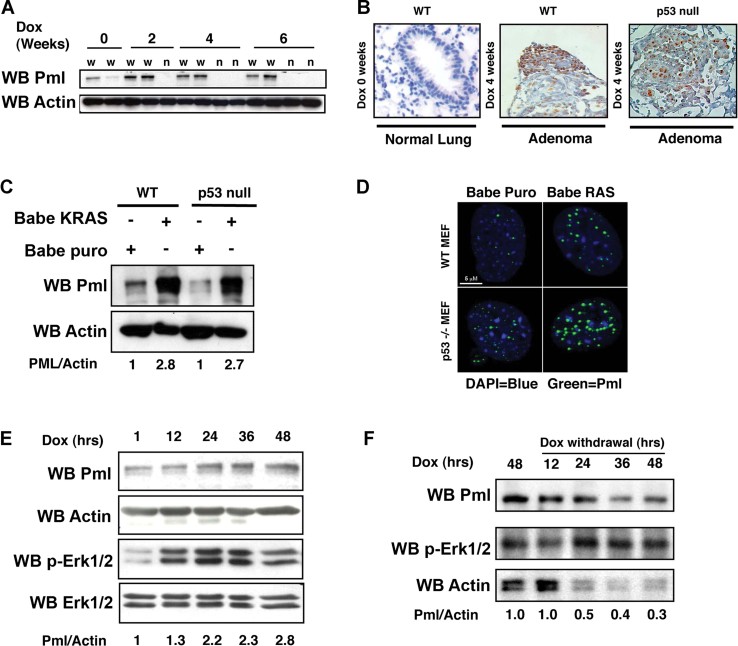
To obtain insights into the networks controlling OIS induction, we tested whether oncogenic K-RAS expression leads to PML protein upregulation in vivo. We determined Pml protein levels in the lungs of CCSP-rtTA/Tet-op-K-Ras mice. Exposure of these mice to doxycycline (dox) leads to expression of oncogenic K-Ras in the respiratory epithelium and development of adenocarcinomas (NSCLC) within weeks of dox administration (Fisher et al, 2001). Western blot (WB) analysis of lung lysates revealed that Pml protein is upregulated in the lungs as early as 2 weeks after starting dox treatment. (Fig 1A). Immunohistochemical (IHC) analysis revealed that Pml protein upregulation occurs preferentially in neoplastic tissue (Fig 1B). It has been suggested that PML is a p53 transcriptional target gene (de Stanchina et al, 2004). Unexpectedly, however, we found that Pml protein is upregulated in oncogenic K-Ras induced NSCLC also in p53 null mice (Fig 1B). These results suggest that PML protein is upregulated in vivo independently of p53 status.
Figure 1. Oncogenic K-RAS upregulates Pml protein in vivo and in vitro independently of p53.
- WB in protein extracts from the lungs of CC10/rtTA/K-Ras mice fed with dox as indicated. W and N indicate WT and null genotype, respectively.
- Pml was detected by IHC in lung sections of CC10/rtTA/K-Ras and CC10/rtTA/K-Ras/p53−/− mice (four mice/group) fed with dox for the indicated lengths of time. Brown staining indicated Pml positive staining after dox treatment.
- WB of MEFs transduced with the indicated retroviruses.
- Immunofluorescence of MEFs transduced with recombinant retroviruses. Genotypes are indicated. Green, endogenous Pml staining; blue staining, nuclear DNA. Size bar: 5 µm.
- WB of Tet-o-K-Ras/p53−/− MEFs transduced with a retroviral vector expressing rtTA, treated with dox as indicated.
- WB of Tet-o-K-Ras/p53−/− MEFs transduced with a retroviral vector expressing rtTA. Dox was added to the cultures for 48 h and then withdrawn for the indicated lengths of time.
Oncogenic K-RAS upregulates PML protein in MEFs independently of p53
Primary mouse embryonic fibroblasts (MEFs) and diploid human fibroblasts are well-established experimental model of oncogenic K-RAS-induced OIS (Campisi & d'Adda di Fagagna, 2007; Ferbeyre et al, 2000; Narita et al, 2006; Pearson et al, 2000; Serrano et al, 1997). Therefore, to understand the mechanism of p53-independent PML upregulation, we tested whether Pml protein upregulation would also occur in primary MEFs lacking p53. Indeed, we found that oncogenic K-RAS induces upregulation of Pml and of the number and size of PML-NBs both in wild-type (WT) and in p53 null MEFs (Fig 1C and D).
We obtained equivalent results when we expressed oncogenic H-RAS (Fig 1A of Supporting Information). We confirmed these results in p53 null MEFs harbouring a dox-regulated oncogenic K-Ras transgene and a reverse tetracycline transactivator (rtTA; p53 null rtTA/K-Ras MEFs; Pao et al, 2003). Incubation of these cells with dox resulted in a time-dependent upregulation of phospho-Erk (P-Erk) together with a concomitant upregulation of Pml (Fig 1E). Conversely, withdrawal of dox resulted in down-regulation of P-Erk and return of Pml protein to basal levels (Fig 1F). Furthermore, oncogenic K-RAS induces PML also in IMR90 and in BJ diploid human fibroblasts (Fig 1B–D of Supporting Information). Finally, oncogenic K-RAS upregulates PML in human fibroblasts also in conditions of OIS bypass (Fig 1E of Supporting Information). These observations indicate that the networks responsible for PML upregulation are conserved between murine and human cells and that their activation occurs also in conditions that evade OIS.
Taken together, these data corroborate our in vivo observations and confirm that oncogenic K-RAS signalling induces PML protein upregulation in a p53-independent manner.
PML upregulation by oncogenic K-RAS is independent of PML transcription and dependent on mTOR and eIF4E
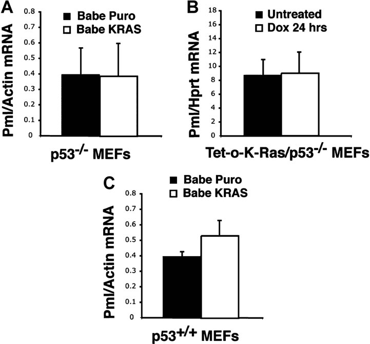
We next investigated whether PML protein upregulation upon oncogenic K-RAS signalling is due to either transcriptional or post-transcriptional mechanisms. We observed no significant differences in Pml mRNA levels by quantitative real-time PCR in p53 null MEFs following transduction with Babe K-RAS or in p53 null rtTA/K-Ras MEFs exposed to dox for 24 h (Fig 2A and B). In WT MEFs (i.e. with a competent p53 response), Pml mRNA was modestly upregulated by oncogenic K-RAS (Fig 2C). We therefore hypothesized that post-transcriptional mechanism(s) support the upregulation of Pml in both p53 null and WT genetic backgrounds.
Figure 2. Pml upregulation upon oncogenic K-Ras does not depend on Pml transcription and is sensitive to rapamycin.
- Histogram showing Pml/Actin mRNA ratio in p53 null MEFs following transduction with Babe Puro (black bar) or Babe Puro RAS (white bar).
- Histogram shows Pml mRNA/HPRT mRNA ratio in Tet-o-K-Ras/p53−/− MEFs expressing rtTA. Black bar, untreated MEFs; white bar, MEFS treated with dox for 24 h.
- Histogram shows Pml mRNA/Actin mRNA ratio in primary MEFs with WT p53 transduced with the indicated retroviruses. Data were obtained from three-independent experiments performed in triplicate. Error bars: standard deviations.
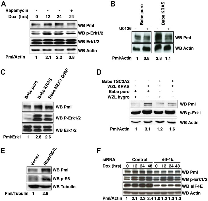
To test whether PML upregulation involves regulation of protein synthesis, we assessed whether PML upregulation is affected by rapamycin, a specific mTOR inhibitor. We found that this agent abrogated PML protein upregulation in p53 null rtTA/K-Ras MEFs exposed to dox (Fig 3A and Fig 2A of Supporting Information).
Figure 3. Oncogenic K-RAS induced PML protein upregulation depends on MEK1 and TSC2.
- WB of Tet-o-K-Ras/p53−/−/rtTA MEFs treated with dox and rapamycin as indicated.
- WB of p53 null MEFs transduced and treated with U0126 as indicated.
- WB of p53 null MEFs transduced with the indicated retroviruses.
- WB analysis of p53 null MEFs transduced as indicated.
- WB of p53 null MEFs following retroviral transduction.
- WB analysis of tet-o-K-Ras/p53−/−/rtTA following siRNA transfection and dox treatment. The intensity of the bands is expressed with the indicated ratios.
Induction of PML by oncogenic K-RAS requires MEK1/2 and TSC2
The RAF/MEK/ERK1/2 signalling cascade is both necessary and sufficient for the establishment of oncogenic K-RAS-induced OIS (Lin et al, 1998). Therefore, we determined whether the MAPK signalling cascade is also involved in mediating PML protein upregulation. We found that treatment of p53 null MEFs expressing oncogenic K-RAS with UO126, a specific MEK1/2 inhibitor, abrogates Pml protein upregulation (Fig 3B). Next, we assessed the effect of MEK1/2 activation on Pml protein level by expressing MEKQ56P, an activated MEK1 mutant, in p53 null MEFs and found that its expression is sufficient to upregulate Pml (Fig 3C).
We have previously demonstrated that ERK1/2 inactivates TSC2 through a direct phosphorylation event, which leads to TSC2 dissociation from TSC1 and impairment of TSC2 inhibition of mTOR signalling (Ma et al, 2005). Thus, an ERK1/2 non-phosphorylatable TSC2 mutant (TSC2A2) significantly hinders the ability of ERK1/2 to activate mTOR (Ma et al, 2005). We tested whether PML upregulation by oncogenic RAS requires TSC2 inactivation by ERK1/2. To this end, we transduced p53 null MEFs with recombinant retroviruses expressing oncogenic K-RAS, TSC2A2 or a combination of the two. We found that TCSC2A2 expression significantly impairs the ability of oncogenic K-RAS to upregulate Pml protein while it did not affect Pml protein levels in the absence of oncogenic K-RAS (Fig 3D). Moreover, we found that expression of RhebQ64L, a constitutively active mutant that inactivates TSC2 and activates mTOR, leads to a dramatic upregulation of Pml protein in p53 null MEFs (Fig 3E). These results strongly suggest that mTOR activation is not only necessary, but also sufficient to mediate PML protein upregulation.
Since mTOR controls protein synthesis through direct regulation of eIF4E and the ribosomal S6 protein (S6K), we tested whether these molecules mediate PML protein upregulation. siRNA knockdown of eIF4E resulted in abrogation of PML protein upregulation by oncogenic RAS (Fig 3F), while siRNA-mediated knockdown of the ribosomal S6 protein had no effect on PML protein upregulation (unpublished observation).
Taken together, these data indicate that PML protein upregulation depends on the RAS/MEK/ERK/mTOR signalling cascade and that eIF4E is the final effector of this signalling cascade when pertaining to PML protein upregulation.
The PML 5′UTR mediates oncogenic K-RAS induced PML protein upregulation
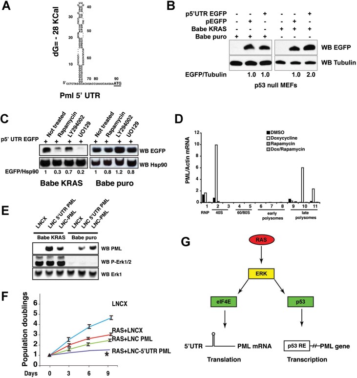
It is well known that eIF4E regulates selective translation of mRNAs though recognition of their 5′-cap structure, therefore, we tested whether the Pml 5′UTR is sufficient to direct PML protein upregulation in the presence of oncogenic RAS. The National Center for Biotechnology Information (NCBI) database lists two Pml mRNAs: NM_178087.2 and NM_008884.2. We used RNA ligase-mediated rapid amplification of 5′ cDNA ends (RLM-5′RACE) to identify the predominant Pml mRNA species in WT and p53 null MEFs. We found that the majority of the transcripts contained a 96 nucleotide (nt.) long 5′UTR identical to NM_178087.2 (Fig 4A). A minor population of transcripts contained heterogeneous 5′ ends that were 62–91 nt. long.
Figure 4. The Pml 5′UTR mediates oncogenic K-RAS-induced PML protein upregulation.
- Primary sequence of the Pml 5′UTR folded by the mFOLD algorithm. Underlined letters indicate Pml ATG. Energy of folding is indicated.
- WB analysis of p53 null MEFs stably transduced with Babe K-RAS or Babe puro. After selection, cells were transfected as indicated. The intensity of the bands is indicated as a ratio between EGFP and tubulin.
- WB analysis of p53 null MEFs transduced and treated as indicated. The ratio between the intensity of EGFP and Hsp90 is indicated.
- Graph shows Pml RNA levels detected by RT-PCR in cytoplasmic cell extracts of p53 null rtTA/K-Ras MEFs fractionated through a sucrose gradient and to obtain polyribosome fractions. Cells were treated with dox for 24 h with or without rapamycin.
- WB analysis of IMR90 cells transduced with LNCX, LNC 5′UTR PML or LNC PML, after selection cells were either transduced with babe puro or babe K-RAS retroviruses.
- The graph shows cell proliferation assays of IMR90 cells transduced as indicated. Asterisk indicates statistical significance between cells transduced with LNC PML and LNC-5′UTR PML. The curve represents the result of three-independent experiments performed in triplicate. Standard deviations were not drawn when too small to appear on graph.
- Molecular mechanisms controlling PML-mediated OIS induction. RE = p53 responsive element.
Notably, the mFOLD program (Zuker, 2003) predicted that the major Pml 5′UTR would contain an ordered stem and loop structure (free energy of folding ΔG = − 28 kcal/mol; Fig 4A).
To directly evaluate the impact of the 5′UTR in the regulation of mouse Pml protein level we generated a plasmid vector in which the CMV immediate-early promoter drives the expression of the 5′UTR of mouse Pml fused to EGFP (p5′UTR EGFP). Transfection of p5′UTR EGFP in p53 null MEFs transduced with the Babe K-RAS retrovirus led to significantly increased levels of EGFP when compared to cells transfected with the pEGFP parental control vector. In contrast, we noted no significant differences in EGFP protein levels following transfection of p53 null MEFs with either p5′UTR EGFP or pEGFP (Fig 4B). Transfection efficiency for the experiments was monitored by adding a Renilla expression vector to the transfection mix (Fig 2B of Supporting Information). We assessed the effects of rapamycin, UO126 and of the PI3K inhibitor LY294002 on the levels of EGFP produced by p5′UTR EGFP following transfection in p53 null MEFs transduced with babe K-RAS or Babe puro. We found that rapamycin or UO126 significantly reduced the levels of EGFP produced after transfection in Babe K-RAS-transduced cells, while the PI3K inhibitor LY294002 had only a minor effect. In contrast, rapamycin or UO129 did not cause any significant effects in Babe puro-transduced cells (Fig 4C). These data indicate that the Pml 5′UTR is sufficient to mediate the effects of oncogenic K-RAS on Pml protein upregulation and that this effect is dependent on the MEK/ERK/mTOR signalling axis specifically in oncogenic K-RAS-expressing cells.
Oncogenic K-RAS expression leads to recruitment of Pml mRNA to polysomes
Next, we directly tested whether oncogenic K-Ras expression leads to recruitment of Pml mRNA to polysomes in p53 null rtTA/K-Ras MEFs exposed to dox, rapamycin, a combination of the two or DMSO. We used a Polymerase Chain Reaction (after reverse transcription) (RT-PCR) assay that tracks the polysomal distribution of mRNAs in cellular extracts along a sucrose gradient (Arava, 2003). We found that Pml mRNA significantly associates with polysomes when oncogenic K-Ras is expressed, but not in control cells or in cells treated with the mTOR inhibitor rapamycin. Importantly, we found that Pml mRNA was distributed mainly to heavy polysomes, as expected for an mRNA that is translated efficiently (Fig 4D). We determined that PML mRNA is recruited to polysomes also in human immortalized fibroblasts with competent p53. As a positive control, we used the mRNA of p27 Kip1, which is recruited to polysomes during oncogenic stress (Fig 2C of Supporting Information; Rajasekhar et al, 2003). These results provide evidence that oncogenic K-RAS-induced PML upregulation is due also to enhanced PML mRNA translation.
The Pml 5′UTR contributes to oncogenic K-RAS-induced cellular senescence
Finally, we tested the role of the PML 5′UTR in directing OIS induction in immortalized human fibroblasts. To this end, we generated retroviruses expressing either the PML open reading frame (LNC PML) or the PML cDNA fused to the 5′UTR (LNC 5′UTR PML). LNC PML, LNC 5′UTR PML, and the parental LCNX viruses were used to transduce IMR90 human fibroblasts. After drug selection with neomycin, cultures were further transduced either with Babe puro or Babe K-RAS retroviruses. PML protein levels were evaluated 6 days after completion of selection with puromycin. This analysis revealed that the PML protein was significantly upregulated by oncogenic K-RAS in IMR90 fibroblasts transduced with LNC 5′UTR PML, when compared to cells transduced with LNC PML (Fig 4E). Moreover, we found that in IMR90 fibroblasts expressing oncogenic K-RAS, transduction with LNC 5′UTR PML further suppresses cellular proliferation as compared to transduction with LNC PML (Fig 4F), which correlated with an increase in the percentage of cells expressing the OIS marker senescence associated β-galactosidase (SA-β-Gal; Fig 2D of Supporting Information).
Next, we determined the effect of a PML cDNA including its 5′UTR on cellular proliferation of p53 null MEFs. The p53 tumour suppressor is essential for OIS (Campisi & d'Adda di Fagagna, 2007). As expected, we did not detect any effects on OIS. However, we noticed that the PML cDNA comprising its 5′UTR was capable of modestly decreasing cell proliferation in p53 null MEFs expressing oncogenic K-RAS (Fig 2E of Supporting Information). We also found that expression of PML cDNA, irrespectively of the presence of its 5′UTR, did not affect oncogenic K-RAS-induced OIS in Pml null MEFs (unpublished observation). This observation is in agreement with the report that ectopic expression of a single PML isoform is not sufficient to induce OIS in Pml null MEFs (Bischof et al, 2002).
Taken together, these data indicate that the PML upregulation mediated by its 5′UTR is of biological significance. These observations are also consistent with the notion that PML has p53-independent antiproliferative effects (Bernardi & Pandolfi, 2007; Wang et al, 1998).
DISCUSSION
Thus far it has been thought that PML upregulation in conditions of cellular stress is due to transcriptional mechanisms (Bernardi & Pandolfi, 2007; Chelbi-Alix et al, 1995; de Stanchina et al, 2004). Our finding that translation initiation mechanisms contribute to PML protein upregulation upon oncogenic stress challenges this view and provides novel insights into the molecular mechanisms regulating oncogenic K-RAS-induced OIS. We also find that PML protein upregulation can also occur independently of transcriptional mechanisms that require p53. Thus, since PML has p53-independent tumour suppressor properties, this mechanism may also provide a back-up tumour suppressor mechanism in cancer cells that are p53-deficient.
Translational control of gene expression provides the cell with a rapid response to external and internal stressors, as opposed to a slower response mediated by nuclear pathways of mRNA synthesis and subsequent nuclear export. For these reasons, we designed our experiments to specifically address the mechanisms regulating PML protein abundance in conditions of acute exposure to oncogenic stress.
Control of translational regulation occurs mostly at the rate-limiting initiation step, which is controlled by the cap-binding protein eIF4E. Our discovery that the 5′UTR sequence of the Pml gene is essential for oncogenic K-Ras-induced upregulation of Pml is consistent with the notion that changes in the 4E-BPs/eIF4E ratio do not alter general translation, but rather affect the translation of a subset of mRNAs that harbour a structured 5′UTR (Hay & Sonenberg, 2004). Significantly, analysis of the human PML 5′UTR sequence listed in the NCBI database revealed a sequence of 87 nt. that folds into a stem and loop structure that closely resembles its murine counterpart (unpublished observation), suggesting that the mechanism of PML upregulation that we have uncovered is not restricted to mouse cells. Accordingly, we have found that PML is upregulated by oncogenic K-RAS both in murine and in human fibroblasts. These observations suggest that the mechanisms leading to PML upregulation in the presence of oncogenic RAS are conserved between murine and human cells.
We propose that 5′UTR-dependent PML upregulation is a mechanism that allows the cell to acutely counteract inappropriate growth stimuli by directly activating protein translation through a MEK/mTOR/eIF4E signalling axis, while slower transcription-dependent mechanisms are activated (Fig 4G). In this regards, our data complement and extend a previous report indicating that PML is a p53 target gene (de Stanchina et al, 2004).
Our report lends support to the emerging notion that translation-dependent mechanisms play a critical role in the OIS response (Petroulakis et al, 2009). We suggest that these mechanisms are not limited to OIS or to PML regulation but may rather be of general significance in cellular responses to stress. Indeed, it has been shown that p53 protein levels are controlled at the translational level upon DNA damage induced by ionizing radiation (Mazan-Mamczarz et al, 2003; Takagi et al, 2005).
Since the initial description that oncogenic K-RAS induces OIS, it has been discovered that the activation of several oncogenic networks can cause OIS. For instance, PTEN inactivation may lead to PTEN-loss-induced cellular senescence (PICS), a type of cellular senescence distinct from OIS, through a mechanism that requires translational upregulation of the p53 (Alimonti et al, 2010). Therefore, it is tempting to speculate that PML, which is a p53 co-factor, may also be upregulated in PICS positively regulating p53, which can be addressed in further studies taking advantage of knockout models. Furthermore, the regulation of OIS involves the integration of several context-dependent signalling pathways and feedback mechanisms. For instance, it has been reported that the activation of the PI3K signalling pathway overcomes oncogenic K-RAS-induced OIS, while others have reported that OIS is due to negative feedback response that potently suppresses oncogenic K-RAS signalling (Courtois-Cox et al, 2006; Kennedy et al, 2011). The former scenario should lead to the upregulation of PML through activation of mTOR, while the second is expected to downregulate it.
The paper explained
PROBLEM:
Oncogene-induced premature cellular senescence (OIS) is a critical tumour suppressor mechanisms that induces a permanent proliferative arrest. Drug therapies that either promote or suppress OIS may significantly impact tumourigenesis in cancer patients.
RESULTS:
In this work, we report that the promyelocytic tumour suppressor (PML), a critical inducer of cellular senescence, is upregulated by oncogenic K-RAS through translation-dependent mechanisms that require mTOR, eIF4E and the PML 5′untranslated region.
IMPACT:
Thus far, induction of oncogene-induced cellular senescence has been largely attributed to transcriptional mechanisms. Thus, these findings unveil a novel mechanism regulating OIS. Moreover, this data raise the concern that inhibitors of the PI3K/mTOR pathway, which are developed as targeted cancer drugs, may blunt OIS causing unexpected and counterproductive effects in cancer patients.
Of note, PML can act as a negative regulator of mTOR (Bernardi et al, 2006). Therefore the mTOR-dependent upregulation of PML in response to stress could not only lead to cellular senescence when cells are unable to cope with aberrant oncogenic signalling, but also represent a way to tune the mTOR-dependent translational output. The role of these possible feedback mechanisms should be ultimately assessed in vivo in mouse cancer models.
Several mTOR inhibitors are used in the treatment of cancer patients. Our work has therefore potential clinical implications since the ability of rapamycin to block PML upregulation in OIS could limit the efficacy of this drug when cellular senescence is a desirable therapeutic outcome. Future studies are therefore warranted to evaluate whether therapies that target the translational apparatus may lead to unintended and counterproductive effects during the treatment of cancers where tumour suppressor proteins are upregulated due to translational mechanisms.
On the other hand, our data suggest that in APL where the PML 5′UTR is fused to the RARα moiety of the PML-RARα oncoprotein, mTOR inhibitors may be extremely beneficial because they would block the translation of the oncogene while globally opposing cell growth and proliferation.
MATERIALS AND METHODS
Mouse strains
P53−/−, CCSP-rtTA/Tet-op-K-Ras, and β-actin TVA transgenic mice were bred to generate compound mutant mice (Fisher et al, 2001; Jacks et al, 1994; Orsulic et al, 2002). Starting at 8 weeks of age, CCSP-rtTA/Tet-op-K-Ras and CCSP-rtTA/Tet-op-K-Ras/p53−/− mice were maintained either on standard or dox containing diet (625 ppm; Harlan-Teklad). Animal studies were performed in accordance to institutional policies.
Cell culture, plasmids, retroviruses and cellular senescence assays
IMR90 and BJ human diploid fibroblasts were obtained from the ATCC and used between passage 10 and 20. These cell lines have the capacity to undergo more than 50 population doublings in culture before undergoing senescence (Nichols et al, 1977; Yi et al, 1999). Day 12–14 mouse embryonic fibroblasts were derived as described previously (Scaglioni et al, 2006). The PML isoform IV expression vector was cloned in the TAG2B expression vector (Agilent Technologies) described previously. pBabe puro, pWzl Hygro, pBabe K-RAS and pBabe H-RAS were obtained from Addgene. LNCX was obtained from Dr. Dusty Miller (Fred Hutchinson Cancer Research Center). The RCAS-rtTA virus was obtained from Dr. Harold E. Varmus. pBabe puro RhebQ64L and pbabe puro MEK1 Q56P were generated by site directed mutagenesis. Recombinant retroviruses were generated as described (Pao et al, 2003; Pear et al, 1993). The p5′UTR EGFP construct was generated by cloning the Pml 5′UTR sequence into pEGFP-N (Clontech). LNC 5′UTR PML was obtained by cloning the murine Pml 5′UTR sequence directly in front of human PML IV in the polylinker of LNCX. Dox was added to cell cultures at a concentration of 1 µg/ml. SA-β-Gal assays were performed as described (Scaglioni et al, 2006). Every experiment was repeated at least three times with equivalent results. Transfections were performed with the effectene reagent (Qiagen). Transfection efficiency for the experiments shown in Fig 4B was monitored by adding the pRL Renilla expression vector (Promega) to the transfection mix.
Immunoblotting, chemicals and antibodies
Immunoblotting was performed with the following antibodies: anti-Flag (Sigma), anti-GFP (Cell Signaling), anti-PML 36.1-104 (Millipore), anti p-ERK1/2, ERK1/2 and anti-eIF4E (Cell Signaling Laboratories), anti-pan RAS (Abcam), anti-actin and anti HSP90 (Sigma). Specific bands were quantified by densitometry. Chemicals were from Calbiochem, Sigma or Invitrogen.
RNA Interference, quantitative RT-real time PCR and RLM-RACE
MEFs were transfected with eIF4E or S6 ribosomal protein siRNA duplexes (eIF4E siRNA/siAb™ Assay Kit-Upstate Cell Signaling Solutions) or nonspecific control SMARTpool siRNA using siRNA DharmaFECT Transfection Reagent. Cells were analysed 48 h after transfection. Quantitative RT real time PCR was performed using Pml, actin and hypoxantine phosphoribosyl transferase specific primers (TaqMan, Gene Expression Assays). RLM-RACE was performed according to the manufacturer instructions (Invitrogen). Every experiment was repeated at least three times with equivalent results.
Immunofluorescence and immunohistochemistry microscopy
Cell cultures were fixed and stained as described (Scaglioni et al, 2006).
Statistical analysis
Prism software was used for the statistical analysis. p-Values were calculated using the unpaired Student's t-test.
Isolation of polyribosomes and RNA
Polyribosome fractionation was performed following standard procedures (Arava, 2003). Every experiment was repeated at least three times with equivalent results.
Acknowledgments
We are grateful to Drs. Harold E. Varmus, William Pao and Katerina Politi for providing the CC10-rtTA/tet-op-K-Ras transgenic mice and reagents. PPS was supported by NIH K08 grant CA 112325, NIH RO1 CA 137195A1, American Cancer Society Institutional Research Grant # 02-196, the Concern Foundation, the Gibson Foundation, Leukaemia Texas Inc. The American-Italian Cancer Foundation supported Andrea Rabellino. GK was supported by a Cancer prevention and Research Institute of Texas training grant RP101496. PPP was supported by NIH grant R01 CA-71692 and RB by K01 CA118259.
Supporting Information is available at EMBO Molecular Medicine online.
The authors declare that they have no conflict of interest.
Author contributions
PPS, AR, TMY, SC, RB, GK performed the experiments described in this paper; CN provided critical reagents; KC performed the polysome profiles; PPS and PPP contributed the overall idea supervised the work and wrote the manuscript.
Supplementary material
Detailed facts of importance to specialist readers are published as ”Supporting Information”. Such documents are peer-reviewed, but not copy-edited or typeset. They are made available as submitted by the authors.
References
- Alimonti A, Nardella C, Chen Z, Clohessy JG, Carracedo A, Trotman LC, Cheng K, Varmeh S, Kozma SC, Thomas G, et al. A novel type of cellular senescence that can be enhanced in mouse models and human tumor xenografts to suppress prostate tumorigenesis. J Clin Invest. 2010;120:681–693. doi: 10.1172/JCI40535. [DOI] [PMC free article] [PubMed] [Google Scholar]
- Arava Y. Isolation of polysomal RNA for microarray analysis. Methods Mol Biol (Clifton, NJ) 2003;224:79–87. doi: 10.1385/1-59259-364-X:79. [DOI] [PubMed] [Google Scholar]
- Bernardi R, Pandolfi PP. Structure, dynamics and functions of promyelocytic leukaemia nuclear bodies. Nat Rev Mol Cell Biol. 2007;8:1006–1016. doi: 10.1038/nrm2277. [DOI] [PubMed] [Google Scholar]
- Bernardi R, Guernah I, Jin D, Grisendi S, Alimonti A, Teruya-Feldstein J, Cordon-Cardo C, Simon MC, Rafii S, Pandolfi PP. PML inhibits HIF-1alpha translation and neoangiogenesis through repression of mTOR. Nature. 2006;442:779–785. doi: 10.1038/nature05029. [DOI] [PubMed] [Google Scholar]
- Bischof O, Kirsh O, Pearson M, Itahana K, Pelicci PG, Dejean A. Deconstructing PML-induced premature senescence. EMBO J. 2002;21:3358–3369. doi: 10.1093/emboj/cdf341. [DOI] [PMC free article] [PubMed] [Google Scholar]
- Campisi J, d'Adda di Fagagna F. Cellular senescence: when bad things happen to good cells. Nat Rev Mol Cell Biol. 2007;8:729–740. doi: 10.1038/nrm2233. [DOI] [PubMed] [Google Scholar]
- Chelbi-Alix MK, Pelicano L, Quignon F, Koken MH, Venturini L, Stadler M, Pavlovic J, Degos L, de The H. Induction of the PML protein by interferons in normal and APL cells. Leukemia. 1995;9:2027–2033. [PubMed] [Google Scholar]
- Chen Z, Trotman LC, Shaffer D, Lin HK, Dotan ZA, Niki M, Koutcher JA, Scher HI, Ludwig T, Gerald W, et al. Crucial role of p53-dependent cellular senescence in suppression of Pten-deficient tumorigenesis. Nature. 2005;436:725–730. doi: 10.1038/nature03918. [DOI] [PMC free article] [PubMed] [Google Scholar]
- Courtois-Cox S, Genther Williams SM, Reczek EE, Johnson BW, McGillicuddy LT, Johannessen CM, Hollstein PE, MacCollin M, Cichowski K. A negative feedback signaling network underlies oncogene-induced senescence. Cancer cell. 2006;10:459–472. doi: 10.1016/j.ccr.2006.10.003. [DOI] [PMC free article] [PubMed] [Google Scholar]
- de Stanchina E, Querido E, Narita M, Davuluri RV, Pandolfi PP, Ferbeyre G, Lowe SW. PML is a direct p53 target that modulates p53 effector functions. Mol Cell. 2004;13:523–535. doi: 10.1016/s1097-2765(04)00062-0. [DOI] [PubMed] [Google Scholar]
- Downward J. Targeting RAS signalling pathways in cancer therapy. Nat Rev. 2003;3:11–22. doi: 10.1038/nrc969. [DOI] [PubMed] [Google Scholar]
- Ferbeyre G, de Stanchina E, Querido E, Baptiste N, Prives C, Lowe SW. PML is induced by oncogenic ras and promotes premature senescence. Genes Dev. 2000;14:2015–2027. [PMC free article] [PubMed] [Google Scholar]
- Fisher GH, Wellen SL, Klimstra D, Lenczowski JM, Tichelaar JW, Lizak MJ, Whitsett JA, Koretsky A, Varmus HE. Induction and apoptotic regression of lung adenocarcinomas by regulation of a K-Ras transgene in the presence and absence of tumor suppressor genes. Genes Dev. 2001;15:3249–3262. doi: 10.1101/gad.947701. [DOI] [PMC free article] [PubMed] [Google Scholar]
- Hay N, Sonenberg N. Upstream and downstream of mTOR. Genes Dev. 2004;18:1926–1945. doi: 10.1101/gad.1212704. [DOI] [PubMed] [Google Scholar]
- Jacks T, Remington L, Williams BO, Schmitt EM, Halachmi S, Bronson RT, Weinberg RA. Tumor spectrum analysis in p53-mutant mice. Curr Biol. 1994;4:1–7. doi: 10.1016/s0960-9822(00)00002-6. [DOI] [PubMed] [Google Scholar]
- Kennedy AL, Morton JP, Manoharan I, Nelson DM, Jamieson NB, Pawlikowski JS, McBryan T, Doyle B, McKay C, Oien KA, et al. Activation of the PIK3CA/AKT pathway suppresses senescence induced by an activated RAS oncogene to promote tumorigenesis. Mol Cell. 2011;42:36–49. doi: 10.1016/j.molcel.2011.02.020. [DOI] [PMC free article] [PubMed] [Google Scholar]
- Lin AW, Barradas M, Stone JC, van Aelst L, Serrano M, Lowe SW. Premature senescence involving p53 and p16 is activated in response to constitutive MEK/MAPK mitogenic signaling. Genes Dev. 1998;12:3008–3019. doi: 10.1101/gad.12.19.3008. [DOI] [PMC free article] [PubMed] [Google Scholar]
- Ma L, Chen Z, Erdjument-Bromage H, Tempst P, Pandolfi PP. Phosphorylation and functional inactivation of TSC2 by Erk implications for tuberous sclerosis and cancer pathogenesis. Cell. 2005;121:179–193. doi: 10.1016/j.cell.2005.02.031. [DOI] [PubMed] [Google Scholar]
- Malumbres M, Barbacid M. RAS oncogenes: the first 30 years. Nat Rev. 2003;3:459–465. doi: 10.1038/nrc1097. [DOI] [PubMed] [Google Scholar]
- Manning BD, Tee AR, Logsdon MN, Blenis J, Cantley LC. Identification of the tuberous sclerosis complex-2 tumor suppressor gene product tuberin as a target of the phosphoinositide 3-kinase/akt pathway. Mol Cell. 2002;10:151–162. doi: 10.1016/s1097-2765(02)00568-3. [DOI] [PubMed] [Google Scholar]
- Mazan-Mamczarz K, Galban S, Lopez de Silanes I, Martindale JL, Atasoy U, Keene JD, Gorospe M. RNA-binding protein HuR enhances p53 translation in response to ultraviolet light irradiation. Proc Natl Acad Sci USA. 2003;100:8354–8359. doi: 10.1073/pnas.1432104100. [DOI] [PMC free article] [PubMed] [Google Scholar]
- Narita M, Krizhanovsky V, Nunez S, Chicas A, Hearn SA, Myers MP, Lowe SW. A novel role for high-mobility group a proteins in cellular senescence and heterochromatin formation. Cell. 2006;126:503–514. doi: 10.1016/j.cell.2006.05.052. [DOI] [PubMed] [Google Scholar]
- Nichols WW, Murphy DG, Cristofalo VJ, Toji LH, Greene AE, Dwight SA. Characterization of a new human diploid cell strain, IMR-90. Science. 1977;196:60–63. doi: 10.1126/science.841339. [DOI] [PubMed] [Google Scholar]
- Orsulic S, Li Y, Soslow RA, Vitale-Cross LA, Gutkind JS, Varmus HE. Induction of ovarian cancer by defined multiple genetic changes in a mouse model system. Cancer cell. 2002;1:53–62. doi: 10.1016/s1535-6108(01)00002-2. [DOI] [PMC free article] [PubMed] [Google Scholar]
- Pao W, Klimstra DS, Fisher GH, Varmus HE. Use of avian retroviral vectors to introduce transcriptional regulators into mammalian cells for analyses of tumor maintenance. Proc Natl Acad Sci USA. 2003;100:8764–8769. doi: 10.1073/pnas.1133333100. [DOI] [PMC free article] [PubMed] [Google Scholar]
- Pear WS, Nolan GP, Scott ML, Baltimore D. Production of high-titer helper-free retroviruses by transient transfection. Proc Natl Acad Sci USA. 1993;90:8392–8396. doi: 10.1073/pnas.90.18.8392. [DOI] [PMC free article] [PubMed] [Google Scholar]
- Pearson M, Carbone R, Sebastiani C, Cioce M, Fagioli M, Saito S, Higashimoto Y, Appella E, Minucci S, Pandolfi PP, et al. PML regulates p53 acetylation and premature senescence induced by oncogenic Ras. Nature. 2000;406:207–210. doi: 10.1038/35018127. [DOI] [PubMed] [Google Scholar]
- Petroulakis E, Parsyan A, Dowling RJ, LeBacquer O, Martineau Y, Bidinosti M, Larsson O, Alain T, Rong L, Mamane Y, et al. p53-dependent translational control of senescence and transformation via 4E-BPs. Cancer Cell. 2009;16:439–446. doi: 10.1016/j.ccr.2009.09.025. [DOI] [PubMed] [Google Scholar]
- Rajasekhar VK, Viale A, Socci ND, Wiedmann M, Hu X, Holland EC. Oncogenic Ras and Akt signaling contribute to glioblastoma formation by differential recruitment of existing mRNAs to polysomes. Mol Cell. 2003;12:889–901. doi: 10.1016/s1097-2765(03)00395-2. [DOI] [PubMed] [Google Scholar]
- Scaglioni PP, Yung TM, Cai LF, Erdjument-Bromage H, Kaufman AJ, Singh B, Teruya-Feldstein J, Tempst P, Pandolfi PP. A CK2-dependent mechanism for degradation of the PML tumor suppressor. Cell. 2006;126:269–283. doi: 10.1016/j.cell.2006.05.041. [DOI] [PubMed] [Google Scholar]
- Serrano M, Lin AW, McCurrach ME, Beach D, Lowe SW. Oncogenic ras provokes premature cell senescence associated with accumulation of p53 and p16INK4a. Cell. 1997;88:593–602. doi: 10.1016/s0092-8674(00)81902-9. [DOI] [PubMed] [Google Scholar]
- Takagi M, Absalon MJ, McLure KG, Kastan MB. Regulation of p53 translation and induction after DNA damage by ribosomal protein L26 and nucleolin. Cell. 2005;123:49–63. doi: 10.1016/j.cell.2005.07.034. [DOI] [PubMed] [Google Scholar]
- Wang ZG, Ruggero D, Ronchetti S, Zhong S, Gaboli M, Rivi R, Pandolfi PP. PML is essential for multiple apoptotic pathways. Nat Genet. 1998;20:266–272. doi: 10.1038/3073. [DOI] [PubMed] [Google Scholar]
- Yi X, Tesmer VM, Savre-Train I, Shay JW, Wright WE. Both transcriptional and posttranscriptional mechanisms regulate human telomerase template RNA levels. Mol Cell Biol. 1999;19:3989–3997. doi: 10.1128/mcb.19.6.3989. [DOI] [PMC free article] [PubMed] [Google Scholar]
- Zuker M. Mfold web server for nucleic acid folding and hybridization prediction. Nucleic Acids Res. 2003;31:3406–3415. doi: 10.1093/nar/gkg595. [DOI] [PMC free article] [PubMed] [Google Scholar]
Associated Data
This section collects any data citations, data availability statements, or supplementary materials included in this article.


