Abstract
The tumour suppressor PTEN is a key negative regulator of the PI3K-Akt pathway, and is frequently either reduced or lost in human tumours. Murine genetic studies have confirmed that reduction of Pten promotes tumourigenesis in multiple organs, and demonstrated dependency of tumour development on the activation of downstream components such as Akt. Insulin-like growth factors (IGFs) act via IGF1R to activate the PI3K-Akt pathway, and are commonly upregulated in cancer. A context-dependent interplay between IGFs and PTEN exists in normal tissue and tumours; increased IGF2 ligand supply induces Pten expression creating an autoregulatory negative feedback loop, whereas complete loss of PTEN may either cooperate with IGF overexpression in tumour promotion, or result in desensitisation to IGF ligand. However, it remains unknown whether neoplasia associated with Pten loss is dependent on upstream IGF ligand supply in vivo. We evaluated this by generation of Pten+/− mice with differing allelic dosage of Igf2, an imprinted gene encoding the potent embryonic and tumour growth factor Igf2. We show that biallelic Igf2 supply potentiates a previously unreported Pten+/− placental phenotype and results in strain-dependent cardiac hyperplasia and neonatal lethality. Importantly, we also show that the effects of Pten loss in vivo are modified by Igf2 supply, as lack of Igf2 results in extended survival and delayed tumour development while biallelic supply is associated with reduced lifespan and accelerated neoplasia in females. Furthermore, we demonstrate that reduction of PTEN protein to heterozygote levels in human MCF7 cells is associated with increased proliferation in response to IGF2, and does not result in desensitisation to IGF2 signalling. These data indicate that the effects of Pten loss at heterozygote levels commonly observed in human tumours are modified by Igf2 ligand, and emphasise the importance of the evaluation of upstream pathways in tumours with Pten loss.
Keywords: PTEN, IGF2, Cowden syndrome, imprinting, placenta, mammary gland
Introduction
PTEN is a dual specificity phosphatase that acts as a key negative regulator of the ligand-activated PI3K-Akt pathway (Carracedo and Pandolfi, 2008). PTEN acts to dephosphorylate phosphatidyl inositol (3,4,5) triphosphate to the diphosphate (4,5), thus reducing activation of Akt. Germline mutations in PTEN cause Cowden syndrome (OMIM 158350), characterised by hamartomas and increased risk of cancer of the breast, thyroid and endometrium, and somatic mutation and epigenetic silencing of PTEN occur at high frequency in sporadic tumours (for example, COSMIC database http://www.sanger.ac.uk/genetics/CGP/cosmic/). Pten+/− mice develop hyperplastic and neoplastic change in multiple organs from 3 months of age and typically die at <15 months from tumours or autoimmune disease (Di Cristofano et al., 1998; Podsypanina et al., 1999; Wang et al., 2010). These phenotypes are potentiated by reduced Pml, Cdkn2a or Tsc2 dosage (Di Cristofano et al., 2001; Ma et al., 2005; Manning, 2005; Trotman et al., 2006) and partially ameliorated by a decrease in downstream components of the PI3K-Akt pathway such as Akt1, Pdk1 and Rictor (Bayascas et al., 2005; Chen et al., 2006; Guertin et al., 2009). In contrast, upstream Grb2 heterozygosity does not modify Pten+/− tumour development (Cully et al., 2004), indicating selective requirement of additional signalling components in Pten+/−-associated tumourigenesis.
Insulin-like growth factor 2 (IGF2) is a potent embryonic growth factor with homology to IGF1 that signals through IGF1R and insulin receptor isoform A to activate the PI3K-Akt and mitogen-activated protein kinase pathways (Foulstone et al., 2005). The supply of IGF2 ligand is tightly regulated, with one mechanism being genomic imprinting of Igf2 both in human and mouse. Imprinting restricts Igf2 expression to the paternal allele with reciprocal maternal expression of a non-coding RNA (ncRNA) H19 (Bartolomei et al., 1991; DeChiara et al., 1991). Coordinate expression of Igf2 and H19 on each allele is regulated by a differentially methylated intergenic imprinting control region (ICR) and competition for shared downstream enhancers between the two loci (Bell and Felsenfeld, 2000). Loss and gain of function of IGF2 contribute to the human growth disorders Silver-Russell (OMIM #180860) and Beckwith-Wiedemann (OMIM #130650) syndromes, respectively. In the mouse, paternal inheritance of an Igf2 null allele (Igf2−p) is associated with approximately 1% wild-type (wt) levels of Igf2 expression and results in foetoplacental growth restriction (60% wt) (DeChiara et al., 1991). In contrast, maternal inheritance of a 13 kb H19/ICR deletion (H19−m) results in both loss of ncRNA and biallelic Igf2 expression, Igf2 mRNA levels of 127–219% wt and foetal (127%) and placental (140%) overgrowth (Leighton et al., 1995). Loss of imprinting of IGF2 is common in human cancers (van Roozendaal et al., 1998; Cui et al., 2002) and murine studies have confirmed that Igf2 is required for tumour progression in the RIP-TAg pancreatic (Christofori et al., 1994), ApcMin intestinal (Hassan and Howell, 2000) and the Ptch+/− medulloblastoma models (Corcoran et al., 2008), whereas biallelic Igf2 supply in combination with ApcMin promotes adenoma progression (Sakatani et al., 2005; Harper et al., 2006). Recently, growth regulatory and tumour suppressive roles for H19 have been established (Yoshimizu et al., 2008; Gabory et al., 2009), and tumour-promoting miRNA (micro RNA) within the Igf2 (miR-483*) and H19 (miR-675-3p) loci reported (Tsang et al., 2010; Veronese et al., 2010).
As a negative regulator of the PI3K-Akt pathway, PTEN performs a pivotal role in the regulation of IGF2 signalling, and several lines of evidence indicate a complex interplay between IGF2 and PTEN in normal tissues and tumours. Pten expression is induced in response to IGF2 ligand supply, generating negative feedback loops dependent on Egr1 and IGFBP2 (Moorehead et al., 2003; Perks et al., 2007). Moreover, evidence in human glioblastomas with PTEN loss indicates that IGF2 overexpression promotes tumour growth, suggesting that the two may cooperate in tumour development (Soroceanu et al., 2007). However, desensitisation of IGF signalling can occur following either PI3 K activation or PTEN loss through feedback mechanisms that include regulation of IGF1R levels, insulin receptor substrate (IRS)-1 stability, regulation of IRS2 and inhibition of IRS-1 by protein kinase C (PKC)ζ (Liu et al., 2001; Ravichandran et al., 2001; Lackey et al., 2007). Collectively, these data suggest that although subtle changes in Igf2 and Pten expression may autoregulate through feedback mechanisms, such compensation is inadequate to prevent the effects of allelic dosage variation of either gene in the mouse (DeChiara et al., 1991; Leighton et al., 1995; Di Cristofano et al., 1998; Podsypanina et al., 1999) and raise the possibility that combined dysregulation may cooperate in tumour promotion. However, at present the in vivo dependency of Pten+/−-associated tumourigenesis on Igf2 supply remains unclear. Here, we sought to address this question by the generation of Pten+/− mice with differing Igf2 allelic dosage, resulting in compound mutants with either normal, substantially decreased or approximately doubled Igf2 expression.
Results
Igf2 and Pten regulate placental growth and organisation
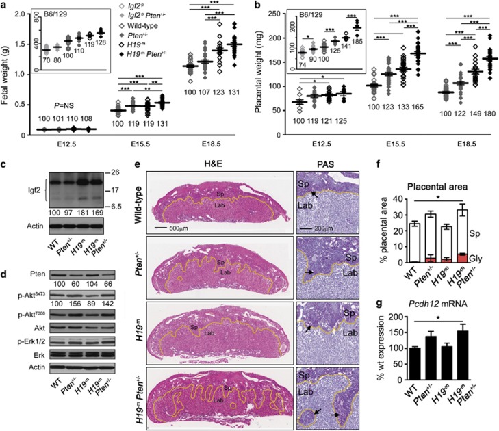
We first evaluated the interactions of Igf2 and Pten by mating mutant mice on a C57BL/6 background to generate wt, Pten+/−, H19−m, and H19−m, Pten+/− compound mutant progeny. The expected foetoplacental overgrowth of H19−m mutants was evident by E12.5 and persisted throughout gestation (Figures 1a and b). Unexpectedly, we also detected an increase in Pten+/− foetal and placental weights by E15.5 (119% and 123% wt, respectively, P<0.001 both comparisons) that subsequently diminished in the foetus by E18.5 (107% wt, P=NS) but persisted in the placenta (122% wt, P<0.001). H19−m, Pten+/− compound mutants were, in turn, larger than either class of single mutant (Figures 1a and b), though the combination was less than additive in the foetus (131% wt at E15.5 and E18.5) and greater than additive in the placenta (165% and 180% wt, P<0.001 vs all groups) (Figures 1a and b).
Figure 1.
Igf2 and Pten regulate foetoplacental growth, Sp and Gly cells. (a, b) Fetal (a) and placental (b) weights by embryonic stage on a C57BL/6 background (main panel) and on an F1 hybrid C57BL/6/129S2 background at E15.5 (insets). Numbers below columns indicate weights as % of wt, numbers on y axis in insets indicate weights in mg. (c) Igf2 immunoblot of littermate E15.5 placental lysates. Multiple bands correspond to the 22 kDa, 156 residue pro-peptide and the 11–17 kDa, 87 and 104 residue ‘big' forms described in human, which undergo variable glycosylation. The mature 7.5 kDa 67-residue peptide was not detected in placental tissue. Numbers below bands indicate % of wt level (quantified by densitometry in Supplementary figure S1b), to the right represent molecular weights in kDa. (d) Immunoblot of littermate E15.5 placental lysates for indicated proteins. Numbers below bands indicate levels as % of wt (quantified by densitometry in Supplementary figure S1d). (e) Littermate placental sections at E18.5. To the left are low magnification haematoxylin and eosin-stained sections, higher magnification panels to the right show staining with periodic acid Schiff. Sp and Lab are indicated, and the Sp/Lab interface highlighted (yellow line). Note disorganisation of Sp/Lab interface in Pten+/− and H19−mPten+/− mutants. periodic acid Schiff staining demonstrates persistent Gly cells in all mutant classes most prominent and abnormally localised in H19−mPten+/− placenta (pink cells indicated by arrows, right panels). Images are representative of ⩾5 placentas examined in each class. (f) Quantification of proportion of Sp and Gly as percentage of placenta determined by point counting. (g) Expression of Gly cell marker Pcdh12 in E15.5 placenta assayed by RT–qPCR. Error bars in (a, b, f, g) indicate ±s.e.m. from ⩾5 biological replicates. *, ** and *** indicate P<0.05, <0.01 and <0.001, respectively.
An approximate doubling of Igf2 mRNA in H19−m and H19−m, Pten+/− mutant placentas was associated with a relatively smaller increase in protein (Figure 1c, Supplementary Figures S1a and b), whereas Pten expression and protein in Pten heterozygotes were half and two thirds wt level, respectively, and independent of Igf2 (Figure 1d, Supplementary Figures S1c and d). As expected, the decrease in Pten protein was accompanied by an increase in Ser473-phosphorylated Akt (p-AktS473) in both Pten+/− and H19−m, Pten+/− mutants (Figure 1d), though importantly, no additional effect of biallelic Igf2 expression was detected on Akt and Erk 1/2 activation. We next evaluated localisation of the placental growth effects. The murine placenta can be divided into three zones; the part-maternally derived decidua; the junctional zone, comprising spongiotrophoblast (Sp) and glycogen cells (Gly); and the labyrinth (Lab), which contains foetal and maternal vessels and is the site of nutrient exchange. Overgrowth of H19−m placentas was largely associated with preservation of placental architecture, whereas Pten+/− mutants displayed tortuosity of the Sp/Lab interface by E15.5 with expansion of the junctional zone (31.48±1.7 vs 25.7±1.68% in wt) (Figures 1e and f).
Biallelic Igf2 supply potentiated both Pten+/− placental phenotypes, with marked abnormality of the Sp/Lab border and greater increase in the size of the junctional zone (34.8±3.49%, P<0.05 vs wt) in H19−m, Pten+/− placentas (Figures 1e and f). Gly cells are known to be Igf2 dependent (Lopez et al., 1996; Carter et al., 2006; Esquiliano et al., 2009) and, as anticipated, were present in increased numbers at E18.5 in H19−m placentas, though interestingly at lower levels than in Pten+/− mutants (Figure 1f). This indication of Gly cell regulation by Pten was supported by an increased expression of the Gly cell marker Pcdh12 (Coan et al., 2006) in Pten+/− placentas (137% wt, P=NS) (Figure 1g). Combined dysregulation in H19−m, Pten+/− placentas was accompanied by further increase in Gly cell number and Pcdh12 expression (154% wt, P<0.05) (Figures 1f and g), and markedly abnormal distribution of Gly cells throughout the Lab and Sp was confirmed with staining by Periodic acid-Schiff (Figure 1e). Immunohistochemistry demonstrated an apparent reciprocal distribution for Pten and Igf2 in subcellular regions of the placenta with increased labelling for activated Akt in the Sp (Supplementary Figures S1e and f) Examination of proliferation in the Lab using ki67 was increased by E18.5 in placentas with biallelic Igf2 supply, consistent with their increased relative overgrowth in late gestation (Supplementary Figure S1g).
We next generated mice with varying allelic dosage of Igf2: null (Igf2−p), monoallelic or biallelic expression and either wt or heterozygous Pten supply on a uniform F1 hybrid C57BL/6/129S2 background. This both reproduced the growth effects and confirmed that the Pten+/− foetoplacental phenotype persisted despite near complete absence of Igf2 (Supplementary Figures S2a–c).
Biallelic Igf2 supply combined with Pten+/− results in cardiac hyperplasia and neonatal lethality
By postnatal day 10 on a C57BL/6 background, a deficit of H19−m,Pten+/− compound mutants was evident (49% of expected, P<0.0001). This was greater in females than males (29% of expected, P<0.001 vs 64%, P=NS) (Supplementary Figure S3a). A significant and disproportionate increase in heart weights in H19−m,Pten+/− female neonates (Supplementary Figure S3b) was absent at postnatal day 5, by which time the deficit was evident. Magnetic resonance imaging at E15.5 (Supplementary Figure S3c) demonstrated that the increased weight of H19−m,Pten+/− hearts was mirrored by changes in biventricular volume and interventricular septal thickness (Supplementary Figures S3d and e). Additionally, two H19−m,Pten+/− foetuses demonstrated a marked dilatation and thinning of the ventricular myocardium (Supplementary Figure S3c). Collectively, these data suggested an association between the cardiac abnormalities and lethality. Quantification of cardiomyocyte density demonstrated growth because of hyperplasia (Supplementary Figure S3f), and analysis confirmed expected Igf2 and Pten mRNA and protein levels in mutant hearts (not shown). As cardiac abnormalities may result from placental defects, we re-examined H19−m,Pten+/− placentas by sex, but found no differences between males and females (not shown). Surviving H19−m,Pten+/− mutants did not display any signs of cardiac failure, and in vivo assay of heart function by dynamic magnetic resonance imaging at postnatal day 30 demonstrated no obvious left ventricular impairment (not shown). However, necropsy of adults (>3 months) revealed cardiomegaly in both Pten+/− and H19−m,Pten+/− animals, though in contrast to neonates this was more marked in compound mutant males (Supplementary Figures S3g–i).
Igf2 modifies Pten+/− survival
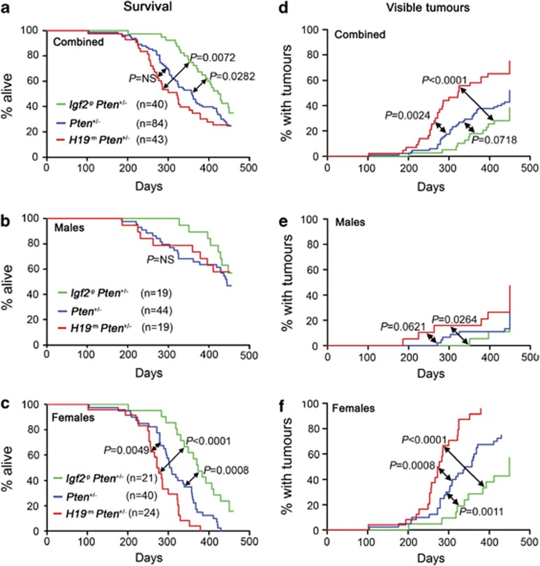
We next examined the effects of Igf2 supply on the survival of adult Pten+/− mice by generation of F1 hybrid C57BL/6/129S2 compound mutants with null, monoallelic or biallelic Igf2 expression. Animals were sacrificed on development of either external tumours of >10 mm diameter, signs of illness or at the study endpoint of 450 days. Lack of Igf2 in Pten+/− mice was associated with significantly extended lifespan (Igf2−p,Pten+/− median 426 days) compared with Pten+/− (359 days) and H19−m,Pten+/− (322 days) animals (Figures 2a–c). This was largely because of effects in females where these differences were greater (373 days, 305.5 days and 274.5 days, respectively, P=0.0008 and P<0.0001), and a significant decrease in survival with biallelic vs monoallelic Igf2 expression was also observed (Figure 2c). Lifespan of males was longer as anticipated (Manning, 2005), and did not vary significantly between groups (Figure 2b). No deaths occurred in any Pten wt animal (n=73) irrespective of Igf2 dose.
Figure 2.
Igf2 allelic dose modifies survival and tumour development in Pten+/− mice. (a–c) Survival of Pten+/− mice by Igf2 allelic dosage. Percentage of total (a), male (b) and female (c) populations alive by time. No deaths occurred in Pten wt controls (n=73). Note the female-predominant effect (c). (d–f) Incidence of large (>10 mm) external tumours. Percentage of total (a), male (b) and female (c) populations with tumours by time. The total includes only external tumours during the study period of 450 days and visceral tumours detected at necropsy at study endpoint. Tumours were predominantly hyperplastic lymph nodes, lymphomas and mammary carcinomas.
Igf2 modifies Pten+/− epithelial tumourigenesis in multiple organs
We analysed the effects of Igf2 on Pten+/− tumourigenesis. Large (>10 mm) external tumours developed significantly earlier in H19−m,Pten+/− (median 323 days) than Pten+/−, or Igf2−p,Pten+/− mice (median not reached, Figures 2d–f). The difference in tumour onset by Igf2 in Pten+/− males (Figure 2e) was significant only for comparison of Igf2 null with biallelic mutants. However, variation in females was larger and highly significant for all comparisons (Figure 2f). External tumours were predominantly hyperplastic lymph nodes, mammary carcinomas and, less frequently, lymphomas. Clinically detectable (>5 mm) Pten+/− lymphadenopathy was accelerated with biallelic Igf2 expression and delayed in the Igf2−p,Pten+/− cohorts (Supplementary Figures S4a–c). Furthermore, histopathological analysis demonstrated a tendency to higher-grade lymphoid lesions with Igf2 gain of function (Supplementary figures S4d–g).
We next focused on the effects of variation of Igf2 allelic supply on epithelial gene expression and tumourigenesis. RT–qPCR (reverse transcriptase-quantitative PCR) on mammary glands, endometria and prostates of virgin animals of 6–8-weeks age confirmed that although Igf2 expression was reduced to approximately 1% of the levels of E9.5 embryos, it remained detectable and was substantially reduced in Igf2−p mutants (1–8% wt), and doubled in H19−m mutants (178.9–200% wt) (Supplementary Figures S5a–c). H19 expression was greatly reduced in H19−m mutants, though no significant change was found in tissues from Igf2−p animals and no alteration in Pten expression with change in Igf2 supply was detected (Supplementary Figures S5d–f).
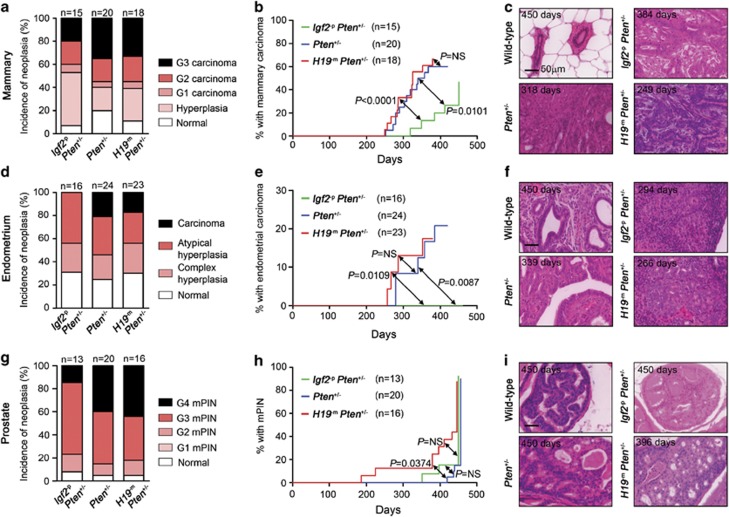
Clinically detectable mammary carcinomas in females occurred significantly later in Igf2−p,Pten+/− mutants than both Pten+/− and H19−m,Pten+/− groups. These data were combined with systematic analysis of macroscopically normal female mammary glands (see Supplementary methods) to determine the true incidence and timing of mammary neoplasia. A trend towards fewer carcinomas in Igf2−p,Pten+/− mutants (Figure 3a) was accompanied by a significant delay in tumour onset (Figure 3b). Carcinomas were histologically similar in all cohorts with prominent stromal component (Figure 3c), with no differences in proliferation by ki67 labelling (not shown). No metastases were detected in any group. No pathological changes were observed in Pten wt female (n=6) or Pten+/− male (n=30) mammary glands, irrespective of Igf2 dose. Endometrial hyperplasia was common in all in Pten+/− groups, though strikingly no case of progression to carcinoma was detected in Igf2−p,Pten+/− females, in contrast to 5/24 (21%) Pten+/− and 4/23 (17%) H19−m,Pten+/− animals (Figure 3d). Thus, lack of Igf2 was associated with a significant delay in carcinoma onset (Figures 3e,f), although these animals were killed for other indications. Although murine prostatic intraepithelial neoplasia (mPIN) was near-universal in all Pten+/− classes, mPIN was detected significantly earlier in H19−m,Pten+/− males and there was a trend towards fewer Grade 4 lesions in the Igf2−p,Pten+/− cohort (Figures 3g–i). Ki67 index in neoplastic endometrial and prostatic lesions did not vary between groups, and patterns of Pten loss and Akt activation were similar (not shown). Though common in all Pten+/− mutants, intestinal polyps were significantly smaller in animals lacking Igf2 (Igf2−p,Pten+/− 3.1±0.3 mm; Pten+/− 4.0±0.2 mm; H19−m,Pten+/− 3.9±0.2 mm, P<0.05 vs Pten+/−). Phaeochromocytoma was fully penetrant in all Pten+/− groups.
Figure 3.
Igf2 allelic dose modifies Pten+/− epithelial neoplasia. (a–c) Mammary pathology in Pten+/− mice by Igf2 supply. Incidence (a) and latency (b) of mammary carcinoma in virgin females. (c) Representative images of normal wt female mammary gland and Grade 3 tumours in each mutant class, illustrating the prominent stromal component of lesions. (d–f) Endometrial pathology in Pten+/− mice by Igf2 supply. Frequency (d) and latency (e) of endometrial hyperplasia/carcinoma with representative images (f) of normal wt endometrium and the highest grade lesions detected in each mutant genotype (Igf2−pPten+/− atypical hyperplasia; Pten+/− G3 serous adenocarcinoma; H19−mPten+/− G2 endometrioid adenocarcinoma). (g–i) mPIN in Pten+/− mice by Igf2 supply. Incidence (g), latency (h) and representative images (i) of normal wt prostate and Grade 4 mPIN in Pten+/− mutants. Numbers in each panel in (c, f and i) indicate the age of animal at the time of killing.
Promotion of mammary tumourigenesis by Igf2 combined with decreased Pten protein
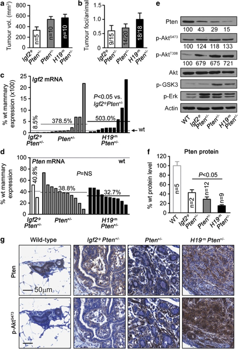
We next sought to examine the effect of Igf2 effect on mammary tumourigenesis. Despite the extended life span of Pten+/− females lacking Igf2, mammary tumours in this group remained smaller than other mutants (Figure 4a). Conversely, tumour multiplicity tended to increase with gain of Igf2 alleles (Figure 4b). Mammary hyperplasia, a preneoplastic lesion (Supplementary Figure S6a), was detected at similar frequency in females of all genotypes (Figure 3a), suggesting that Igf2 may promote the transition to carcinoma in Pten+/− females. RT–qPCR confirmed that Igf2 expression in mammary carcinomas corresponded with Igf2 allelic dose (Figure 4c). As in the normal mammary gland, H19 expression was substantially decreased in H19−m,Pten+/− mutant carcinomas (Supplementary Figure S6b). Levels of Pten mRNA in tumours relative to wt mammary gland were reduced by a similar degree in all genotypes (P=NS) (Figure 4d). There was no evidence of feedback downregulation of Igf1r secondary to this (Supplementary Figure S6c), though interestingly Igf1 expression was low in Pten+/− and H19−m,Pten+/− mammary carcinomas (Supplementary Figure S6d). Notably, despite similar Pten expression, immunoblot analysis demonstrated a significant progressive decrease in tumour Pten protein with increasing Igf2 allelic dose (Figures 4e and f). Associated Akt phosphorylation was universal (Figure 4e, Supplementary Figures S6e and f), and a trend to greater Akt activation with Igf2 gain of function was mirrored by a greater nuclear exclusion of FoxO1, a readout of PI3K-Akt pathway activity (Supplementary Figure 6g). Immunohistochemistry demonstrated foci of epithelial Pten loss in a minority of advanced lesions only (Figure 4g), insufficient in extent to fully explain the relative reduction in total protein.
Figure 4.
Igf2 promotes Pten+/− mammary tumourigenesis. (a, b) Mean mammary tumour volume (a) and mean number of tumour foci per animal (b) by Igf2 allelic dose. (c, d) Waterfall plots showing expression of Igf2 (c) and Pten (d) in mammary carcinomas (only two tumours in Igf2−pPten+/− mutants were of sufficient size to obtain tissue for RNA and protein analysis in addition to fixation). Horizontal lines indicate wt level (light grey) and mean expression of each mutant class (black), with percentage values relative to wt mammary gland. Expression was elevated >2-fold in 4/11 Pten+/− and 6/10 H19−mPten+/− mutants. (e, f) Immunoblot of mammary tumor lysates (e) showing progressive decrease in Pten protein with increasing Igf2 allelic dose, quantified by densitometry in (f). Numbers below bands in (e) indicate protein level as % of wt mammary gland from densitometry in (f) and Supplementary Figures S6e and f. Note the increase in p-AktT308 relative to p-AktS473. (g) Immunohistochemistry demonstrated infrequent foci of complete epithelial Pten loss in advanced lesions only. Error bars in (a,b and f) represent ±s.e.m. Statistical comparisons in (c, d and f) are between Pten+/− mutant classes.
Perturbation of miR-483* and miR-675-3p in Igf2−p and H19−m mutants
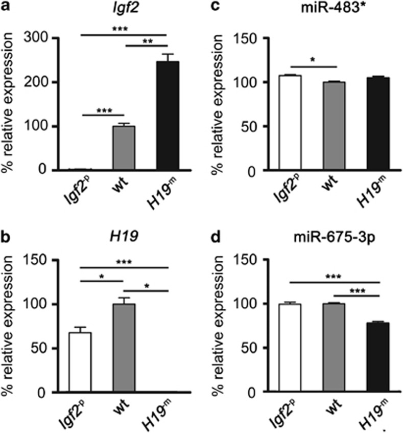
Given the recent demonstration of tumour-promoting functions for miR-483 and miR-675, we next assessed whether their perturbation in Igf2−p and H19−m mutants was likely to have contributed to the modification of phenotype. Using E9.5 embryos, selected for their high expression of Igf2 and H19 (Burns and Hassan, 2001), we confirmed the expected changes in Igf2 expression in Igf2−p and H19−m embryos (0.8%, and 246.6% wt, respectively) (Figures 5a and b). We next examined changes in miRNA expression in both classes of mutant using Ilumina 96-assay universal array matrix miRNA arrays.
Figure 5.
Perturbation of miR-483* and miR-675-3p associate with Igf2 and H19/DMR deletions. Quantification of (a) Igf2, (b) H19, (c) miR-483* and (d) miR-675-3p expression in E9.5 embryos by RT–qPCR (a, b) and Illumina array (c, d). Results are shown as percentage of wt levels. Error bars represent ±s.e.m. of 14–28 biological replicates per group. *, ** and *** indicate P<0.05, <0.01 and <0.001, respectively.
miR-483* is located within the second intron of Igf2 in a region of the gene spared by the targeting construct used in this study (DeChiara et al., 1990). We anticipated an increase in miR-483* levels in H19−m mutants of similar magnitude to that of Igf2, but surprisingly this was not the case (105.1% wt, P=NS). Also unexpected was an upregulation of miR-483* in Igf2−p mutants (107.3% wt) (Figure 5c). Importantly, no correlation between Igf2 and miR-483* expression was detected (R2=0.365, P=NS). miR-675-3p is located within the first exon of H19, which is deleted along with 10 kb of 5′ flanking sequence containing the DMR in the ΔH19/DMR mutant allele (Leighton et al., 1995). As expected, H19−m mutants showed a significant decrease in miR-675-3p expression (78.1% wt, P<0.001), though this was smaller than the reduction in H19 ncRNA (Figure 5d). Notably, miR-675-3p expression was unchanged in Igf2−p mutants (99.6% wt, P=NS) (Figure 5d). Changes in the expression of 377 additional miRNAs analysed were modest and are listed in Supplementary Table S1. Aside from miRNA perturbations that we could attribute to expression of the neomycin resistance gene, we observed a number of miRNAs deregulated to similar extents in mutants as miR-483* and miR-675-3p. Importantly, no miRNA significantly downregulated in Igf2−p embryos demonstrated significant upregulation in H19−m embryos, or vice versa.
Reduction of PTEN protein to heterozygote levels in human breast cancer cells associated with increased IGF2-mediated proliferation
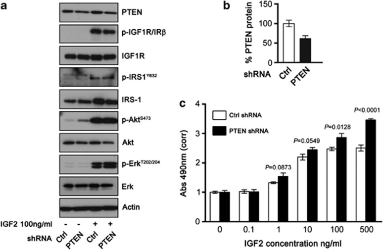
PTEN loss has previously been shown to cause desensitisation to IGF1 and insulin signalling (Lackey et al., 2007). To reconcile these results with our data, and to examine the interaction between IGF2 and PTEN loss in a system free from H19 ncRNA, miR-483* and miR-675-3p perturbation, we used shRNA (small hairpin RNA) to reduce PTEN expression to heterozygote levels in MCF7 human breast cancer cells. PTEN protein in MCF7 cells with partial PTEN knockdown (MCF7PTEN KD) was 61% that of MCF7 cells expressing control shRNA (MCF7CTRL) (Figures 6a and b). As anticipated, reduced PTEN in MCF7PTEN KD cells was accompanied by increased Akt activation (Figure 6a). Importantly, no changes in levels of total IGF1R, total IRS1 (Figure 6a) or inhibitory Ser-612 phosphorylated IRS1 (not shown) were detected. Following IGF2 stimulation, both MCF7CTRL and MCF7PTEN KD cells demonstrated similar IGF1R-PI3K-Akt and mitogen-activated protein kinase pathway activation as demonstrated by comparable levels of activated IGF1R (p-IGF1RY1131), IRS1 (p-IRS-1Y632), Akt (p-AktS473) and Erk1/2 (p-ErkT202/204) (Figure 6a).
Figure 6.
Reduction of PTEN protein to heterozygote levels sensitises MCF7 cells to mitogenic effect of IGF2. (a) MCF7 human breast cancer cells with stable expression of either scrambled control (Ctrl) or anti-PTEN shRNAs were serum starved overnight and then stimulated with recombinant human IGF2 at 100 ng/ml. Lysates were prepared at baseline and after 2 hours following treatment, and immunoblotting for the proteins indicated performed. (b) Densitometry quantification of the reduction in PTEN protein (without IGF2 stimulation) from (a). (c) Proliferation of MCF7 cells expressing either control or anti-PTEN shRNA in serum-free medium following stimulation by IGF2. Exogenous ligand was indicated at the concentrations indicated, and absorbance measured after 48 h by MTS assay. Results are normalised to the values in controls treated with vehicle alone. Error bars indicate ±s.e.m. of at minimum six replicates per dose. Data in (a) and (b) are representative of experiments performed in duplicate and triplicate, respectively.
It has also been shown in MCF7 cells that high concentrations of IGF2 result in relative inhibition of cellular proliferation through feedback induction of PTEN (Perks et al., 2007). We next assessed whether partial PTEN knockdown abrogated this autoregulation. Stimulation of MCF7CTRL and MCF7PTEN KD cells with increasing concentrations of IGF2 confirmed the mitogenic effect of recombinant ligand at concentrations of >1 ng/ml, and as anticipated in MCF7CTRL cells this plateaued at 100 ng/ml (Figure 6c). Notably, MCF7PTEN KD cells demonstrated increased proliferation in response to IGF2, most strikingly at high ligand concentrations, with no evidence of a plateau in proliferation (Figure 6c).
Human stromal interaction of IGF2 and PTEN
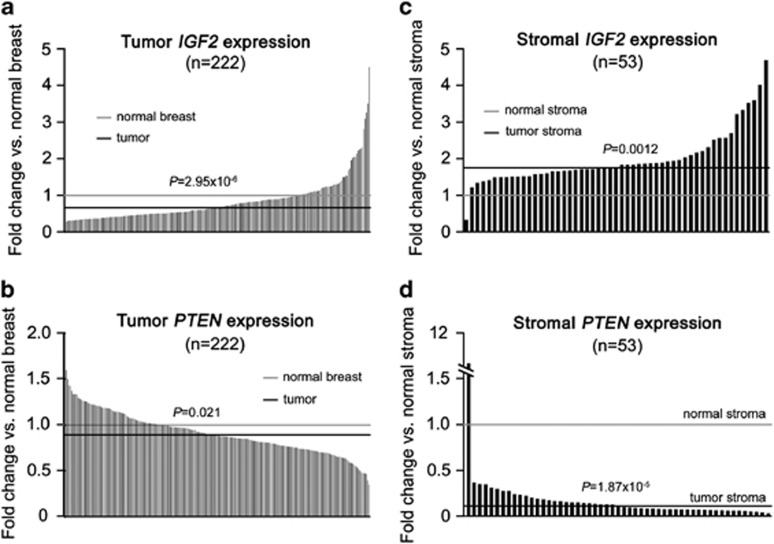
Finally, in light of the apparent promotion of stroma-predominant Pten+/− mammary tumours by Igf2 in the mouse, we evaluated the expression of IGF2 and PTEN in human breast carcinomas. We analysed two microarray data sets with normal and tumour samples, the first comprising a cohort of whole breast tumours (Camps et al., 2008) and the second a published breast cancer micro-dissected stroma data set (Finak et al., 2008). In whole tumours, IGF2 overexpression was relatively infrequent with a >2-fold increase in mRNA detected in 5.6% of tumours (Figure 7a). Unlike the mouse mutants, miR-483 expression closely correlated with that of IGF2 (R2=0.6, P<2.2 × 10⁻¹⁶) and was in fact reduced in tumours compared with normal breast (88.9%, P<2.44 × 10⁻¹³), whereas miR-675 expression was not significantly altered (98.6%, P=NS). Interestingly, the decrease in PTEN expression in breast tumours was modest (median 89.1% normal breast), with few tumours showing levels <50% of normal breast (Figure 7b). In contrast, stromal-specific analysis demonstrated near-universal increase in IGF2 (median 176%, P=0.0012) and decrease in PTEN expression (median 11.1%, P=1.87 × 10⁻⁵) in tumour stroma compared with normal stroma (Figures 7c and d).
Figure 7.
IGF2 and PTEN microarray expression in human breast carcinoma. Human breast cancer data sets suggest stromal role of IGF2 and PTEN dysregulation. IGF2 (a, c) and PTEN (b, d) expression in whole breast carcinomas compared with normal breast tissue (222 tumours and 10 normal breast pools) (a, b), and tumour stroma compared with normal stroma (53 tumour and 12 normal samples) (c, d). Grey lines show median expression in normal breast/breast stromal samples and black lines median expression in breast tumour/breast tumour stroma.
Discussion
We sought to determine whether variation in Igf2 supply secondary to change in Igf2 allelic dosage modifies Pten+/− tumourigesis in order to evaluate the extent to which the effects of Pten loss in vivo are dependent upon upstream signalling. We report several novel findings with respect to development and tumourigenesis.
The Pten+/− placental phenotype we describe demonstrates tissue-specific requirements for biallelic Pten expression in mammalian development. Although both Pten+/− placental overgrowth and Sp disorganisation were increased by biallelic Igf2 supply, they were not ameliorated in Igf2 null mutants, and the discrete localisation of Igf2, Pten and p-AktS473 within the placental Lab are consistent with a complex interaction (Coan et al., 2006). However, when combined with the Akt1 null placental phenotype of small size, Sp and Gly cell deficiency (Yang et al., 2003), our data indicate that an Igf2-Pten-Akt axis regulates placental development (Lopez et al., 1996; Carter et al., 2006; Esquiliano et al., 2009). Although placental abnormalities in mouse mutants are frequently more severe on a C57BL/6 background, we confirmed that this is strain independent (Frank et al., 2002). We failed to detect quantitative differences in proliferation to explain the placental overgrowth in mid-gestation, and it is plausible that our findings result either from subtle cumulative changes or variation earlier than E12.5 (Burns and Hassan, 2001). The use of conditional Igf2 and Pten alleles may facilitate mechanistic insights into the relationship between upstream Igf2 expression and localisation and Pten loss in the placenta.
It is noteworthy that similar to the placenta, Igf2 is expressed at high levels in neonatal heart, and recent data suggest an important role for Igf2 in cardiac development (Li et al., 2011). Though conditional deletion of both Pten alleles results in cardiac hypertrophy (Crackower, 2002), murine cardiac effects of germline heterozygosity have to the best of our knowledge not previously been described. However, a case of ventricular dilatation has been reported in the human (Reardon et al., 2001). The cardiac hyperplasia and neonatal lethality with combined Igf2 and Pten dysregulation we report is consistent with an interaction in heart development, though confirmation of this will require the use of conditional genetics to exclude placental effects.
Igf2 has previously been shown to function as a progression factor or second signal in tumour development (Christofori et al., 1994; Hassan and Howell, 2000; Harper et al., 2006; Corcoran et al., 2008). We demonstrate that the marked reduction of Igf2 expression (1–8% wt) in Igf2−p and more modest increase (179–200% wt) in H19−m mutants are associated with respectively a substantial amelioration and lesser potentiation of Pten+/− tumourigenesis, consistent with a similar role for Igf2 in the context of Pten haploinsufficiency. It is noteworthy that these changes occurred at relevant levels of Igf2 expression, in contrast to previous transgenic non-physiological studies (Bates et al., 1995). Pten+/− mammary tumours typically retain heterozygote levels of Pten protein (Alimonti et al., 2010). The progressive decrease in tumour Pten protein with gain of Igf2 is suggestive of additional post-transcriptional regulatory mechanisms, and contrasts with the previous report of Pten upregulation in response to increased Igf2 (Moorehead et al., 2003). With respect to other relevant feedback regulation secondary to PTEN loss, such as desensitisation to IGF (Lackey et al., 2007), decreased Pten protein in mammary carcinomas was not accompanied by downregulation of Igf1r expression. Moreover, reduction of PTEN to heterozygote levels in MCF7 human breast cancer cells resulted in potentiation of the mitogenic effects of IGF2. These changes were greatest at high doses of ligand, consistent with abrogation of the feedback mechanism of PTEN upregulation previously demonstrated (Moorehead et al., 2003; Perks et al., 2007). Collectively, these data suggest that the effects generated by heterozygote levels of PTEN may still be dependent on upstream ligand.
As noted previously, the H19 ncRNA has recently been demonstrated to have tumour suppressive activity in vivo (Yoshimizu et al., 2008), whereas miRNAs in the IGF2 and H19 loci; miR-483 and miR-675 respectively, have both been ascribed tumour-promoting effects (Tsang et al., 2010; Veronese et al., 2010). Although Igf2 expression across the allelic series increased from 1 to 247%, there was no similar variation of any miRNA. Importantly, loss of H19 (<1% of wt) was not mirrored by equivalent reduction in miR-675-3p in H19−m mutants, and miR-483* expression appeared relatively unchanged. Disruption of miR-483* and miR-675-3p was minimal in Igf2−p animals, with no consistent pattern of H19 dysregulation detected. In addition to these changes, our global analysis shows that many independent miRNA are deregulated in both Igf2−p and H19−m mutants. Our data cannot exclude selective dysregulation of miR-483* and miR-675-3p in tumours, as the small number of Igf2−p,Pten+/− tumours available precluded meaningful analysis. Definitive in vivo assessment of the contribution of the miR-483* and miR-675-3p to phenotype may only be provided by specific conditional deletion of respective loci that modify miRNA expression independent of Igf2. However, our in vitro data demonstrate the proliferative effects of IGF2 at heterozygote levels of PTEN protein in a system free from H19, miR-483* and miR-675-3p perturbation.
IGF2 overexpression by human breast cancer stroma has been postulated to promote neoplastic growth by a paracrine mechanism (Singer et al., 1995). We demonstrate that increased Igf2 mRNA in stroma-prominent Pten+/− murine mammary tumours is mirrored by near-universal upregulation of IGF2 and downregulation of PTEN in human breast cancer stroma. The role of stromal Pten in suppression of mammary tumourigenesis has recently been demonstrated (Trimboli et al., 2009). Our data suggest that the role of stromal IGF2 in breast cancers merits further examination.
In summary, we show that variation of Igf2 ligand supply is associated with modification of development, survival and tumourigenesis in Pten+/− mice, that these changes are unlikely to result from perturbation of the H19 ncRNA, miR-483* or miR-675-3p, and that IGF2 ligand cooperates with partial PTEN loss to promote breast cancer cell proliferation in vitro. Collectively, these data indicate that the effects of Pten loss at heterozygote levels commonly observed in human tumours are modified by Igf2, and emphasise the importance of the evaluation of upstream pathways in tumours with Pten loss.
Materials and methods
Mice
Igf2tm1Rob (paternal allele disruption, Igf2−p) H19tm1Tilg (maternal allele disruption H19−m) and Ptentm1Rps (heterozygote Pten+/−) mice were housed and genotyped by PCR as have been described previously (DeChiara et al., 1991; Leighton et al., 1995; Podsypanina et al., 1999) (see Supplementary Information for details of breeding and primer sequences). All procedures were approved by the UK Home Office and performed under a Home Office Project license following the University of Oxford ethical committee approval.
Tissue analysis
RT–qPCR, immunoblotting, histology and immunohistochemistry were performed by standard techniques. Full methods are provided in Supplementary Information.
3D Magnetic resonance microscopy
Formalin-fixed foetuses obtained at E15.5 were suspended in phosphate-buffered saline doped with gadolinium diethylenetriaminepentaacetic acid (DTPA) and imaged by high-resolution magnetic resonance imaging microscopy by DJS Cardiac measurements were made using ImageJ (image processing and analysis in Java, http://rsbweb.nih.gov/ij/ (NIH)). Full details are provided in Supplementary information.
Histopathology
Haematoxylin and eosin-stained sections of mammary, endometrial and prostate tumours were analysed blinded to genotype by two experienced histopathologists (BRP and SM). Grading of neoplastic change was made according to published criteria (see Supplementary Information).
miRNA analysis of embryos
RNA was extracted from embryos at E9.5 and hybridised to Illumina mouse microRNA assay pool of 380 mouse microRNAs from Sanger miRBase v9.1 (http://www.mirbase.org/) as described in Supplementary Information.
Cell culture
MCF7 cells were grown in Dulbecco's modified Eagle's medium supplemented with 10% foetal calf serum, 1% glutamine, penicillin and streptomycin in a humidified atmosphere with 5% CO2 concentration. Stable expression of shRNAs targeting PTEN or a scrambled control sequence in MCF7 cells was achieved by standard retroviral methods as using the P-SuperRetro Puro vector (Oligoengine Seattle, WA, USA), and Phoenix packaging cells (Gentaur Europe, Kamenhout, Belgium, described fully in Supplementary Information). Experiments were performed in serum free Ham's/Dulbecco's modified Eagle's medium 1:1 medium, using recombinant human IGF2 (GroPep, Adeleide, Australia).
Human microarray analysis
Human data sets were downloaded from GEO (Gene Expression Omnibus) and analysed using R (http://cran.r-project.org/) as described in Supplementary Information (FMB).
Statistical analysis
Data were compiled in Excel and analysed using GraphPad Prism 5.0 (Graphpad Software Inc, La Jolla, CA, USA). Comparisons between genotypes were made by one-way analysis of variance with Tukey's post-test to adjust for multiple comparisons, with the exception of tumours, which were analysed by non-parametric Kruskal-Wallis test with Dunn's post-test to adjust for multiple comparisons. Comparisons of proportions were made by the χ-square test, or Fisher's exact test when numbers in groups were <5. Survival curves/tumour-free curves were plotted according to the method of Kaplan-Meier with comparison between groups made by the log-rank test. Statistical methods used for analysis of human microarray data are provided in Supplementary Information.
Acknowledgments
Igf2 knockout mice were a kind gift from Argiris Efstratidiadis, H19/ICR knockout mice from Shirley Tilghman and Pten knockout mice from Ramon Parsons. PTEN shRNA sequences were generously provided by Nick Leslie. We also thank Richard Stillion (histology), Miguel Constancia, Ionel Sandovici, Elisabeth Robertson, Yoichi Sekita and Shuomo Bhattacharya for their helpful discussions. This work was funded by a Cancer Research UK (CRUK) Clinical Research Training Fellowship (DC) and CRUK Programme Grant C429 (ABH).
The authors declare no conflict of interest.
Footnotes
Supplementary Information accompanies the paper on the Oncogene website (http://www.nature.com/onc)
Supplementary Material
References
- Alimonti A, Carracedo A, Clohessy JG, Trotman LC, Nardella C, Egia A, et al. Subtle variations in Pten dose determine cancer susceptibility. Nat Genet: United States. 2010;42:454–458. doi: 10.1038/ng.556. [DOI] [PMC free article] [PubMed] [Google Scholar]
- Bartolomei MS, Zemel S, Tilghman SM. Parental imprinting of the mouse H19 gene. Nature. 1991;351:153–155. doi: 10.1038/351153a0. [DOI] [PubMed] [Google Scholar]
- Bates P, Fisher R, Ward A, Richardson L, Hill DJ, Graham CF. Mammary cancer in transgenic mice expressing insulin-like growth factor II (IGF-II) Br J Cancer. 1995;72:1189–1193. doi: 10.1038/bjc.1995.484. [DOI] [PMC free article] [PubMed] [Google Scholar]
- Bayascas JR, Leslie NR, Parsons R, Fleming S, Alessi DR. Hypomorphic mutation of PDK1 suppresses tumorigenesis in PTEN(+/-) mice. Current Biology: CB. 2005;15:1839–1846. doi: 10.1016/j.cub.2005.08.066. [DOI] [PubMed] [Google Scholar]
- Bell AC, Felsenfeld G. Methylation of a CTCF-dependent boundary controls imprinted expression of the Igf2 gene. Nature. 2000;405:482–485. doi: 10.1038/35013100. [DOI] [PubMed] [Google Scholar]
- Burns JL, Hassan AB. Cell survival and proliferation are modified by insulin-like growth factor 2 between days 9 and 10 of mouse gestation. Development. 2001;128:3819–3830. doi: 10.1242/dev.128.19.3819. [DOI] [PubMed] [Google Scholar]
- Camps C, Buffa FM, Colella S, Moore J, Sotiriou C, Sheldon H, et al. hsa-miR-210 Is induced by hypoxia and is an independent prognostic factor in breast cancer. Clin Cancer Res: United States. 2008;14:1340–1348. doi: 10.1158/1078-0432.CCR-07-1755. [DOI] [PubMed] [Google Scholar]
- Carracedo A, Pandolfi PP. The PTEN-PI3K pathway: of feedbacks and cross-talks. Oncogene. 2008;27:5527–5541. doi: 10.1038/onc.2008.247. [DOI] [PubMed] [Google Scholar]
- Carter AM, Nygard K, Mazzuca DM, Han VK. The expression of insulin-like growth factor and insulin-like growth factor binding protein mRNAs in mouse placenta. Placenta. 2006;27:278–290. doi: 10.1016/j.placenta.2005.01.014. [DOI] [PubMed] [Google Scholar]
- Chen ML, Xu PZ, Peng XD, Chen WS, Guzman G, Yang X, et al. The deficiency of Akt1 is sufficient to suppress tumor development in Pten+/− mice. Genes Dev. 2006;20:1569–1574. doi: 10.1101/gad.1395006. [DOI] [PMC free article] [PubMed] [Google Scholar]
- Christofori G, Naik P, Hanahan D. A second signal supplied by insulin-like growth factor II in oncogene-induced tumorigenesis. Nature. 1994;369:414–418. doi: 10.1038/369414a0. [DOI] [PubMed] [Google Scholar]
- Coan PM, Conroy N, Burton GJ, Ferguson-Smith AC. Origin and characteristics of glycogen cells in the developing murine placenta. Dev Dyn. 2006;235:3280–3294. doi: 10.1002/dvdy.20981. [DOI] [PubMed] [Google Scholar]
- Corcoran RB, Bachar Raveh T, Barakat MT, Lee EY, Scott MP. Insulin-like growth factor 2 is required for progression to advanced medulloblastoma in patched1 heterozygous mice. Cancer Res. 2008;68:8788–8795. doi: 10.1158/0008-5472.CAN-08-2135. [DOI] [PMC free article] [PubMed] [Google Scholar]
- Crackower MA. Regulation of myocardial contractility and cell size by distinct PI3K-PTEN signaling pathways. Cell. 2002;110:737–749. doi: 10.1016/s0092-8674(02)00969-8. [DOI] [PubMed] [Google Scholar]
- Cui H, Onyango P, Brandenburg S, Wu Y, Hsieh CL, Feinberg AP. Loss of imprinting in colorectal cancer linked to hypomethylation of H19 and IGF2. Cancer Res. 2002;62:6442–6446. [PubMed] [Google Scholar]
- Cully M, Elia A, Ong SH, Stambolic V, Pawson T, Tsao MS, et al. grb2 heterozygosity rescues embryonic lethality but not tumorigenesis in pten+/- mice. Proc Natl Acad Sci USA. 2004;101:15358–15363. doi: 10.1073/pnas.0406613101. [DOI] [PMC free article] [PubMed] [Google Scholar]
- DeChiara TM, Efstratiadis A, Robertson EJ. A growth-deficiency phenotype in heterozygous mice carrying an insulin-like growth factor II gene disrupted by targeting. Nature. 1990;345:78–80. doi: 10.1038/345078a0. [DOI] [PubMed] [Google Scholar]
- DeChiara TM, Robertson EJ, Efstratiadis A. Parental imprinting of the mouse insulin-like growth factor II gene. Cell. 1991;64:849–859. doi: 10.1016/0092-8674(91)90513-x. [DOI] [PubMed] [Google Scholar]
- Di Cristofano A, De Acetis M, Koff A, Cordon-Cardo C, Pandolfi PP. Pten and p27KIP1 cooperate in prostate cancer tumor suppression in the mouse. Nat Genet. 2001;27:222–224. doi: 10.1038/84879. [DOI] [PubMed] [Google Scholar]
- Di Cristofano A, Pesce B, Cordon-Cardo C, Pandolfi PP. Pten is essential for embryonic development and tumour suppression. Nat Genet. 1998;19:348–355. doi: 10.1038/1235. [DOI] [PubMed] [Google Scholar]
- Esquiliano DR, Guo W, Liang L, Dikkes P, Lopez MF. Placental glycogen stores are increased in mice with H19 null mutations but not in those with insulin or IGF type 1 receptor mutations. Placenta. 2009;30:693–699. doi: 10.1016/j.placenta.2009.05.004. [DOI] [PMC free article] [PubMed] [Google Scholar]
- Finak G, Bertos N, Pepin F, Sadekova S, Souleimanova M, Zhao H, et al. Stromal gene expression predicts clinical outcome in breast cancer. Nat Med. 2008;14:518–527. doi: 10.1038/nm1764. [DOI] [PubMed] [Google Scholar]
- Foulstone E, Prince S, Zaccheo O, Burns JL, Harper J, Jacobs C, et al. Insulin-like growth factor ligands, receptors, and binding proteins in cancer. J Pathol. 2005;205:145–153. doi: 10.1002/path.1712. [DOI] [PubMed] [Google Scholar]
- Frank D, Fortino W, Clark L, Musalo R, Wang W, Saxena A, et al. Placental overgrowth in mice lacking the imprinted gene Ipl. Proc Natl Acad Sci USA. 2002;99:7490–7495. doi: 10.1073/pnas.122039999. [DOI] [PMC free article] [PubMed] [Google Scholar]
- Gabory A, Ripoche MA, Le Digarcher A, Watrin F, Ziyyat A, Forne T, et al. H19 acts as a transregulator of the imprinted gene network controlling growth in mice. Development. 2009;136:3413–3421. doi: 10.1242/dev.036061. [DOI] [PubMed] [Google Scholar]
- Guertin DA, Stevens DM, Saitoh M, Kinkel S, Crosby K, Sheen J-H, et al. mTOR complex 2 is required for the development of prostate cancer induced by Pten loss in mice. Cancer Cell. 2009;15:148–159. doi: 10.1016/j.ccr.2008.12.017. [DOI] [PMC free article] [PubMed] [Google Scholar]
- Harper J, Burns JL, Foulstone EJ, Pignatelli M, Zaina S, Hassan AB. Soluble IGF2 receptor rescues Apc(Min/+) intestinal adenoma progression induced by Igf2 loss of imprinting. Cancer Res. 2006;66:1940–1948. doi: 10.1158/0008-5472.CAN-05-2036. [DOI] [PubMed] [Google Scholar]
- Hassan AB, Howell JA. Insulin-like growth factor II supply modifies growth of intestinal adenoma in Apc(Min/+) mice. Cancer Res. 2000;60:1070–1076. [PubMed] [Google Scholar]
- Lackey J, Barnett J, Davidson L, Batty IH, Leslie NR, Downes CP. Loss of PTEN selectively desensitizes upstream IGF1 and insulin signaling. Oncogene. 2007;26:7132–7142. doi: 10.1038/sj.onc.1210520. [DOI] [PMC free article] [PubMed] [Google Scholar]
- Leighton PA, Ingram RS, Eggenschwiler J, Efstratiadis A, Tilghman SM. Disruption of imprinting caused by deletion of the H19 gene region in mice. Nature. 1995;375:34–39. doi: 10.1038/375034a0. [DOI] [PubMed] [Google Scholar]
- Li P, Cavallero S, Gu Y, Chen TH, Hughes J, Hassan AB, et al. IGF signaling directs ventricular cardiomyocyte proliferation during embryonic heart development. Development. 2011;138:1795–1805. doi: 10.1242/dev.054338. [DOI] [PMC free article] [PubMed] [Google Scholar]
- Liu YF, Paz K, Herschkovitz A, Alt A, Tennenbaum T, Sampson SR, et al. Insulin stimulates PKCzeta -mediated phosphorylation of insulin receptor substrate-1 (IRS-1). A self-attenuated mechanism to negatively regulate the function of IRS proteins. J Biol Chem. 2001;276:14459–14465. doi: 10.1074/jbc.M007281200. [DOI] [PubMed] [Google Scholar]
- Lopez MF, Dikkes P, Zurakowski D, Villa-Komaroff L. Insulin-like growth factor II affects the appearance and glycogen content of glycogen cells in the murine placenta. Endocrinology. 1996;137:2100–2108. doi: 10.1210/endo.137.5.8612553. [DOI] [PubMed] [Google Scholar]
- Ma L, Teruya-Feldstein J, Behrendt N, Chen Z, Noda T, Hino O, et al. Genetic analysis of Pten and Tsc2 functional interactions in the mouse reveals asymmetrical haploinsufficiency in tumor suppression. Genes Dev. 2005;19:1779–1786. doi: 10.1101/gad.1314405. [DOI] [PMC free article] [PubMed] [Google Scholar]
- Manning BD. Feedback inhibition of Akt signaling limits the growth of tumors lacking Tsc2. Genes Dev. 2005;19:1773–1778. doi: 10.1101/gad.1314605. [DOI] [PMC free article] [PubMed] [Google Scholar]
- Moorehead RA, Hojilla CV, De Belle I, Wood GA, Fata JE, Adamson ED, et al. Insulin-like growth factor-II regulates PTEN expression in the mammary gland. J Biol Chem. 2003;278:50422–50427. doi: 10.1074/jbc.M306894200. [DOI] [PubMed] [Google Scholar]
- Perks CM, Vernon EG, Rosendahl AH, Tonge D, Holly JM. IGF-II and IGFBP-2 differentially regulate PTEN in human breast cancer cells. Oncogene. 2007;26:5966–5972. doi: 10.1038/sj.onc.1210397. [DOI] [PubMed] [Google Scholar]
- Podsypanina K, Ellenson LH, Nemes A, Gu J, Tamura M, Yamada KM, et al. Mutation of Pten/Mmac1 in mice causes neoplasia in multiple organ systems. Proc Natl Acad Sci USA. 1999;96:1563–1568. doi: 10.1073/pnas.96.4.1563. [DOI] [PMC free article] [PubMed] [Google Scholar]
- Ravichandran LV, Esposito DL, Chen J, Quon MJ. Protein kinase C-zeta phosphorylates insulin receptor substrate-1 and impairs its ability to activate phosphatidylinositol 3-kinase in response to insulin. J Biol Chem. 2001;276:3543–3549. doi: 10.1074/jbc.M007231200. [DOI] [PubMed] [Google Scholar]
- Reardon W, Zhou XP, Eng CC. A novel germline mutation of the PTEN gene in a patient with macrocephaly, ventricular dilatation, and features of VATER association. J Med Genet. 2001;38:820–823. doi: 10.1136/jmg.38.12.820. [DOI] [PMC free article] [PubMed] [Google Scholar]
- Sakatani T, Kaneda A, Iacobuzio-Donahue CA, Carter MG, de Boom Witzel S, Okano H, et al. Loss of imprinting of Igf2 alters intestinal maturation and tumorigenesis in mice. Science. 2005;307:1976–1978. doi: 10.1126/science.1108080. [DOI] [PubMed] [Google Scholar]
- Singer C, Rasmussen A, Smith HS, Lippman ME, Lynch HT, Cullen KJ. Malignant breast epithelium selects for insulin-like growth factor II expression in breast stroma: evidence for paracrine function. Cancer Res. 1995;55:2448–2454. [PubMed] [Google Scholar]
- Soroceanu L, Kharbanda S, Chen R, Soriano RH, Aldape K, Misra A, et al. Identification of IGF2 signaling through phosphoinositide-3-kinase regulatory subunit 3 as a growth-promoting axis in glioblastoma. Proc Natl Acad Sci USA. 2007;104:3466–3471. doi: 10.1073/pnas.0611271104. [DOI] [PMC free article] [PubMed] [Google Scholar]
- Trimboli AJ, Cantemir-Stone CZ, Li F, Wallace JA, Merchant A, Creasap N, et al. Pten in stromal fibroblasts suppresses mammary epithelial tumours. Nature. 2009;461:1084–1091. doi: 10.1038/nature08486. [DOI] [PMC free article] [PubMed] [Google Scholar]
- Trotman LC, Alimonti A, Scaglioni PP, Koutcher JA, Cordon-Cardo C, Pandolfi PP. Identification of a tumour suppressor network opposing nuclear Akt function. Nature. 2006;441:523–527. doi: 10.1038/nature04809. [DOI] [PMC free article] [PubMed] [Google Scholar]
- Tsang WP, Ng EK, Ng SS, Jin H, Yu J, Sung JJ, et al. Oncofetal H19-derived miR-675 regulates tumor suppressor RB in human colorectal cancer. Carcinogenesis. 2010;31:350–358. doi: 10.1093/carcin/bgp181. [DOI] [PubMed] [Google Scholar]
- van Roozendaal CE, Gillis AJ, Klijn JG, van Ooijen B, Claassen CJ, Eggermont AM, et al. Loss of imprinting of IGF2 and not H19 in breast cancer, adjacent normal tissue and derived fibroblast cultures. FEBS Lett. 1998;437:107–111. doi: 10.1016/s0014-5793(98)01211-3. [DOI] [PubMed] [Google Scholar]
- Veronese A, Lupini L, Consiglio J, Visone R, Ferracin M, Fornari F, et al. Oncogenic role of miR-483-3p at the IGF2/483 locus. Cancer Res. 2010;70:3140–3149. doi: 10.1158/0008-5472.CAN-09-4456. [DOI] [PMC free article] [PubMed] [Google Scholar]
- Wang H, Karikomi M, Naidu S, Rajmohan R, Caserta E, Chen HZ, et al. Allele-specific tumor spectrum in pten knockin mice. Proc Natl Acad Sci USA: United States. 2010;107:5142–5147. doi: 10.1073/pnas.0912524107. [DOI] [PMC free article] [PubMed] [Google Scholar]
- Yang ZZ, Tschopp O, Hemmings-Mieszczak M, Feng J, Brodbeck D, Perentes E, et al. Protein kinase B alpha/Akt1 regulates placental development and fetal growth. J Biol Chem. 2003;278:32124–32131. doi: 10.1074/jbc.M302847200. [DOI] [PubMed] [Google Scholar]
- Yoshimizu T, Miroglio A, Ripoche MA, Gabory A, Vernucci M, Riccio A, et al. The H19 locus acts in vivo as a tumor suppressor. Proc Natl Acad Sci USA. 2008;105:12417–12422. doi: 10.1073/pnas.0801540105. [DOI] [PMC free article] [PubMed] [Google Scholar]
Associated Data
This section collects any data citations, data availability statements, or supplementary materials included in this article.


