Abstract
The American Academy of Sleep Medicine (AASM) Sleep Apnea Definitions Task Force reviewed the current rules for scoring respiratory events in the 2007 AASM Manual for the Scoring and Sleep and Associated Events to determine if revision was indicated. The goals of the task force were (1) to clarify and simplify the current scoring rules, (2) to review evidence for new monitoring technologies relevant to the scoring rules, and (3) to strive for greater concordance between adult and pediatric rules. The task force reviewed the evidence cited by the AASM systematic review of the reliability and validity of scoring respiratory events published in 2007 and relevant studies that have appeared in the literature since that publication. Given the limitations of the published evidence, a consensus process was used to formulate the majority of the task force recommendations concerning revisions.
The task force made recommendations concerning recommended and alternative sensors for the detection of apnea and hypopnea to be used during diagnostic and positive airway pressure (PAP) titration polysomnography. An alternative sensor is used if the recommended sensor fails or the signal is inaccurate. The PAP device flow signal is the recommended sensor for the detection of apnea, hypopnea, and respiratory effort related arousals (RERAs) during PAP titration studies. Appropriate filter settings for recording (display) of the nasal pressure signal to facilitate visualization of inspiratory flattening are also specified. The respiratory inductance plethysmography (RIP) signals to be used as alternative sensors for apnea and hypopnea detection are specified. The task force reached consensus on use of the same sensors for adult and pediatric patients except for the following: (1) the end-tidal PCO2 signal can be used as an alternative sensor for apnea detection in children only, and (2) polyvinylidene fluoride (PVDF) belts can be used to monitor respiratory effort (thoracoabdominal belts) and as an alternative sensor for detection of apnea and hypopnea (PVDFsum) only in adults.
The task force recommends the following changes to the 2007 respiratory scoring rules. Apnea in adults is scored when there is a drop in the peak signal excursion by ≥ 90% of pre-event baseline using an oronasal thermal sensor (diagnostic study), PAP device flow (titration study), or an alternative apnea sensor, for ≥ 10 seconds. Hypopnea in adults is scored when the peak signal excursions drop by ≥ 30% of pre-event baseline using nasal pressure (diagnostic study), PAP device flow (titration study), or an alternative sensor, for ≥ 10 seconds in association with either ≥ 3% arterial oxygen desaturation or an arousal. Scoring a hypopnea as either obstructive or central is now listed as optional, and the recommended scoring rules are presented. In children an apnea is scored when peak signal excursions drop by ≥ 90% of pre-event baseline using an oronasal thermal sensor (diagnostic study), PAP device flow (titration study), or an alternative sensor; and the event meets duration and respiratory effort criteria for an obstructive, mixed, or central apnea. A central apnea is scored in children when the event meets criteria for an apnea, there is an absence of inspiratory effort throughout the event, and at least one of the following is met: (1) the event is ≥ 20 seconds in duration, (2) the event is associated with an arousal or ≥ 3% oxygen desaturation, (3) (infants under 1 year of age only) the event is associated with a decrease in heart rate to less than 50 beats per minute for at least 5 seconds or less than 60 beats per minute for 15 seconds. A hypopnea is scored in children when the peak signal excursions drop is ≥ 30% of pre-event baseline using nasal pressure (diagnostic study), PAP device flow (titration study), or an alternative sensor, for ≥ the duration of 2 breaths in association with either ≥ 3% oxygen desaturation or an arousal. In children and adults, surrogates of the arterial PCO2 are the end-tidal PCO2 or transcutaneous PCO2 (diagnostic study) or transcutaneous PCO2 (titration study). For adults, sleep hypoventilation is scored when the arterial PCO2 (or surrogate) is > 55 mm Hg for ≥ 10 minutes or there is an increase in the arterial PCO2 (or surrogate) ≥ 10 mm Hg (in comparison to an awake supine value) to a value exceeding 50 mm Hg for ≥ 10 minutes. For pediatric patients hypoventilation is scored when the arterial PCO2 (or surrogate) is > 50 mm Hg for > 25% of total sleep time. In adults Cheyne-Stokes breathing is scored when both of the following are met: (1) there are episodes of ≥ 3 consecutive central apneas and/or central hypopneas separated by a crescendo and decrescendo change in breathing amplitude with a cycle length of at least 40 seconds (typically 45 to 90 seconds), and (2) there are five or more central apneas and/or central hypopneas per hour associated with the crescendo/decrescendo breathing pattern recorded over a minimum of 2 hours of monitoring.
Commentary:
A commentary on this article appears in this issue on page 621.
Citation:
Berry RB; Budhiraja R; Gottlieb DJ; Gozal D; Iber C; Kapur VK; Marcus CL; Mehra R; Parthasarathy S; Quan SF; Redline S; Strohl KP; Ward SLD; Tangredi MM. Rules for scoring respiratory events in sleep: update of the 2007 AASM Manual for the Scoring of Sleep and Associated Events. J Clin Sleep Med 2012;8(5):597-619.
Keywords: AASM Manual for the Scoring of Sleep and Associated Events, scoring respiratory events in sleep, sleep apnea definitions, apnea and hypopnea, respiratory effort related arousals, hypoventilation, Cheyne-Stokes breathing
1.0. INTRODUCTION
In 2007 the American Academy of Sleep Medicine (AASM) published rules for scoring respiratory events in the AASM Manual for the Scoring of Sleep and Associated Events, 1st ed.1 (hereafter referred to as the 2007 scoring manual). Widespread use of the rules has resulted in questions about rule interpretation and application. The 2007 scoring manual steering committee has addressed a number of questions concerning the respiratory rules on the scoring manual frequently asked questions (FAQs) page of the AASM website. Since 2007 several publications have addressed the impact of the respiratory scoring rules on the diagnosis of obstructive sleep apnea in children and adults.2–5 Additional publications concerning the technology of respiratory monitoring have also appeared.6,7 Given these developments, the Board of Directors of the AASM considered the need for reappraisal of the scoring rules almost five years after publication. The Board of Directions subsequently appointed the Sleep Apnea Definitions Task Force (hereafter referred to as the task force) to consider possible revisions to the scoring rules and to make recommendations concerning changes.
2.0. METHODS
The task force consisted of nine of the original thirteen individuals who authored the review8 of the evidence used to develop the 2007 respiratory scoring rules and four additional individuals with clinical experience in the application of the respiratory scoring rules. The task force met by conference call on several occasions and once face to face. The goals of the task force were: (1) to clarify and simplify the respiratory scoring rules, (2) to review evidence for new monitoring technologies relevant to the scoring rules, and (3) to strive for greater concordance between adult and pediatric rules. It is hoped that the discussion in this publication will prove useful in the clinical realm and stimulate further research concerning the existing knowledge gaps for which more evidence is needed.
The task force reviewed the 1999 sleep related breathing disorders in adults consensus publication,9 the comprehensive scoring of respiratory events review that provided evidence for the 2007 scoring manual,8 and the International Classification of Sleep Disorders, 2nd edition.10 A PubMed search for relevant articles published since 2005 was performed. The following terms were paired with numerous terms for respiratory events and relevant technology: scoring, interpretation, definition, validity, reliability, precision, measurement. Additional articles were pearled from relevant evidence papers.
The strength of evidence for the task force recommendations includes (standard), (guideline), (consensus), or (adjudication).11 Standard recommendations are based on level 1 evidence or overwhelming level 2 evidence. Guideline recommendations are based on level 2 evidence or consensus of level 3 evidence. Consensus recommendations are based on consensus of the task force. Adjudication reflects consensus of the AASM Board of Directors when the task force was unable to make a recommendation. When there was an absence of high-level evidence,11 recommendations were based on consensus. A modified RAND consensus process12 was followed. The task force drafted respiratory definitions ballot items with a wide spectrum of possible definitions including the 2007 definitions. After initial voting on definitions, there was discussion and editing of items that failed to reach consensus. Voting and editing of definitions continued until a consensus was reached. All task force members disclosed potential conflicts of interest. Individual members abstained from voting on ballot questions concerning technology when there was a question of a potential conflict of interest based on prior research funding. The Board of Directors of the AASM reviewed the recommendations of the task force and requested clarification or suggested reappraisal of certain respiratory rules based on recent publications. Following further voting and editing, the Board of Directors approved a set of revised respiratory rules.
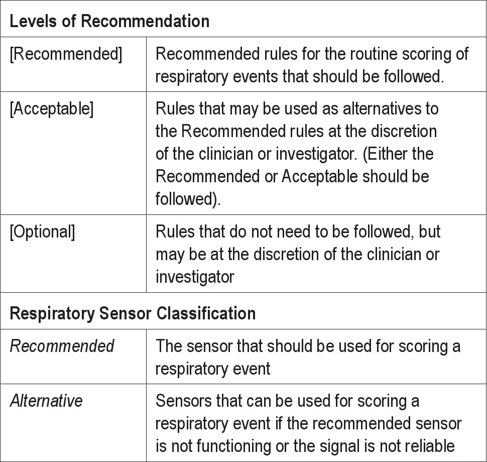
Although proposed revisions to the rules are shown here at the conclusion of each section to make the discussion more understandable, the final and complete set of rules can be found in the online AASM Manual for the Scoring of Sleep and Associated Events, Version 2.0, which is a web-based document, amenable to updates as new literature emerges. This manuscript reviews the issues confronted by the task force during their review as well as the rationale behind the revisions. In the 2007 scoring manual, the levels of recommendation were: Recommended, Alternative, Optional. In this document the level “Alternative” is changed to “Acceptable” to correspond with terminology in the new scoring manual (Version 2.0) (Table 1). In the 2007 scoring manual, sensors were specified (recommended) for detection of apnea, hypopnea, and respiratory effort. Alternative sensors were specified for use if the recommended sensor failed or was not accurate. This terminology will be continued in this document (Table 1).
Table 1.
Levels of recommendation and sensor classification
3.0. RECOMMENDATIONS FOR ADULT AND PEDIATRIC PATIENTS
3.1. Technical Considerations for Adult and Pediatric Patients
In considering the definitions of respiratory events, the task force recognized that most of the 2007 scoring manual definitions include a recommendation for the sensors to be used for event detection. While the major focus of the task force was to update the definitions of respiratory events, it was also necessary to consider sensor technology as it relates to event definitions. It must be recognized that the information obtained from any sensor depends critically on the proper placement of the sensor and appropriate adjustment of gain and filters for viewing the signal. To be accurate, some sensors may require calibration procedures. Filter settings were recommended for most signals of interest in the 2007 AASM scoring manual.1 It is also important to consider that validation of a sensor type manufactured by one company may not invariably generalize to other brands of the same type of sensor.
3.1.1. Detection of Apnea and Hypopnea—General Considerations
The task force recommends a few changes and clarifications in the technical considerations section of the respiratory scoring rules chapter (Tables 2, 3, and 4). The 2007 scoring manual did not specify the sensor for detection of apnea and hypopnea during positive airway pressure (PAP) titration. PAP devices used for titration during polysomnography (PSG) have the ability to output an analog or digital signal from the internal flow sensor.13 Use of this signal to detect apnea and hypopnea during PAP titration is recommended in both the positive airway pressure and noninvasive positive pressure ventilation (NPPV) titration clinical guidelines.14,15 Flattening of the inspiratory portion of the flow waveform provides evidence of airflow limitation and increased upper airway resistance.13,16 Based on consensus and clinical evidence, the task force recommends that the PAP device flow signal should be used to score apneas or hypopneas during PAP titration. Of note, the magnitude of oral airflow, if present, during a PAP titration with a nasal mask is not estimated by the PAP flow signal.
Table 2.
Recommended sensors for routine respiratory monitoring
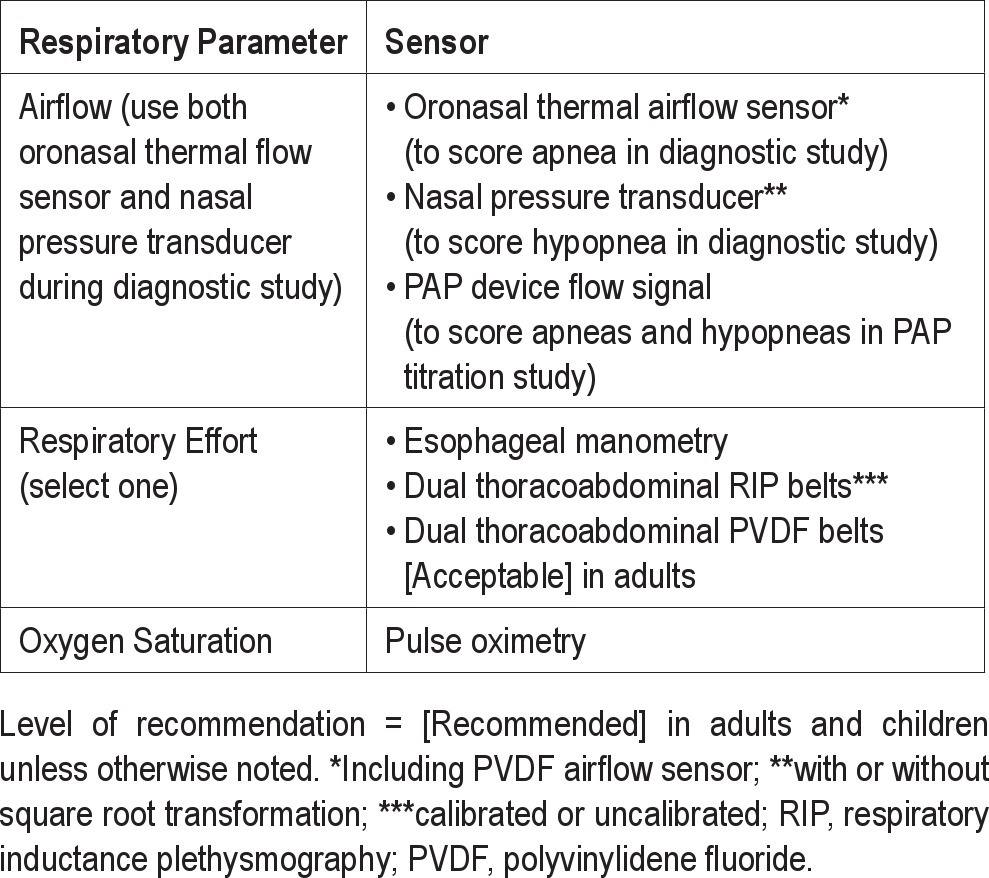
Table 3.
Alternative sensors for scoring respiratory events during diagnostic study
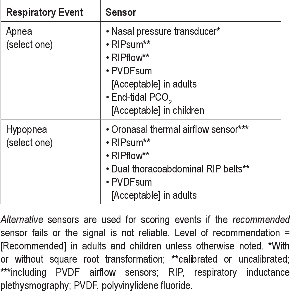
Table 4.
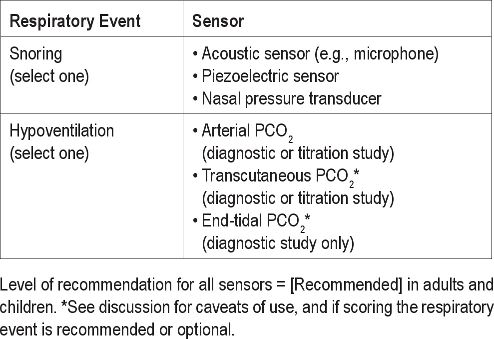
Other sensors for respiratory monitoring
While the 2007 scoring manual lists the use of respiratory inductance (inductive) plethysmography (RIP) sensors17–19 as alternative sensors for scoring apnea and hypopnea, the specific RIP signals to be used were not clearly specified. The available RIP signals include the dual RIP belt signals (thorax and abdomen), the RIPsum (sum of the thorax and abdomen belt signals) and the RIPflow (the time derivative of the RIPsum signal).18–20 Deflections in the RIPsum signal provide an estimate of tidal volume when RIP is calibrated.18,19,21 In uncalibrated RIP, deflections in the RIPsum signal allow detection of a relative change in tidal volume compared to baseline breathing.9,19,22 If the RIPsum signal is not available, a reduction in tidal volume can be inferred if there is a reduction in the excursions of the thoracic and abdominal belts.9,22 Of note, the pattern of undiminished excursions in the signals from the thoracoabdominal belts that are out of phase during an event is also consistent with a reduction in tidal volume (RIPsum). The RIPflow signal is a semi-quantitative estimate of airflow in calibrated RIP, and relative airflow in uncalibrated RIP.19,20,23–25 Calibration of the RIP signal is usually not performed in routine clinical PSG unless the technology for calibration during natural breathing26 is available. During apnea, the RIPsum and RIPflow signals show absent or minimal excursions, and during hypopnea, the excursions are diminished compared to baseline breathing.18,19 Of note, airflow limitation can be inferred from subtle qualitative changes in the inspiratory portion of the thorax RIP, abdominal RIP, and RIPsum signals,27 or from flattening of the inspiratory portion of the RIPflow waveform.19,20,28 The recommended RIP signals for scoring apnea and hypopnea events are specified in Tables 2 and 3.
The 2007 scoring manual recommends use of the nasal pressure signal for scoring hypopnea in both adults and children. While the detection of hypopnea depends on the reduction in the amplitude of the signal, the inspiratory portion of the nasal pressure waveform provides additional useful information. Flattening of the shape of the inspiratory nasal pressure waveform is a surrogate for airflow limitation19,24,29–31 and is included in the respiratory effort related arousal (RERA) rules in the 2007 scoring manual.1 Visualization of flattening of the signal requires that the nasal pressure signal be recorded either as a DC signal or an AC signal with a low-frequency filter setting (cutoff frequency) that is sufficiently low (frequency cutoff 0.03 Hz or lower) (Figure 1).30 Snoring can also be detected as oscillations superimposed on the unfiltered nasal pressure signal32 if an appropriate high-frequency filter setting is used (100 Hz).1 The task force recommends that appropriate high and low filters settings be specified for nasal pressure recording in future revisions of the scoring manual.
Figure 1. Nasal pressure signal displayed as a DC signal and as an AC signal with various low frequency filter settings (Hz).
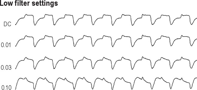
The direction of inspiration is upward. At a low filter setting of 0.1 Hz, the ability to demonstrate airflow flattening is impaired.
3.1.2. Sensors for Apnea Detection
In the 2007 scoring manual the recommended sensor for detecting apnea in both adults and children is an oronasal thermal sensor (Table 1 for definition of recommended). Oronasal thermal sensors have the advantage of being able to detect both nasal and oral airflow. Thermal sensors detect a change in temperature between inhaled and exhaled gas. Here thermal airflow sensors include thermistors, thermocouples, or polyvinylidene fluoride (PVDF) sensors.19,33–35 The task force found no evidence to change this recommendation for diagnostic sleep studies, although it broadened the definition of thermal sensors to include PVDF sensors.
The 2007 scoring manual recommends somewhat different alternative sensors for apnea detection in adults (nasal pressure transducer or RIP) and children (nasal pressure transducer, end-tidal PCO2, and summed RIP) (Table 1 for definition of alternative). The nasal pressure signal is not the recommended sensor for apnea detection as the signal may show decrease excursions (decreased amplitude) during mouth breathing.32 Due to the non-linear characteristics of the nasal pressure signal (proportional to the flow squared), the signal underestimates low flow rates and could result in a hypopnea appearing to be an apnea.36 A square root transformation of the nasal pressure signal more closely approximates flow and minimizes this problem.
As noted above, the excursions of the RIPsum and RIPflow signals usually have minimal amplitude during apnea.18,19,23 However, during obstructive apnea continued excursions in the RIPsum or RIPflow signals may be seen if the thorax and abdominal belt signals do not precisely sum to zero. This problem is minimized by calibration of the RIP signals; however, even the calibrated RIPsum may not remain accurate due to belt movement or changes in patient position.37
Studies have evaluated the accuracy of RIPsum or RIPflow as a surrogate of tidal volume/airflow to detect apneas and hypopneas in adults23,38 and children.25 These studies usually analyzed the combination of apneas and hypopneas.23,38 That is, a separate analysis for apneas and hypopneas was not performed. In one study of calibrated RIP, the use of the RIPsum and RIPflow signals to determine the apnea hypopnea index (AHI) showed good agreement with a pneumotachograph (accurate flowmeter).23 Another study using uncalibrated RIP to determine the AHI found the intermeasurement agreement between use of the RIPsum and pneumotachograph to be considerably lower than between nasal pressure and pneumotachograph.38 A separate analysis for apnea and hypopnea detection was not performed. Respiratory belts utilizing a PVDF sensor can also provide a sum signal as well as thoracoabdominal signals. One study in adult patients being evaluated for suspected obstructive sleep apnea (OSA) suggests that the PVDFsum signal may have utility as a method for apnea/hypopnea detection independent of direct airflow monitoring (nasal pressure or thermistry).6 The PVDFsum signal identified apnea based on a reduction in signal amplitude to 10% of baseline and hypopnea by a 50% reduction in signal. There was good agreement between classification of patients with an AHI ≥ 5/hour using PVDFsum compared to detection of airflow by thermistry and nasal pressure. In a separate part of the study, ten normal subjects simulated central and obstructive apneas while monitored with a pneumotachograph, RIP belts, and PVDF belts. Respiratory events defined as > 50% drop in signal amplitude were identified and compared. The PVDF sensor performed as well as the RIP when compared against the pneumotachograph in terms of the total number of respiratory events that were detected. Further evidence for the utility of the PVDF signals (thoracoabdominal belts or sum) to detect apnea/hypopnea is needed.
In summary, there is evidence that RIPflow or RIPsum (in adults and children) or PVDFsum (in adults only) may be used as an alternative sensor for apnea detection with the understanding that most studies analyzed the combination of apneas and hypopneas. Calibration of RIP may improve the accuracy of RIPflow and RIPsum, but a head-to-head comparison of AHI results using the same sensor, with and without calibration, has not been performed. Further comparisons between PVDFsum, RIPsum, or RIPflow, and an oronasal thermal sensor for detection of apneas (both central and obstructive) are needed.
The end-tidal PCO2 signal is listed as an alternative sensor for apnea detection in pediatric patients in the 2007 scoring manual. A more accurate description of the signal is exhaled PCO2, but the phrase end-tidal PCO2 monitoring is widely used. Monitoring of exhaled PCO2 is routinely performed during pediatric PSG, and the absence of signal deflections (no CO2 exhaled) has been used to score apneas (Figure 2).1,39 The side stream method is most commonly used and consists of gas suctioned via a nasal cannula to an external sensor at bedside. Mouth breathing and occlusion of the nasal cannula can impair the ability of end-tidal PCO2 monitoring to detect apnea. One must remember that the magnitude of signal excursion depends entirely on the highest value of PCO2 in the exhaled breath rather than the magnitude of tidal volume or flow. Signal excursions can persist during inspiratory apnea if small expiratory puffs with a high PCO2 are present.40 A large study in infants compared the ability of the RIPsum, end-tidal PCO2, and oronasal thermistor monitoring to detect apnea.39 End-tidal PCO2 detected 182 of 196 apneas detected by either a thermistor or RIPsum.
Figure 2. Use of the CO2 waveform to detect apnea.
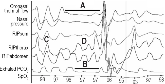
Here RIPsum, RIPthorax, RIPabdomen are the summed, thorax, and abdominal signals from respiratory inductance plethysmography. The exhaled PCO2 is the capnography signal. The presence of apnea is documented by (A) oronasal thermal flow and (B) capnography. Note that the capnography signal lags behind the flow signal. The event depicted is an obstructive apnea. Thoracoabdominal paradox (D) is noted during the event but not during unobstructed breathing (C).
After review of the existing evidence, the task force decided upon recommended (Table 2) and alternative (Table 3) sensors for apnea detection. The task force concluded that the recommended sensor for apnea detection during diagnostic study should continue to be an oronasal thermal sensor in adults and children [Recommended] (Consensus). The task force also reached consensus on specification of the nasal pressure signal or the RIPsum or RIPflow signals from calibrated or uncalibrated RIP as the alternative (sensor) signals for apnea detection during diagnostic study in adults and children [Recommended] (Consensus). In adults the PVDFsum signal may also be used as an alternative sensor for apnea detection, although the ability to differentiate obstructive apneas versus hypopneas has not been defined [Acceptable] (Adjudication). The end-tidal PCO2 is another alternative apnea sensor in children if other sensors are not functioning or not available [Acceptable] (Consensus). As noted in the previous section, the PAP device flow signal is the recommended signal for apnea detection during PAP titration. Alternative sensors for apnea detection during PAP titration studies are not specified.
3.1.3. Sensors for Hypopnea Detection
Hypopnea detection requires a sensor to reliably detect a reduction in airflow or tidal volume. The gold standard for airflow detection is a pneumotachograph, usually placed in the outlet of a mask over the nose and mouth, which measures the pressure drop across a linear resistance.9,19,23,24,40 However, this technology is not practical for clinical studies. The 2007 scoring manual recommends a nasal pressure transducer with or without square root transformation as the recommended sensor for detection of airflow for identification of hypopnea in adults.1 In children the untransformed nasal pressure signal is recommended. The 2007 scoring manual also recommends somewhat different alternative hypopnea sensors for adults (calibrated or uncalibrated RIP, oronasal thermal sensor) and children (oronasal thermal sensor).
As noted above, nasal pressure monitoring (nasal cannula connected to a pressure transducer) provides a signal proportional to the square of the flow.36 A square root transformation of the signal provides a more accurate estimate of flow, but the accuracy of the transformed nasal pressure signal typically deteriorates over a night of monitoring due to factors such as changes in catheter position.23 The effect of using a transformed rather than untransformed nasal pressure signal on the apnea hypopnea index is usually small; the AHI based on the transformed signal is slightly lower.1,23 The utility of nasal pressure monitoring has been documented in a significant number of publications19,23–25,29–32 and is sensitive to even subtle changes in airflow. The inspiratory portion of the nasal pressure waveform can display flattening, a surrogate of airflow limitation when using appropriate filter settings. As noted above, the major disadvantage of nasal pressure monitoring is the inability to detect or estimate the magnitude of oral airflow.32
Oronasal thermistors and thermocouples detect the presence of airflow due to a change in sensor temperature, as exhaled gas is warmed to body temperature. The signal from these thermal devices is not proportional to flow33,41 and often overestimates flow as flow rates decrease.33 Excursions in the signal typically show some decrement during hypopnea, although not as prominent as those in the nasal pressure signal.19 Thermal sensors using polyvinylidene fluoride (PVDF) film produce a signal that is roughly proportional to the temperature difference between the two sides of the film and have a faster response time than thermistors or thermocouples.34,35 One study comparing the ability of an oronasal PVDF airflow sensor to a pneumotachograph in ten patients with OSA found that the output of a PVDF airflow sensor tracked the magnitude of changes in flow with reasonable accuracy.34 Although this study did not directly compare the PVDF sensor to traditional thermal sensors, it does suggest that PVDF sensors more accurately estimate the magnitude of airflow.34 While the inspiratory PVDF waveform may not routinely exhibit flattening during airflow limitation, PVDF sensors have the advantage over nasal pressure sensors of being able to detect oral airflow. One can also argue that hypopnea definitions are based on changes in amplitude rather than signal contour. A limitation of the evidence for using PVDF airflow sensors for hypopnea detection is the small number of patients that have been studied. A study of PVDF airflow sensors in children has yet to be published.
As noted above, the calibrated or uncalibrated RIP signals (RIPsum, RIPflow, thorax and abdominal belt excursions) also decrease during hypopnea.18–20,22–24 Studies of uncalibrated9,22,38 and calibrated RIP23,24 have shown reasonable accuracy in detection of hypopnea. However, in some very obese patients inspiration is associated with small thoracoabdominal excursions making use of RIP for detection of hypopnea more difficult. Heitman et al.38 found the event by event device agreement between a pneumotachograph and the nasal pressure signal to be higher than between the pneumotachograph and the RIPsum (uncalibrated RIP). Clark and coworkers found the nasal pressure signal to be more sensitive for detection of airflow limitation than RIPflow (calibrated RIP).25 Thurnheer et al.23 found the bias in AHI between nasal pressure (transformed or untransformed) and a pneumotachograph to be similar to that between RIPflow (calibrated RIP) and the pneumotachograph. One might expect calibration of RIP to improve the accuracy of hypopnea detection, but this is rarely performed in clinical PSG. Another issue is that not all commercially available PSG systems provide a RIPsum and/or a RIPflow signal. As noted in the discussion of apnea sensors, the PVDFsum signal (from PVDF effort belts) appears to have utility for detection of apneas and hypopneas based on a single study in adults. As with other hypopnea sensors, the PVDFsum does not appear to be as sensitive for detecting events (based entirely on flow) as nasal pressure.6 The ability of PVDFsum to detect hypopnea when combined with arterial oxygen desaturation remains to be determined.
Given the above considerations, the task force was able to reach consensus on the recommended (Table 2) and alternative (Table 3) sensors for hypopnea detection in adults and children during diagnostic polysomnography. The task force recommends that the recommended sensor for detection of airflow for identification of hypopnea in adults and children continue to be a nasal pressure transducer (with or without square root transformation) [Recommended] (Consensus). The simplicity and sensitivity of nasal pressure and the ability for scorers to easily recognize changes in flow based on changes in shape as well as amplitude are distinct advantages. Alternative sensors for identification of hypopnea in adults and children include oronasal thermal sensors (including PVDF airflow sensors) or calibrated or uncalibrated RIP (RIPsum, RIPflow, dual thoracoabdominal RIP belts) [Recommended] (Consensus). The PVDFsum is an alternative hypopnea sensor to be used in adults only [Acceptable] (Adjudication). As noted earlier, the PAP device flow signal is the recommended signal for hypopnea detection during PAP titration. Alternative sensors for hypopnea detection during PAP titration studies are not specified.
3.1.4. Sensors for Detection of Respiratory Effort
The 2007 scoring manual recommends esophageal manometry or calibrated or uncalibrated respiratory inductance plethysmography for detection of respiratory effort in adults and children. Esophageal manometry is the gold standard for detection of respiratory effort, and the signal excursions provide an estimate of the magnitude of effort.8,9,42 However, esophageal manometry is rarely used in clinical practice due to its invasiveness and patient discomfort. Instead, the monitoring of thoracoabdominal excursions is used to detect the presence of respiratory effort. The magnitude of these excursions may or may not be proportional to esophageal pressure excursions, yet for routine clinical sleep monitoring, the detection of respiratory effort to differentiate central and obstructive apnea is the major concern. Failure to detect respiratory effort when present may result is the incorrect classification of an obstructive apnea as central. In one study of 22 patients with OSA using strain gauge sensors positioned on the chest and abdomen, 422 events were classified as central apneas; however, 156 of the events were reclassified as obstructive based on esophageal pressure tracings.43
The 2007 scoring manual did not specify the use of dual effort belts (thoracoabdominal belts) for detection of respiratory effort. Nevertheless, this is standard clinical practice for a number of reasons. Some patients have larger excursions in either the thorax or abdominal belts during the night, and this can vary with body position. If one effort belt fails, the other still provides information about respiratory effort. During biocalibration the polarity of belt signals is adjusted so that belt distension results in signal excursion in the same direction for both belts. Use of dual belts has the additional advantage of the ability to demonstrate paradoxical motion of the thorax and abdomen and adds the ability to identify events as obstructive18,19,44 (Figure 2).
The technology available for respiratory effort belts includes strain gauges, impedance plethysmography, inductance plethysmography (RIP), and belts with piezoelectric or PVDF sensors.6,18,19,43 An advantage of the RIP technology is that inductance of the band and ultimately the signal output depends on the entire surface area enclosed by the band. Effort belts with piezoelectric or PVDF sensors typically utilize a single sensor between belt material surrounding the thorax or abdomen. The signal depends on variations in the tension on the sensor which may or may not reflect the magnitude of thoracoabdominal excursions. Studies have shown that RIP belts are able to detect subtle changes in respiratory effort27,28 and out of phase (paradoxical) motion of the thorax and abdomen excursions is often noted during obstructive apnea or hypopnea.18,44 Calibration of RIP signals should improve the accuracy of this sensor technology for detection of respiratory effort.18 However, even calibrated RIP may not detect feeble respiratory effort in some patients with misclassification of obstructive apneas as central.44 Of note, many of the studies documenting the accuracy of RIP used belts from one or two manufacturers, yet RIP belts are currently supplied by many different manufacturers, and information on the specific technology used for a given belt is not available to the clinician. Validation studies for each type of RIP belt would increase confidence that their performance is similar to the belts used in previous investigations.
Prior to publication of the 2007 scoring manual, many sleep centers used piezoelectric effort belts. Today use of RIP belts has replaced piezoelectric belts in most sleep centers. The task force found scant evidence directly comparing RIP and piezoelectric effort belts with respect to the AHI or detection of central apnea.45,46 Montserrat et al. did find that subtle changes in piezoelectric belt signals were useful in detecting subtle increases in respiratory effort.16 A recent study directly compared RIP belts and effort belts using a PVDF sensor.6 Monitoring was performed with both types of belts in place in 50 adult patients referred for evaluation of possible obstructive sleep apnea (OSA). Respiratory events were scored using montages with either RIP or PVDF belt signals visible to detect respiratory effort. Although there were differences in the AHI values between the sensor types in some patients, overall the results obtained by both technologies were very similar. The average number of central apneas, obstructive apneas, hypopneas, and the overall AHI as determined using RIP versus PVDF belts for respiratory effort detection were almost identical and showed a high level of agreement as assessed by the κ statistic. Thus, PVDF sensor effort belts appear to adequately detect respiratory effort in adults.6 Unlike RIP signals, there have been no studies of PVDF belts in children. More validation studies directly comparing effort belts with different technology (RIP versus piezoelectric, RIP versus PVDF) are needed.
The 2007 scoring manual includes surface diaphragmatic/intercostal electromyography (EMG) as an alternative sensor for detection of respiratory effort. Bursts of the diaphragmatic/intercostal EMG signal are noted with each inspiration. Similar monitoring methods as those used for recording of anterior tibial muscle EMG can be used. However, unlike leg EMG, the diaphragmatic/intercostal EMG signal is often contaminated with prominent electrocardiographic activity. There continues to be scant literature on the use of surface EMG to detect respiratory effort.47,48 Although diaphragmatic/intercostal EMG monitoring is potentially useful as an adjunct to other methods of detecting respiratory effort, the task force felt that more research in this area is needed.
Given the above considerations, the task force decided to uphold the 2007 manual recommendation that esophageal manometry or calibrated or uncalibrated dual thoracoabdominal RIP belts be used for detection of respiratory effort in adults and children (Table 2) [Recommended] (Consensus). It was concluded that dual thoracoabdominal PVDF belts may be used to detect respiratory effort in adult patients but with a lower level of recommendation due to limited published evidence (Table 2) [Acceptable] (Adjudication).
3.1.5. Detection of Blood Oxygen
The task force did not find evidence to change the 2007 recommended sensor for estimation of arterial oxygen saturation which is pulse oximetry (SpO2) with an appropriate averaging time (Table 2) [Recommended] (Consensus). It was noted that the presence of carboxyhemoglobin (e.g., in heavy smokers) may result in the SpO2 being higher than the true fraction of total hemoglobin bound to oxygen.49 Accurate measurement of the amount of carboxyhemoglobin requires use of a co-oximeter that uses the absorption of four or more wavelengths of light (compared to two wavelengths in routine oximetry). The presence of carboxyhemoglobin also shifts the oxygen hemoglobin saturation curve to the left causing a given SpO2 to be associated with a lower than expected partial pressure of oxygen.
Because of the sigmoid shape of the oxyhemoglobin dissociation curve, a much greater drop in arterial partial pressure of oxygen (PaO2) occurs in the setting of a drop of 4% from a baseline saturation of 96% to 92% (PaO2 change ~18 mm Hg) compared to the same drop of 4% from a baseline saturation of 92% to 88% (PaO2 change ~ 9 mm Hg). Therefore, linking respiratory event definitions to a specific change in saturation for event detection requires a greater fall in the PaO2 for patients with a high baseline saturation (e.g., 98%) than those with lower baseline saturations. The desaturation associated with a respiratory event is defined as a drop from a baseline SpO2 preceding the event to the nadir in the SpO2 following the event. While identification of the nadir in the SpO2 following a respiratory events is usually straightforward, selecting a “baseline” SpO2 in a patient with back-to-back respiratory events is more difficult. The highest SpO2 following a respiratory event can exceed values present during stable breathing. Defining a “baseline SpO2” during sleep may be difficult in such patients. While the above ambiguities in SpO2 measurement were recognized, the task force did not recommend changes in terminology or measurement.
3.1.6. Detection of Snoring
The 2007 AASM scoring manual did not recommend a sensor for snoring. There is a paucity of published data on snoring sensors. Optimal visualization of snoring requires a high frequency filter setting that permits recording/display of rapid oscillations (100 Hz recommended in the 2007 scoring manual).1 Snoring may be visualized in the nasal pressure signal as high-frequency oscillations32 superimposed on the slower varying flow signal but is not seen in the PAP device flow signal which is either filtered or too under-sampled to show the high-frequency vibrations. Snore sensors are typically piezoelectric sensors that detect vibration of the neck or microphones that record the sound of snoring. The AASM guidelines for continuous positive airway pressure (CPAP) and for NPPV titration14,15 both listed a snore signal as an option for recording. Based on limited information, the task force recommends several sensors as options for snore detection: the unfiltered nasal pressure signal, piezoelectric sensors to detect vibration, or acoustic sensors (e.g., microphone) to record sound (Table 4) [Recommended] (Consensus). The ability of the sensor to detect simulated snoring should be demonstrated before sleep recording. The task force concurs with the 2007 manual that whether or not to monitor snoring is at the discretion of the clinician or investigator [Optional] (Consensus).
3.1.7. Detection of Hypoventilation
The gold standard method for documenting hypoventilation is the processing of an arterial sample for determination of the arterial partial pressure of carbon dioxide (PaCO2). Given the difficulty of drawing an arterial sample during sleep, the 2007 scoring manual states that finding an elevated PaCO2 obtained immediately after waking would provide evidence of hypoventilation during sleep. Regardless of whether this value underestimates the sleeping PaCO2, the ability to draw or process an arterial blood gas sample is rarely available in sleep centers. In neonates and some research studies, capillary blood samples have been used to provide an estimate of the PaCO2 when sampling of arterial blood is difficult.50 Although capillary sampling requires less expertise than obtaining an arterial blood gas, processing of the sample still requires equipment rarely available in most sleep centers.
Given the difficulties in obtaining a direct measurement of PaCO2, surrogate measures such as end-tidal PCO2 (PETCO2) and transcutaneous PCO2 (PTCCO2) are commonly used during PSG. A 2007 scoring manual note for the hypoventilation rule in adults states that there is insufficient evidence to allow specification of sensors for direct or surrogate measures of PaCO2. It goes on to clarify that “both end-tidal CO2 and transcutaneous CO2 may be used as surrogate measures of PaCO2 if there is demonstration of reliability and validity within laboratory practices.”1 In contrast, the pediatric hypoventilation scoring rules state “acceptable methods for assessing alveolar hypoventilation are either transcutaneous or end-tidal PCO2 monitoring.”1 The task force examined the literature to see if the recommendations for hypoventilation sensors in adults and children could be brought into closer concordance.
Monitoring of exhaled CO2 (capnography) is widely used in pediatric PSG. In children, respiratory events are often associated with increases in PETCO2 with minimal changes in the SpO2.51 In most sleep centers, a bedside device containing the CO2 measuring sensor continuously suctions gas through a nasal cannula worn by the patient (side stream method). During inhalation, room air is suctioned (PCO2 = 0), and during exhalation, the PCO2 in the exhaled gas is measured. The end-tidal PCO2 provides an estimate of the arterial value (usually PaCO2 > PETCO2). The PaCO2 – PETCO2 difference is usually 2 to 7 mm Hg, and the difference is higher in patients with lung disease.52 The PETCO2 is not an accurate estimate of the PaCO2 during mouth breathing or with low tidal volume and fast respiratory rates. Some manufacturers make a sampling nasal cannula with a “mouth guide” to allow sampling of gas exhaled through the mouth. To be considered accurate, a definite plateau in the exhaled PETCO2 versus time waveform should be observed.1,9,51,52 End-tidal PCO2 measurements are often inaccurate during application of supplemental oxygen or during mask ventilation.53 The exhaled gas sample is diluted by supplemental oxygen flow or PAP device flow. Some clinicians use a small nasal cannula under the mask to sample exhaled gas at the nares in order to minimize dilution during PAP titration. However, the accuracy of measurements using this approach has not been documented.
Transcutaneous CO2 monitoring (PTCCO2)54 is also used during PSG to estimate the PaCO2, but the signal has a longer response time than the PETCO2 to acute changes in ventilation. In adults, one study using an earlobe sensor found that the PTCCO2 lagged behind PaCO2 by about 2 minutes.55 Newer transcutaneous device technology has allowed more rapid response time and enabled the devices to work at a lower skin temperature. The advantage of PTCCO2 monitoring compared to PETCO2 is that the accuracy of transcutaneous measurements is not degraded by mouth breathing, supplemental oxygen, or mask ventilation. However, PTCCO2 will not provide data for breath-by-breath changes, e.g., changes in the first few breaths after an apnea.
The task force considered the literature on the accuracy of end-tidal CO2 and transcutaneous PCO2 as surrogates for determination of arterial PCO2. Sanders et al. found that both PETCO2 and PTCCO2 monitoring did not provide an accurate estimation of PaCO2 during sleep53; however, this study was limited by the technology used, i.e., PETCO2 was measured using a loose-fitting mask, which would be unlikely to result in an adequate waveform. A study of PETCO2 during the awake postoperative state in both non-obese and obese patients found the average PETCO2 values to differ from the PaCO2 by around 8 mm Hg (nasal cannula) or 6 mm Hg (nasal cannula + oral guide).56 Another study evaluated the utility of PETCO2 for determining daytime hypoventilation in a group of patients with neuromuscular disease. Although fairly accurate in some patients, in others there was a large difference between the PETCO2 and PaCO2.57
A number of studies54–59 have evaluated the accuracy of PTCCO2 compared to PaCO2 or capillary PCO2. Maniscalco et al. found the average PaCO2 – PTCCO2 difference to be −1.4 mm Hg (95% confidence interval −1.7 to 7.5 mm Hg) in a group of obese adult patients with various disease processes.58 In a recent study, Storre et al.59 compared capillary blood gas PCO2 and PTCCO2 from 3 different devices in a group of patients being started on nocturnal noninvasive ventilation. The bias (PaCO2 – PTCCO2 drift uncorrected) for one device was as low as 0.8 mm Hg, with the limits of agreement extending from −4.9 to 6.5 mm Hg. Pavia and coworkers60 used PTCCO2 monitoring during sleep in 50 children receiving chronic NPPV. Twenty-one patients had nocturnal hypoventilation (without desaturation on oximetry). Of these 21 patients, 18 did not have daytime hypoventilation (capillary blood gas). The authors concluded that PTCCO2 monitoring was useful for detection of unsuspected nocturnal hypoventilation. Senn and coworkers61 compared transcutaneous and arterial PCO2 measurement in a group of patients undergoing mechanical ventilation. The mean difference ± 2 SDs between PTCCO2 and PaCO2 during stable ventilation was 3 ± 7 mm Hg.
A number of investigations compared values obtained from simultaneous measurements of PETCO2 and PTCCO2. Kirk et al.62 analyzed the findings of PSG with simultaneous monitoring of PETCO2 and PTCCO2 in 609 children. In 437 patients, the difference between the mean nocturnal PETCO2 and PTCCO2 was between −4 and +4 mm Hg. Of note, interpretable results were found in 61% of PSGs for PETCO2 and 71.5% for PTCCO2. Thus, obtaining interpretable data using either PETCO2 or PTCCO2 is often challenging. Hirabayashi and coworkers63 studied awake, spontaneously breathing adults and found the bias ± 2 SD of end-tidal and transcutaneous PCO2 measurements compared to PaCO2 to be 0.48 ± 8.2 for PTCCO2 and −6.3 ± 9.8 mm Hg for PETCO2. The authors concluded that transcutaneous monitoring was more accurate. Casati and colleagues compared PETCO2 and PTCCO2 to PaCO2 in a group of anesthetized mechanically ventilated patients. The exhaled CO2 was sampled between the endotracheal tube and the ventilator circuit. The PTCCO2 was within 3 mm Hg of the PaCO2 in 21 of 45 pairs sampled, and the PETCO2 was within 3 mm Hg of the PaCO2 in 7 of 45 pairs sampled. The authors concluded that PTCCO2 was more accurate than PETCO2.64
In summary, it appears that both PTCCO2 and PETCO2 have clinical utility as surrogates of PaCO2 during diagnostic studies. At least in some settings, these surrogates may be more useful for following trends in the PaCO2 than providing a highly accurate estimate of the exact PaCO2 value. Use of both methodologies requires careful review of tracings to determine if artifacts are present. PTCCO2 is the preferred technology in patients with lung disease, significant mouth breathing, or those who are using supplemental oxygen or mask ventilation. The clinician should recognize that PTCCO2 values can occasionally be quite spurious and clinical judgment is needed. When readings do not match the clinical setting, a change in sensor site or recalibration may be needed. PETCO2 is preferred where breath-to-breath changes in PaCO2 need to be detected. In this setting, the ability to detect an increase in PCO2 associated with a respiratory event is clinically useful.
The task force concurred with the 2007 manual that whether or not to monitor hypoventilation in adults during diagnostic study or PAP titration is at the discretion of the clinician or investigator [Optional] (Consensus). Monitoring hypoventilation in children during diagnostic study is [Recommended] (Consensus) and during PAP titration is [Optional] (Consensus). The task force recommends that arterial PCO2, transcutaneous PCO2, or end-tidal PCO2 be used for detecting hypoventilation during diagnostic study in both adults and children (Table 4) [Recommended] (Consensus). During PAP titration in both adults and children, either arterial PCO2 or transcutaneous PCO2 is the recommended method to detect hypoventilation (Table 4) [Recommended] (Consensus). There are a number of caveats that accompany these recommendations: (1) Sensors should be properly calibrated according to manufacturer specifications. (2) Clinical judgment is essential when assessing the accuracy of end-tidal PCO2 and transcutaneous PCO2 readings. The values should NOT be assumed to be accurate surrogates of the arterial PCO2 when the values do not fit the clinical picture. (3) Transcutaneous PCO2 should be calibrated with a reference gas according to the manufacturer's recommendations and when the accuracy of the reading is doubtful. (4) End-tidal PCO2, to be accurate, a plateau in the PCO2 versus time wave form should be present. Validation of the surrogate PCO2 with a simultaneous PaCO2 or capillary gas is ideal but not required.
3.1.8. Summary of Sensor Recommendations
As a reminder, sensors noted here as recommended have corresponding alternative sensors that may only be used if the recommended fails or is inaccurate. At this time alternative sensors are only named for identifying apneas and hypopneas in diagnostic studies. For monitoring of airflow during diagnostic studies, both an oronasal thermal sensor and a nasal pressure transducer should be used [Recommended] (Table 2). For apnea detection, the oronasal sensor is the recommended sensor and the nasal pressure can serve as an alternative sensor [Recommended]. Other alternative apnea sensors include use of RIPsum, RIPflow [both Recommended] and PVDFsum (adults only) or end-tidal PCO2 (children only) [both Acceptable] (Table 3). During PAP titration studies, the PAP device flow is the signal that should be used for apnea detection [Recommended]. For hypopnea monitoring during diagnostic studies, nasal pressure is the recommended sensor [Recommended]. Alternative hypopnea sensors include an oronasal thermal sensor, RIPsum, RIPflow, dual thoracoabdominal RIP belts [all Recommended], or PVDFsum (adults only) [Acceptable]. During PAP titration studies, the PAP device flow is the sensor to be used for hypopnea detection [Recommended]. For respiratory effort monitoring (Table 2), use esophageal manometry or thoracoabdominal RIP belts (adults and children) [both Recommended] or thoracoabdominal PVDF effort belts (adults only) [Acceptable]. Pulse oximetry is the sensor to be used for oxygen saturation [Recommended]. Detecting snoring is [Optional], and a nasal pressure transducer, piezoelectric sensor, or acoustic sensor (e.g., microphone) should be used [Recommended] (Table 4). Detection of hypoventilation is [Optional] in adults, [Recommended] in children during diagnostic study only, and the following may be used for diagnostic studies: arterial PCO2, transcutaneous PCO2, or end-tidal PCO2 [Recommended] (Table 4). For PAP titration studies, arterial PCO2 or transcutaneous PCO2 may be used [Recommended].
3.2. Event Duration Rules for Adult and Pediatric Patients
The 2007 scoring manual states that the event duration for scoring either apnea or hypopnea is measured from the nadir preceding the first breath that is clearly reduced to the beginning of the first breath that approximates baseline breathing amplitude. The only recommended revision to the 2007 scoring manual is to explicitly mention the recommended signal to be used for measurement. For apnea duration, the oronasal thermal sensor signal (diagnostic study) or PAP device flow signal (PAP titration study) should be used to determine the event duration [Recommended] (Consensus). For hypopnea event duration, the nasal pressure signal (diagnostic study) or PAP device flow signal (PAP titration study) should be utilized [Recommended] (Consensus). If the recommended sensor fails or the signal is inaccurate an alternative sensor signal can be used. The ability to determine baseline breathing is a problem in patients that have nearly continuous events. The AASM “Chicago consensus paper”9 states, “Baseline is defined as the mean amplitude of stable breathing and oxygenation in the 2 minutes preceding onset of the event (in individuals who have a stable breathing pattern during sleep) or the mean amplitude of the 3 largest breaths in the 2 minutes preceding onset of the event (in individuals without a stable breathing pattern).” The 2007 scoring manual1 states, “When baseline breathing amplitude cannot be easily determined (and when underlying breathing variability is large), events can be terminated when either there is a clear and sustained increased in breathing amplitude, or in the case where an oxygen desaturation has occurred, there is event-associated oxygen re-saturation of at least 2%.” The task force recommends that the 2007 manual guideline for determining baseline breathing be upheld [Recommended] (Consensus).
3.3. Definition of RDI and ODI for Adult and Pediatric Patients
Both the 1999 Chicago consensus paper8 and the ICSD-29 recommended that the sum of apnea, hypopneas, and RERAs per hour of sleep be used to diagnose OSA (along with symptoms). Neither of these documents, the practice parameters for PSG94 or the 2007 scoring manual, defines the metric “RDI” (respiratory disturbance index). Others have defined RDI as the sum of the AHI (apneas plus hypopneas per hour of sleep) and RERA index (RERAs per hour of sleep).95 The literature is very confusing, with many articles defining RDI as the number of apneas and hypopneas per hour of sleep. The Centers for Medicare and Medicaid defines the term RDI as the number of apneas and hypopneas per hour of monitoring. The task force reached consensus on the definition of RDI as the sum of the AHI and RERA index. However, reporting of the RDI metric should be considered [Optional] (Consensus) as scoring RERAs is also considered [Optional]. Some task force members also felt that if a hypopnea definition is used that includes arousal, that an oxygen desaturation index (ODI) defined as the number of ≥ 3% arterial oxygen desaturations per hour of sleep should be reported. This metric reflects the respiratory events per hour associated with desaturation. The task force did reach consensus on whether this should be required and thus recommended that reporting ODI remains [Optional] (Consensus).
Definitions of RDI and ODI [Recommended] (Consensus)
RDI = AHI + RERA index
ODI = ≥ 3% arterial oxygen desaturations/hour
4.0. ADULT SCORING RULES
4.1. Apnea Rule for Adults
The task force carefully examined the apnea rule in the 2007 scoring manual for possible revisions or clarifications. It reads, “Score an apnea when all the following criteria are met: (1) There is a drop in the peak thermal sensor excursion by ≥ 90% of baseline, (2) The duration of the event lasts at least 10 seconds, and (3) At least 90% of the event's duration meets the amplitude reduction criteria for apnea.”1 Notably, the 2007 scoring manual rule for apnea does NOT require an associated arterial oxygen desaturation. The task force found no evidence to support a change in the 2007 scoring manual recommendation for a ' 90% drop in oronasal thermal flow lasting at least 10 seconds (adults). Of note, the basis for a 90% drop in the thermal sensor signal is entirely arbitrary, but is an attempt to operationalize the requirement of “absent or nearly absent airflow” that often appears in the literature for the definition of an apnea. As previously discussed, the task force did clarify that the PAP device flow sensor be used for scoring apnea during PAP titration and name alternative apnea sensors for diagnostic study (Tables 2 and 3).
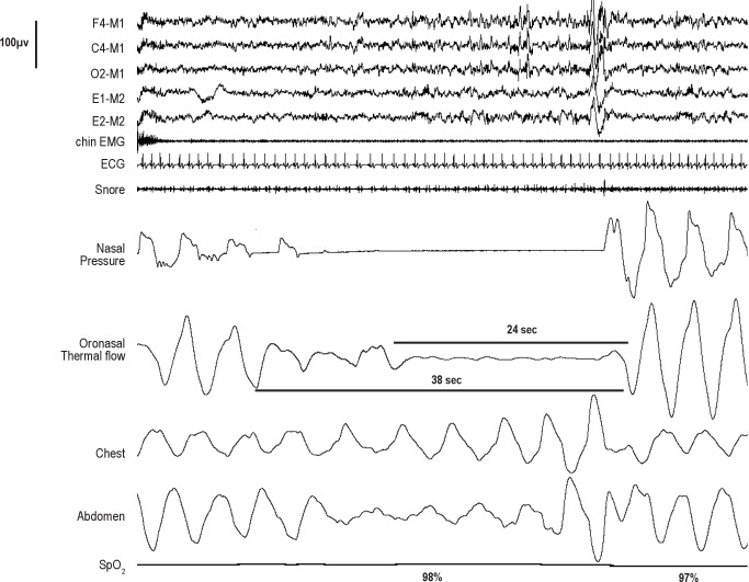
The 2007 scoring manual apnea rule included a third requirement to address situations where the duration of the qualifying drop in airflow is significantly shorter than the event duration as defined by the event duration rule. This stipulation has resulted in numerous questions to the 2007 Scoring Manual Steering Committee for clarification. In one interpretation of the rule, only 9 contiguous seconds of the event duration must meet amplitude criteria. The requirement that 90% of event duration must meet amplitude criteria was not a part of the Chicago consensus paper9 respiratory event definitions and does not appear in apnea definitions elsewhere in the literature prior to 2007. There are cases where there is a drop ≥ 90% of baseline in airflow that lasts for longer than 10 seconds but much less than 90% of the event duration (see Figure 3). Using the 2007 apnea rule, one cannot score such an event as an apnea. This event may not meet hypopnea criteria and therefore cannot be scored as either an apnea or hypopnea. In addition, the length of a respiratory event as defined by the event duration rule may be more difficult to define than the duration of the drop in airflow meeting amplitude criteria. For these reasons, the task force eliminated requirement 3 and added a note addressing the situation in which a shorter portion of a longer hypopnea event qualifies as an apnea.
Figure 3. The event duration (based on a drop in oronasal thermal flow) is 38 seconds as defined by the event duration rule.
An apnea cannot be scored using the apnea rule in the 2007 scoring manual as 24 seconds (the duration of the ≥ 90% drop in oronasal flow) is not 90% of the event duration. A hypopnea cannot be scored based on the drop in nasal pressure, as there is no associated desaturation or arousal. Using the proposed revised apnea definition this event would be scored as an apnea as there is a ≥ 90% reduction in the peak excursions of the oronasal thermal signal compared to baseline that lasts ≥ 10 seconds. The respiratory effort (thoracoabdominal excursions) during the entire apnea indicates that this is an obstructive apnea.
Apnea Rule for Adults [Recommended] (Consensus)
Score a respiratory event in adults as an apnea if both of the following are met:
There is a drop in the peak signal excursion by ≥ 90% of pre-event baseline using an oronasal thermal sensor (diagnostic study), positive airway pressure device flow (titration study), or an alternative apnea sensor.
-
The duration of the ≥ 90% drop in sensor signal is ≥ 10 seconds.
Note: If a portion of a respiratory event that would otherwise meet criteria for a hypopnea meets criteria for apnea, the entire event should be scored as an apnea. The duration of the event is from the nadir in flow preceding the first breath that is clearly reduced to the start of the first breath that approximates baseline breathing.
It should be noted that the task force also considered the possibility of combining both apneas and hypopneas into a single respiratory event. The numbers of both event types are ultimately combined to compute an apnea-hypopnea index. The Chicago consensus paper defined obstructive/hypopnea events and central apnea/hypopnea events.9 However, the task force felt that the definitions of an apnea and apnea subtypes are relatively straightforward compared to hypopnea. In addition, there is a large literature and historical precedent of separating events types into apnea and hypopnea. Use of a combined apnea/hypopnea event also provides some challenges. For example, how would one define a mixed apnea/hypopnea event? Given these considerations the task force did not recommend a change in the current practice of scoring apneas and hypopneas separately.
4.1.1. Classification of Apnea
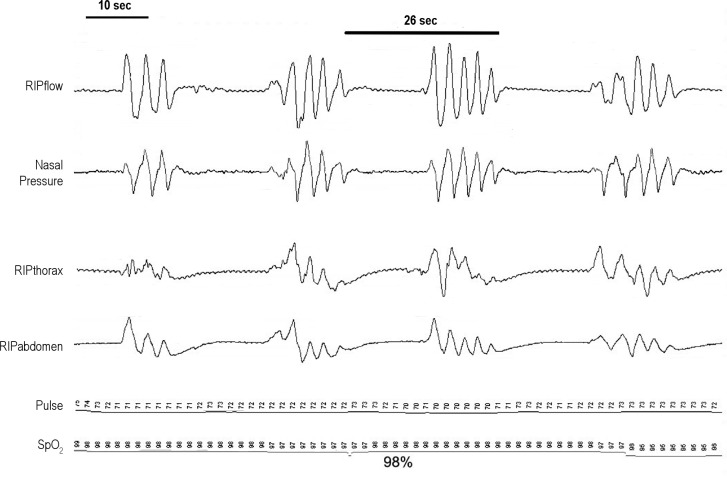
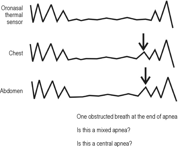
The classification of apnea as obstructive, mixed, or central in the 2007 scoring manual is based on respiratory effort. The definitions are consistent with those appearing in previous literature. The Chicago consensus paper did not specifically define mixed apnea because of the lack of reliability in scoring these events. The task force considered combining obstructive and mixed events as well as a revision of the definition of central and mixed apneas to address events where there are only 1 or 2 obstructive breaths (see Figure 4). If one changes the central apnea definitions to allow 1 or 2 obstructed breaths, then a change in the mixed apnea definition must follow. The choice of how many obstructive breaths to allow and still consider an event to be central seems entirely arbitrary. After discussion, the task force recommended that the definitions of obstructive, central, and mixed apnea in the 2007 scoring manual not be revised (Consensus).
Figure 4. A mixed apnea event is illustrated that contains a single obstructed breath at the end of what would otherwise be scored as a central apnea.
4.2. Hypopnea Rules for Adults
The definition of a hypopnea (a reduction rather than absence in airflow) continues to be an area of considerable controversy.1,5,8,9,65–68 The concept of hypopnea was originally introduced to address those situations in which a drop in arterial oxygen saturation was associated with a change in airflow rather than absence of airflow. Block et al. scored a hypopnea if “flows in the nose and mouth decreased and chest movement decreased and desaturation occurred.”69 The technology used included airflow detection by thermistors attached to mouth and lip and chest movement detection by impedance plethysmography. Gould et al. defined hypopnea based on a reduction in uncalibrated RIP thoracoabdominal belt excursions (a surrogate estimate of tidal volume).22 The 1999 consensus conference defined an apnea-hypopnea as a 10-second or longer event characterized by either a clear decrease (> 50%) of a valid measure of breathing or a clear amplitude reduction (but < 50% decrease) of a validated measure of breathing associated with either an arousal or ≥ 3% oxygen desaturation occurring near the termination of the putative event.9
The 2007 scoring manual provides two hypopnea definitions (recommended and alternative, also known as “4A” and “4B”).1 The need for two definitions was a product of controversy concerning the most appropriate hypopnea definition as well as the fact that the Centers of Medicare and Medicaid Services (CMS) currently accepts only the recommended definition. The recommended hypopnea definition requires a 30% or greater drop in flow for 10 seconds or longer associated with ≥ 4% oxygen desaturation. The alternative hypopnea definition requires a ≥ 50% in flow for 10 seconds or longer associated with a ≥ 3% oxygen desaturation OR an arousal. The hypopnea rule for children is similar to the adult rule except for the minimum duration.
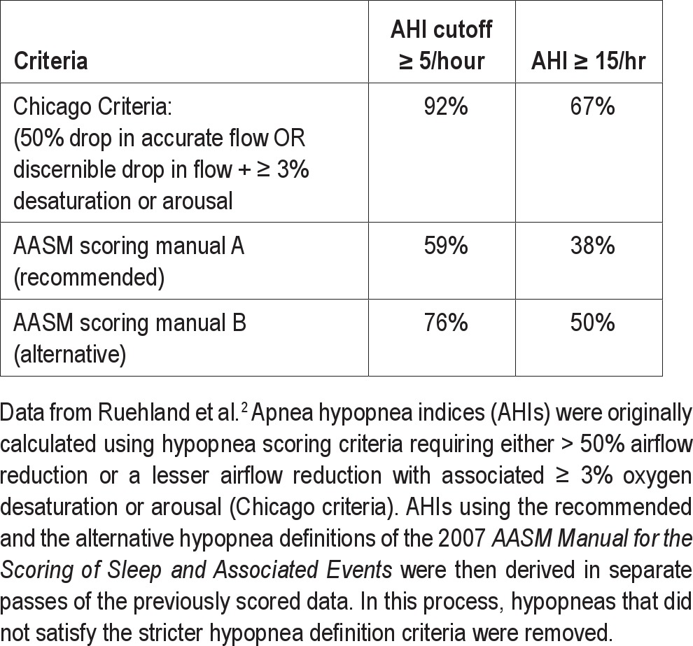
The task force noted that different hypopnea definitions can result in considerably different AHI values.2,65–68 Ruehland et al. retrospectively scored studies of 320 consecutive adult patients evaluated in the sleep center for suspected OSA.2 This is a population that should have an increased likelihood of having OSA. An AHI was determined using the Chicago consensus paper criteria9 and the two 2007 scoring manual hypopnea definitions (Table 5).1 Adding arousal to the hypopnea definition (as done in the alternative hypopnea definition of the 2007 scoring manual) added a fairly modest increase to the percentage diagnosed with OSA compared to use of the recommended hypopnea definition. Furthermore, even using the more liberal 2007 scoring manual alternative hypopnea definition, 24% of the patients being evaluated would not have been diagnosed with OSA using an AHI ≥ 5/hour.
Table 5.
Effect of hypopnea definitions on the AHI in a group of patients undergoing polysomnography for suspected obstructive sleep apnea
Given the above considerations, the task force readdressed the hypopnea definition issue with respect to: (1) the required arterial oxygen desaturation, (2) the inclusion of arousal criteria, and (3) the qualifying drop in flow (30% versus 50%).
3% Versus 4% Oxygen Desaturation
The recommended hypopnea definition requires a 30% or greater drop in nasal pressure excursions for 10 seconds or longer associated with ≥ 4% oxygen desaturation. At the time that the 2007 scoring manual task force examined the literature (pre 2006) there was considerable evidence that respiratory events linked to arterial oxygen desaturation of either ≥ 3 or 4% identified individuals at increased risk of cardiovascular consequences.8,9,70 The current task force found further evidence to support this conclusion. For example, Punjabi et al.71 found respiratory events based on a desaturation of at least 4% were associated with an increased risk of cardiovascular consequences. Further analysis of Wisconsin cohort data and Sleep Heart Health study data suggests that the use of ≥ 3% desaturation criterion yields an AHI that is as predictive of adverse outcomes as an AHI based on ≥ 4% oxygen desaturation criterion.8,9 This is to be expected, given the very high correlation of > 0.95 between the AHIs determined using 3% versus 4% oxygen desaturation.66 Mehra et al. used a definition of hypopnea based on ≥ 3% desaturation and found significant associations between AHI and the risk of atrial fibrillation or complex ventricular ectopy in older men without self-reported heart failure.72 A study by Stamatakis et al. found that levels of desaturation of 2% or 3% were associated with fasting hyperglycemia.73 A recent study using a hypopnea definition requiring ' 3% oxygen desaturation found an association between incident stroke and obstructive sleep apnea.74 Recognizing the apparent equivalence of hypopnea definitions requiring ≥ 3% or ≥ 4% desaturation, the task force has recommended adoption of the 3% criterion. However, it should be noted that using ' 3% instead of ≥ 4% desaturation requirement for defining hypopnea does increase the AHI substantially (Table 6), with median AHI in a general community sample being almost twice as great using a 3% as a 4% criterion.66,75 Therefore, thresholds for identification of the presence and severity of OSA, and for inferring health-related consequences of OSA, must be calibrated to the hypopnea definition employed.
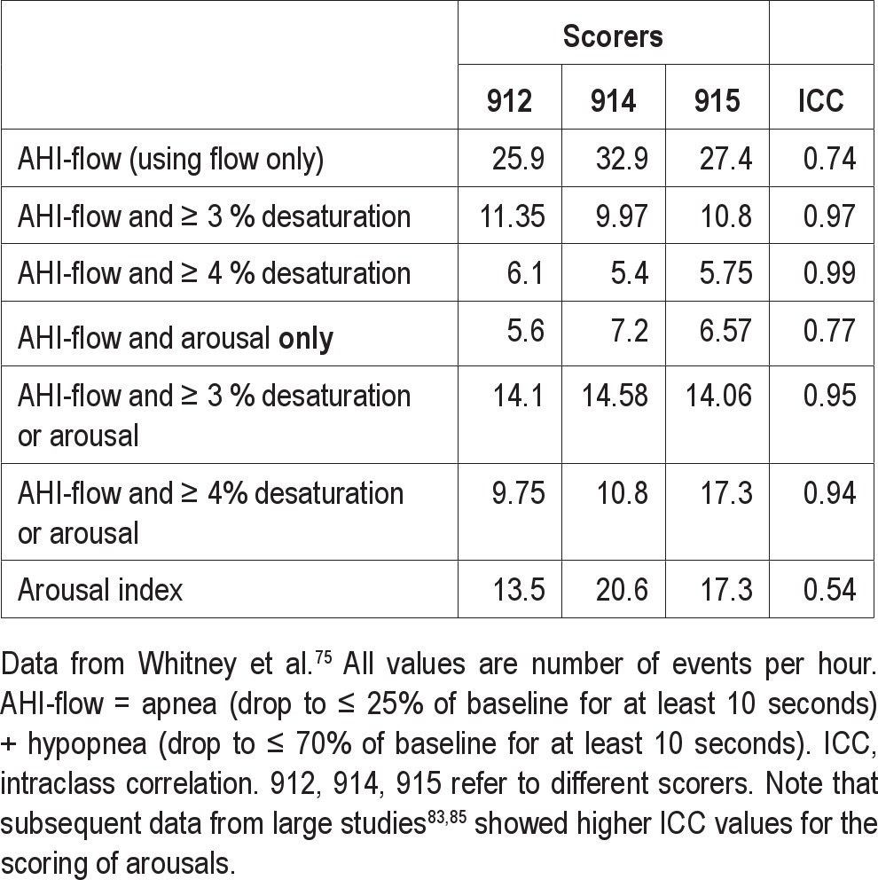
Table 6.
Scoring reliability of different respiratory indices (N = 20)
Inclusion of Arousal in Hypopnea Definition
The alternative definition of hypopnea for adults in the 2007 scoring manual requires a 50% or greater drop in nasal pressure excursions for 10 seconds or longer associated with either ' 3% desaturation or an arousal. Whether or not to include arousal as part of the hypopnea definition remains controversial. Opponents of inclusion of arousal in the hypopnea definition cite the fact that the majority of studies have not found an association between arousal frequency and adverse cardiovascular outcomes (independent of arterial oxygen desaturation). Another argument against inclusion of arousal in the hypopnea definition is that the scoring of arousals is said to be less reliable and would consequently reduce the reliability of scoring respiratory events.75 Opponents of inclusion of arousal criteria in the hypopnea definition also argue that the current Medicare/Medicaid hypopnea definition does not consider arousals and that a hypopnea definition based only on flow and oxygen saturation would be applicable to limited channel sleep testing (i.e., out-of-center sleep testing). One can also argue that milder sleep apnea patients would not be excluded if diagnosis of obstructive sleep apnea syndrome was based on the number of apnea, hypopneas, and respiratory effort related arousals (RERAs) per hour of sleep. The ICSD-2 diagnostic criteria for OSA are based on this metric (≥ 15/hour or ≥ 5/hour with symptoms).10
Proponents of inclusion of arousal in the hypopnea definition cite evidence that sleep fragmentation without arterial oxygen desaturation can be associated with symptoms76,77 (e.g., daytime sleepiness) and that treatment with CPAP can improve symptoms and objective sleepiness.5,76 For symptomatic patients with milder OSA and a significant proportion of events associated with arousal but not ≥ 3% desaturation, the AHI may be ≥ 5/hour using the alternative hypopnea definition but not the recommended definition. While a metric based on the AHI + RERA index may be greater than 5/hour in such patients, RERAs are not scored in many sleep centers and not recognized by Medicare. Of interest, if one uses a hypopnea definition based on desaturation or arousal there are relatively few RERA events.78,79 There is a paucity of data concerning symptomatic patients with an AHI < 5/hour using the recommended hypopnea definition. Guilleminault et al.5 analyzed a cohort of 35 lean subjects diagnosed with OSA based on the Chicago consensus paper definition of hypopnea (50% drop in flow OR discernible drop in flow + ≥ 3% desaturation or arousal). The cohort was selected based on demonstrated improvement of OSA after treatment with CPAP or surgery (based on repeat sleep study) and improvement in subjective sleepiness. The original diagnostic studies were rescored, and 40% of the patients had an AHI < 5/hour using a hypopnea definition based only on flow and ≥ 4% oxygen desaturation. Therefore, in the admittedly carefully selected cohort, a significant number of patients (40%) who benefited from treatment would NOT meet diagnostic criteria for OSA based on the recommended hypopnea definition and therefore not qualify for CPAP treatment. There is a paucity of data demonstrating a relationship between increased arousals and adverse cardiovascular outcomes. Supporters of the inclusion of arousal in the hypopnea definition cite biological plausibility of arousals leading to sympathetic nervous system activation and data showing sympathetic activation related to arousals with increased chin EMG activity (movement arousals).80 Cortical arousal stimuli sufficient to induce a K complex have been shown to elicit sympathetic activity.81 Of interest, an analysis of the Cleveland Family Study by Sulit et al.82 did find the arousal index correlated with the risk of hypertension whereas desaturation did not. This study did not evaluate if an AHI definition including arousal or desaturation had a higher association with the presence of hypertension than a hypopnea definition based solely on desaturation. Another study found an association between the arousal index and white matter disease in older adults.83 Using a hypopnea definition that included arousal criteria, a recent study found that the combination of an elevated AHI and daytime sleepiness was predictive of increased mortality in older adults.84 No analyses using a hypopnea definition based on oxygen desaturation alone were reported.
Regarding the issue of hypopnea definition and portable monitoring, proponents of the alternative hypopnea definition point out that this would simply underscore that PSG is more sensitive for detection of significant respiratory events than limited channel testing. PSG would permit scoring of hypopneas based on arousal as well as arterial oxygen saturation. Respiratory events that cause arousal but are associated with relatively minor drops in the arterial oxygen saturation could be identified as hypopneas. This would allow identification and treatment of a wider spectrum of symptomatic patients.
Concerning the scoring reliability of hypopneas, proponents of a hypopnea definition based on arousal as well as desaturation present several arguments. First, a 2007 AASM review concluded that the scoring of arousals when scorers were trained had moderate reliability.77 The review found evidence that visualization of information other than the EEG and EMG during scoring (e.g., respiratory channels) can improve the reliability of arousal scoring. The frequently quoted study of Whitney et al.75 provides information on the reliability of hypopneas associated with arousal. Twenty randomly chosen studies of good quality were scored by each of 3 scorers. The scorers first identified candidate apnea and hypopnea based only on flow (although oximetry tracings were visible) and then combined events with the presence or absence of arousals (based on a single EEG derivation) and various degrees of arterial oxygen desaturation (Table 6). Arousals were scored based on EEG without regard to respiration. The intraclass correlation (ICC) was highest when respiratory events were linked to oxygen desaturation. The ICC correlation was much lower for the scoring of arousals. However, respiratory events based on flow and the presence of arousal had a better ICC (0.77). Moreover, the reliability of events based on flow, oxygen desaturation, or arousal was even higher (Table 6). Of note, arousal scoring was based on a single EEG derivation from data acquired during an unattended study. In the discussion section of the paper, the authors stated that when the two most experienced scorers performed the analysis, the arousal scoring ICC correlation was much higher (0.72). Indeed, subsequent tracking of reliability showed that, over the course of the Sleep Heart Health Study, the ICC for arousal scoring varied between 0.72 and 0.78.83,84 A recent analysis of the data from the Osteoporotic Fractures in Men Sleep Study85 found the interscorer reliability (intraclass correlations coefficients [ICC]) for the arousal index based on central derivations to be 0.80. Of note, in current practice arousals are scored based on frontal, central, and occipital derivations with montages typically showing respiratory channels. The above considerations suggest that adding arousals to the hypopnea definition will not significantly reduce scoring reliability of hypopneas.
30% Versus 50% Drop in Flow
The task force considered the use of different flow criteria in hypopnea definitions. The 2007 scoring manual definitions of hypopnea for adults use either a 30% drop in flow (recommended definition) or 50% drop (alternative definition). The single pediatric definition requires a 50% drop in flow. Given the difficulty of accurately measuring flow or tidal volume in clinical settings, linking a change in flow or tidal volume to a physiological consequence would help identify an event as physiologically relevant. The degree of oxygen desaturation for a given reduction in airflow varies widely between individuals and depends on baseline arterial oxygen desaturation, oxygen stores (lung volumes), obesity, and the presence or absence of lung disease.86,87 Therefore a less than 50% drop in flow could result in significant oxygen desaturations in some individuals, while in others a desaturation may not occur.
Proponents of using the 30% drop argued that a 30% drop should identify a clear change in breathing from baseline. Furthermore, they argue that the associated consequences (desaturation or arousal) are more important for estimation of an event's physiological significance than the magnitude of drop in flow (as a percentage of baseline). That is, a 30% drop in flow associated with 4% desaturation likely has physiological significance but would not be scored if one required a 50% drop in flow. Proponents of a 50% drop cite the current use of this value in the adult alternative definition and the pediatric hypopnea definition.
Hypopnea Summary
The above discussion outlines the difficulties in choosing a single definition for hypopnea. Although there were dissenters, the task force reached consensus on a definition of a hypopnea rule in adults using a 30% drop in the nasal pressure excursion for 10 seconds or greater associated with ≥ 3% desaturation OR an arousal. The majority of the task force felt that a hypopnea definition based only on desaturation would result in misdiagnosis of some patients in whom respiratory events fragment sleep but result in minor drops in the SpO2. While there seems little doubt that cardiovascular morbidity is associated with oxygen desaturation, the goals of OSA treatment address a much wider range of symptoms including daytime sleepiness, insomnia, and non-restorative sleep. The task force also recognizes that the proposed definition of hypopnea is not currently accepted by the Centers for Medicare and Medicaid Services (CMS) reimbursement. For Medicaid and Medicare patients the use of a hypopnea definition based on a 30% drop in flow and 4% or greater desaturation will need to be used to ensure reimbursement until reimbursement policies are changed to reflect the new hypopnea definition. Following the logic of the proposed revised apnea definition, the requirement that the qualifying drop in flow must occupy > 90% of the event duration was removed from the hypopnea definition.
Hypopnea Rule for Adults [Recommended] (Consensus)
Score a respiratory event as a hypopnea if all of the following are met:
The peak signal excursions drop by ≥ 30% of pre-event baseline using nasal pressure (diagnostic study), PAP device flow (titration study), or an alternative hypopnea sensor.
The duration of the ≥ 30% drop in signal excursions is ≥ 10 seconds.
-
There is ≥ 3% oxygen desaturation from pre-event baseline or the event is associated with an arousal.
Note: If necessary, the number of hypopneas using a definition requiring ≥ 30% drop in flow for ≥ 10 seconds that is associated with ≥ 4% desaturation may additionally be reported to qualify a patient for PAP reimbursement (e.g., Medicaid or Medicare patients).
4.2.1. Classification of Hypopnea
The task force discussed the clinical utility of scoring hypopneas as either obstructive or central events. In the 2007 scoring manual, the definition of Cheyne-Stokes breathing (CSB) does mention central hypopnea. The CMS criteria for reimbursement of a PAP device with a backup rate requires that 50% of events be central in nature.88 Some patients with CSB or complex sleep apnea have a large proportion of central hypopneas. Scoring central hypopneas would allow them to qualify for a device with a backup rate. In addition, as will be discussed below, many of the publications on CSB in heart failure include central hypopneas in computing an AHI.89–93 As the scoring of hypopneas as central or obstructive is clinically useful, the task force sought to define these two events, taking note of the definitions used in other publications. The Chicago consensus paper9 defined central apnea/hypopnea events as those events with a reduction in airflow and a clear reduction in esophageal pressure swings from baseline that parallels chronologically the reduction in airflow. The 2007 scoring manual only states that “classification of a hypopnea as obstructive, central, or mixed should not be performed without a quantitative assessment of ventilatory effort (esophageal manometry, calibrated RIP, or diaphragmatic/intercostal EMG).”1 This presents a problem as calibrated RIP excursions do not always reflect the magnitude of respiratory effort (as measured by esophageal pressure excursions) and esophageal manometry is rarely used.
In general, an obstructive hypopnea is one in which the reduction in airflow is mainly due to increased upper airway resistance, and a central hypopnea is one in which the reduction in flow is mainly due to a reduction in ventilatory effort. The fact that there can be some overlap should be noted as obstructive hypopneas may be characterized by an initial reduction in effort followed by a progressive increase until the event is terminated (Figure 5). Obstructive hypopneas are usually associated with flattening of the inspiratory portion of the nasal pressure (or PAP device flow) waveform, often associated with snoring, and sometimes associated with thoracoabdominal paradox (Figure 6). Central hypopneas are typically characterized by absence of flattening of the inspiratory portion of the nasal pressure or PAP flow wave form (or flattening is present but unchanged from baseline breathing) and absence of thoracoabdominal paradox in the thoracic and abdominal RIP band excursions (Figure 7).
Figure 5. Examples of a central (A) and an obstructive (B) hypopnea are shown.
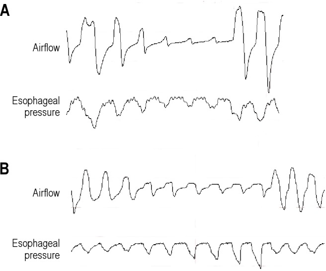
(A) A central hypopnea is characterized by lack of flattening in the airflow (nasal pressure) and a reduction in respiratory effort (esophageal pressure excursions). The reduction in flow is chronologically parallel to the reduction in effort. (B) An obstructive hypopnea is characterized by airflow limitation (flattening of the nasal pressure waveform) and increasing respiratory effort without an increase in airflow (nasal pressure). In this figure inspiration is upward.
Figure 6. An example of an obstructive hypopnea with snoring, flattening of the nasal pressure (NP) waveform, and paradoxical motion of the chest and abdominal (ABD) respiratory inductance plethysmography excursions.
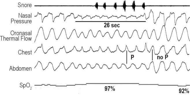
SpO2 is the pulse oximetry. Inspiration is upward in the figure. P denotes paradox during the hypopnea and no P the absence of paradox during unobstructed breathing.
Figure 7. A central hypopnea in a patient with Cheyne-Stokes breathing is illustrated.
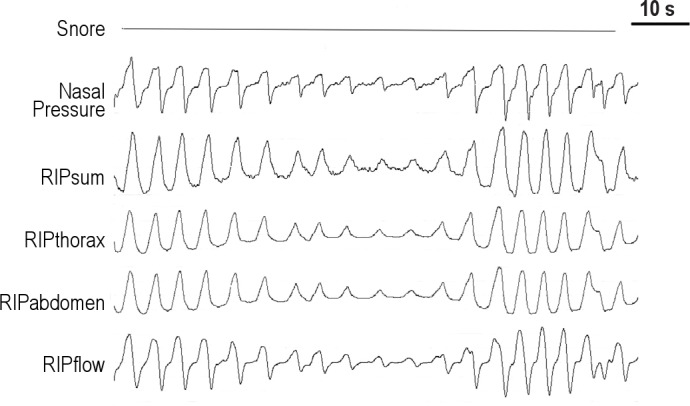
NP is the nasal pressure signal. There is no evidence of snoring or thoracoabdominal paradox in the RIP bands (RIPthorax and RIPabdomen). There is no evidence of airflow limitation (flattening of the nasal pressure signal). The direction of inspiration is upward in this figure.
The task force reviewed definitions of central and obstructive hypopnea in the literature with an emphasis on articles discussing CSB.89–93 One study defined a hypopnea as obstructive versus central if paradoxical thoracoabdominal excursions were noted or when the reduction in flow was out of proportion to the decrease in thoracoabdominal excursions (e.g., in central hypopnea the decrease in flow was proportional to the decrease in effort).90 Most articles have defined a central hypopnea on the basis of a lack of paradox in thoracoabdominal RIP belts and/or absence of flattening in the nasal pressure signal.89,91 Of note, in some studies hypopnea was defined based on a 50% drop in tidal volume (using RIP) without a requirement for associated desaturation or arousal.91 Lanfranchi et al. defined central hypopnea as ≥ 50% decrease in RIPsum lasting 10 seconds or longer followed by ≥ 2% desaturation.92 In this study, subjects with an obstructive AHI ≥ 5/hour were excluded. Ryan and coworkers defined central hypopnea as ≥ 50% decrease in RIPsum lasting 10 seconds or longer with in-phase thoracoabdominal motion and absence of flow limitation on the nasal pressure signal.93 In a study of CPAP and heart failure, Javaheri classified hypopnea as obstructive if paradoxical thoracoabdominal excursions occurred or if the airflow decreased out of proportion to the reduction in the thoracoabdominal excursions; otherwise hypopneas were classified as central.90
Some members of the task force felt that some mention of respiratory effort should be made in the definitions of central and obstructive hypopnea. Others noted that thoracoabdominal movements (RIP excursions) are not a direct measure of the amount of respiratory effort (e.g., esophageal pressure excursions). Task force members pointed out that a decrease in RIP excursions cannot differentiate obstructive and central hypopneas because the excursions may decrease in both types of hypopneas (see Figures 6 and 7). Although a disproportionate increase in effort when compared to flow can be indicative of obstruction, this is difficult to operationalize. The task force recommended the following definitions for scoring hypopneas as obstructive or central [Recommended] (Consensus) but also recommended that performing such scoring be [Optional] (Consensus). Such a separation of hypopneas into central or obstructive is not clinically indicated in the majority of patients.
Classifying Hypopnea in Adults [Recommended] (Consensus)
If electing to score obstructive hypopneas, score a hypopnea as obstructive if ANY of the following criteria are met:
Snoring during the event
Increased inspiratory flattening of the nasal pressure or PAP device flow signal compared to baseline breathing
Associated thoracoabdominal paradox occurs during the event but not during pre-event breathing
If electing to score central hypopneas, score a hypopnea as central if NONE of the following criteria are met:
Snoring during the event
Increased inspiratory flattening of the nasal pressure or PAP device flow signal compared to baseline breathing
Associated thoracoabdominal paradox occurs during the event but not during pre-event breathing
4.3. Respiratory Effort-Related Arousal Rule for Adults
The utility of scoring RERAs (an option in the 2007 scoring manual) is greatest when using a hypopnea definition not based on arousal. As noted above, if a definition of hypopnea is used which requires an associated desaturation OR arousal, then there are relatively few events scored as RERAs.78,79 The task force recommended only minor changes to the current RERA definition. One change includes the use of PAP device flow flattening rather than nasal pressure flattening during PAP titration. The task force acknowledges that “RERA” events are usually scored based on changes in nasal pressure (or PAP flow) rather than esophageal manometry. In this case, an increase in respiratory effort is inferred rather than being directly documented, leading some investigators to coin such an event as a “flow limitation arousal.”31 Nevertheless, the term RERA is widely used, and the task force members did not feel a change in terminology was needed. Task force members recommend that the scoring of RERA events remains [Optional] (Consensus).
RERA Rule for Adults [Recommended] (Consensus)
If electing to score respiratory effort-related arousals, score a respiratory event as a RERA if there is a sequence of breaths lasting at least 10 seconds characterized by increasing respiratory effort or by flattening of the inspiratory portion of the nasal pressure (diagnostic study) or PAP device flow (titration study) waveform leading to arousal from sleep when the sequence of breaths does not meet criteria for an apnea or hypopnea.
4.4. Hypoventilation Rule for Adults
A common definition of awake hypoventilation is an arterial PCO2 (PaCO2) > 45 mm Hg. In choosing a respiratory definition of hypoventilation for sleep studies there are two considerations. The first consideration is to identify a greater than normal increase in PaCO2 from wake to sleep, that is, hypoventilation that is “sleep-related.” The second is to identify an abnormal PaCO2 during sleep. In the 1999 Chicago consensus paper,9 it was stated that the normal increase in PaCO2 from wakefulness to sleep was from 2 to 7 mm Hg based on studies of arterial PaCO2 during sleep in normal subjects.96,97 “Sleep hypoventilation” was defined as a ≥ 10 mm Hg increase in PaCO2 from wake to sleep. The 2007 scoring manual also defined hypoventilation as ≥ 10 mm Hg increase in PaCO2 during sleep compared to an awake supine value. In neither definition was a minimum duration for the increased PaCO2 specified. In Table 7, patient A has a 10 mm Hg increase in PaCO2 and would meet the criteria for hypoventilation according to the 2007 AASM scoring manual. Patient B also has a 10 mm Hg increase, but many would not consider a PaCO2 of 45 mm Hg during sleep to represent hypoventilation. Patient C presents with awake hypoventilation and only a small increase in PaCO2 with sleep onset; yet, most would consider this patient to have hypoventilation during sleep. Of interest, local carrier determinations (LCDs) for the Center for Medicare and Medicaid services (CMS) have recently added a hypoventilation category for patient qualification for a respiratory assist device (e.g., bilevel PAP, bilevel PAP with a backup rate).88 The criteria include a daytime PaCO2 ≥ 45 mm Hg, and either a PaCO2 during sleep or immediately on awakening that is ≥ 7 mm Hg greater than the awake PaCO2 or a facility-based PSG demonstrates SpO2 ≤ 88% for at least 5 minutes of nocturnal recording time (minimum 2 hours of recording time) not caused by obstructive upper airway events.88
Table 7.
Three patients with possible “sleep hypoventilation”
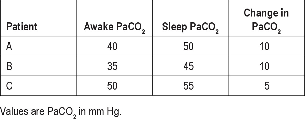
As noted above, end-tidal PCO2 and transcutaneous PCO2 rather than PaCO2 are usually measured in the sleep center. There are normative data for PETCO2 in pediatric patients.98–100 However, there is a paucity of normative data for adult PETCO2 and for PTCCO2 in all age groups. Midgren et al.101 studied normal adults and found an average awake PTCCO2 of 46 mm Hg with the highest value during sleep being 52 mm Hg. These data are from a 1987 article, and transcutaneous technology has advanced since this study. Morrell et al.102 found that PETCO2 increased from 38.7 mm Hg to 40.7 mm Hg during the wake-sleep transition in a group of normal subjects. Chin et al.103 found that the PTCCO2 increased during sleep by about 11 mm Hg in hypercapnic and 6 mm Hg in normocapnic OSA patients.
Based on data that normal individuals rarely have a PaCO2 > 55 mm Hg during sleep, the task force chose this threshold for sleep hypoventilation, with a minimum duration of 10 minutes, based on consensus. The task force considered the addition of a change in PaCO2 (or surrogate PCO2) from wakefulness to sleep with the proviso that the absolute sleeping PaCO2 (or surrogate) value should reach a value that clearly represents hypoventilation. The duration of 10 minutes is admittedly arbitrary; however, normative data for the amount of total sleep time at different PaCO2 values does not exist in sleeping adults. As noted in the earlier section on signals for detection of hypoventilation, scoring hypoventilation during sleep in adults is at the discretion of the clinician or investigator [Optional]. If reporting hypoventilation, the duration of hypoventilation as a percentage of total sleep time should be reported.
Hypoventilation Rule for Adults [Recommended] (Consensus)
If electing to score hypoventilation, score hypoventilation during sleep if either of the below occur:
There is an increase in the arterial PaCO2 (or surrogate) to a value > 55 mm Hg for ≥ 10 minutes
-
There is ≥ 10 mm Hg increase in PaCO2 (or surrogate) during sleep (in comparison to an awake supine value) to a value exceeding 50 mm Hg for ≥ 10 minutes.
Note: [Recommended] surrogates include end-tidal PCO2 or transcutaneous PCO2 for diagnostic study or transcutaneous PCO2 for PAP titration study.
4.5. Cheyne-Stokes Breathing Rule for Adults
Cheyne-Stokes breathing (CSB) is a specific form of periodic breathing (waxing and waning amplitude of flow or tidal volume) characterized by a crescendo-decrescendo pattern of respiration between central apneas or central hypopneas.1,9 The pattern of CSB is important to note as it may reflect unrecognized congestive heart failure and is a risk factor for early mortality or the need for heart transplant in patients with known heart failure.91,92,104 The 2007 scoring manual definition of CSB requires a minimum of 3 consecutive cycles for a run of central apneas or hypopneas to be considered CSB (Figure 8). An AHI ≥ 5/hour (duration of monitoring not specified) due to CSB OR a minimum duration of 10 consecutive minutes of this pattern of breathing was also required. Interestingly, the ICSD-2 diagnostic criteria for CSB requires 10 central apneas per hour of sleep.10
Figure 8.
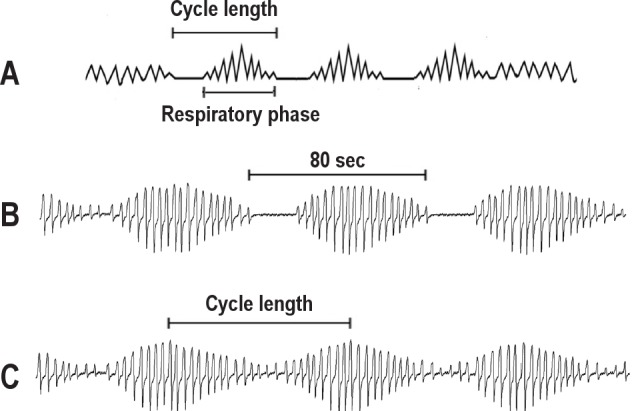
(A) Schematic of Cheyne-Stokes breathing (airflow shown) with a minimum of 3 consecutive central apneas (effort not shown) separated by a crescendo-decrescendo pattern of breathing. (B) Cheyne-Stokes breathing with central apneas (only airflow shown) with a long cycle time of 80 seconds. (C) Cheyne-Stokes breathing with central hypopneas (airflow shown). Although respiratory effort is not shown, these are central hypopneas with no evidence of airflow limitation (no flattening). As it is difficult to identify a beginning or end of the hypopnea, cycle time is defined as the time from one zenith in airflow during the respiratory phase to the next zenith in airflow.
A longer cycle length as well as the crescendo-decrescendo breathing pattern differentiate CSB from other forms of cyclic central apnea, but the specifics of defining cycle length vary between publications concerning CSB. All authors define cycle length as the duration of the central apnea (or hypopnea) + the duration of a respiratory phase (Figure 8). More specifically, Hall et al.105 defined cycle length as the time from the start of the respiratory phase to the end of the subsequent apnea (start of next respiratory phase). Wedewardt and coworkers106 defined cycle length as the time from beginning of a central apnea to the end of the next crescendo-decrescendo respiratory phase (start of the next apnea). Given the requirement of at least 3 consecutive central apneas, the task force adopted the latter definition of cycle length (Figure 8). If central hypopneas occur, the cycle length may be more ambiguous but can be defined as the time from the zenith in the respiratory phase preceding the central hypopnea to the zenith of the next respiratory phase.
Patients with a number of disorders including primary central sleep apnea and narcotic induced central apnea can exhibit periodic breathing with a waxing and waning of respiration. A typical pattern is central apnea – respiratory phase (breathing) – central apnea. Unlike CSB, the respiratory phase (between central apneas) of patients with primary central apnea or narcotic induced central apnea does NOT usually have a crescendo-decrescendo pattern, and the duration of the respiratory phase is typically shorter than in CSB. However, a minority of these patients may exhibit a respiratory phase with a crescendo-decrescendo pattern (Figure 9).
Figure 9. The tracings illustrate periodic breathing in a 35-year-old male with no evidence of cardiac disease who is not taking narcotic medication.
Central apneas are separated by respiration that sometimes shows a crescendo-decrescendo pattern. However, three consecutive ventilatory periods with a crescendo-decrescendo pattern are not present. In addition, the cycle length is only about 26 seconds. The cycle length is defined as the time from the beginning of a central apnea to the end of the subsequent crescendo-decrescendo respiratory phase.
What cycle length or length of breathing between consecutive apneas (respiratory phase) is required to score as CSB (Figure 10)? As few as three breaths could show a crescendo-decrescendo pattern. When CSB is associated with systolic heart failure the respiratory phase is long and the cycle length is approximately 60 seconds. Hall et al.105 compared the patterns of respiration in patients with idiopathic central sleep apnea (primary CSA) and CSB due to systolic heart failure. Patients with CSB had a longer cycle length due to a longer respiratory phase between central apneas (data shown in Table 8). The duration of central apnea was similar in the two groups of patients. A longer cycle length (and respiratory phase) was associated with more impaired cardiac function.
Figure 10. Various possibilities of periodic breathing with a crescendo-decrescendo pattern.
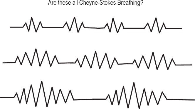
Table 8.
Cycle length of periodic breathing in patients with and without (systolic) heart failure
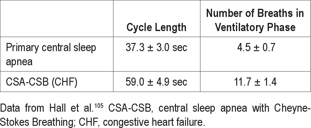
CSB has been described in patients after cerebrovascular accidents107 and in patients with diastolic heart failure (normal ejection fraction).108 In general, one might expect the cycle lengths to be shorter in these patients. While some might disagree with classifying the pattern of breathing exhibited by these groups of patients as CSB, there are no guidelines available regarding the minimum cycle length or respiratory phase duration to score CSB. A study of patients with CSB and various degrees of left ventricular dysfunction (Table 9) found considerable variation in the cycle length. Those individuals with a normal left ventricular ejection fraction exhibited a mean cycle length of 49.1 ± 17.4 seconds.106 Based on the above data, one might choose a minimum cycle length of 40 seconds to score CSB (or at least 5 to 6 breaths in the respiratory phase between apneas or hypopneas).
Table 9.
Variation in cycle length in Cheyne-Stokes breathing with different severities of heart failure

How many CSB events must be present to consider the patient as having CSB? Some patients have a transition from obstructive to central events during the same night (believed to be associated with increasing left ventricular filling pressure and ventilatory drive).109 Patients with both obstructive sleep apnea and heart failure may not exhibit CSB until late in the night or when in the supine position. CSB can also appear during a PAP titration after elimination of the obstructive component of breathing events. In the Outcomes of Sleep Disorders in Older Men (MrOS) Study, CSB was defined as the presence of at least 5 consecutive minutes of breathing with a CSB pattern.72 Using this definition for CSB, Mehra and colleagues found an association between the presence of CSB and complex ventricular ectopy independent of self-reported heart failure.72 Mared et al. defined the presence of CSB in patients when this breathing pattern occupied more that 10% of the recording time.110 As noted above, the CSB scoring rule in the 2007 scoring manual requires at least 3 consecutive central apneas and/or central hypopneas interspersed with a CSB pattern of breathing and either a central AHI of 5/hour or 10 consecutive minutes of CSB. The monitoring period for computation of the AHI was not specified. If one assumes a cycle time of 60 seconds, requiring 10 consecutive minutes equates to about 10 consecutive central events. Do the events have to be consecutive? Would two runs of five CSB events not be as convincing as a 10-event run?
There is also evidence that the presence91,104 or amount of CSB could have some prognostic significance in patients with heart failure. Lanfranchi and coworkers92 followed a group of patients with chronic heart failure documented as having CSB by PSG. A central AHI > 30/hour was a bad prognostic sign for survival. Non-survivors had a greater portion of the night in periodic breathing. Amir et al.111 studied a group of patients with advanced systolic heart failure and found that a longer duration of CSB was associated with higher mortality and a higher NT-proBNP, a marker of poor cardiac function. In this study the mean duration of CSB time was about one hour. Based on these studies, it may be useful to specify the amount of CSB. The task force recommends that a parameter reflecting the CSB duration (absolute or percentage of total sleep time) or the number of CSB events should be specified in the sleep study report if possible [Recommended] (Consensus).
Given the above considerations, the task force proposed a revised CSB definition. The minimum amount of CSB respiration that must be present is arbitrary but was chosen as an AHI of ≥ 5/hour (associated with CSB) with a minimum monitoring period of 2 hours. For most patients this is equivalent to requiring about 10 CSB respiratory events. The wording clearly specifies that central apneas OR central hypopneas can separate periods of crescendo-decrescendo respiration. It was felt that specifying a minimum AHI over a minimum monitoring time would replace the need to specify a minimum total duration of CSB.
Cheyne-Stokes Breathing Rule for Adults [Recommended] (Consensus)
Score a respiratory event as Cheyne-Stokes breathing if both of the following are met:
There are episodes of at least 3 consecutive central apneas and/or central hypopneas separated by a crescendo and decrescendo change in breathing amplitude with a cycle length of at least 40 seconds (typically 45 to 90 seconds).
-
There are 5 or more central apneas and/or central hypopneas per hour associated with the crescendo/decrescendo breathing pattern recorded over a minimum of 2 hours of monitoring.
Note: The duration of CSB (absolute or as a percentage of total sleep time) or the number of CSB events should be presented in the study report.
5.0. PEDIATRIC SCORING RULES
5.1. Ages for which Scoring Rules for Children Should Be Used
The 2007 AASM scoring manual specifies that the respiratory scoring rules for children can be used for infants and children < 18 years. There is the option of using adult respiratory scoring rules for children ≥ 13 years. The task force considered a change in the current rule to one recommending that pediatric rules be used for all children younger than 18 years. When the AASM pediatric scoring rules were developed, there were no data available specifically pertaining to adolescents; therefore, it was suggested that adolescents aged 13-18 years could be scored using either pediatric or adult criteria. Since then, two studies have shown significant differences in respiratory parameters when the PSGs of adolescents aged 13-18 years were scored using pediatric versus adult criteria.3,4 A study of normal adolescents showed that they had a significantly higher AHI when pediatric scoring rules were used.3 Another study of adolescents with suspected OSA also showed a significant difference in AHI using pediatric versus adult scoring rules, especially between the pediatric rule and the recommended adult rule for hypopneas.112 In addition, significantly more children would have been diagnosed with OSA using pediatric versus adult rules. Some members of the task force felt strongly that all patients younger than 18 years should be scored according to pediatric scoring rules. However, the task force was unable to reach a consensus to change the current AASM scoring rule concerning the age range for use of respiratory scoring rule for children. The current AASM scoring rule does allow the clinician the option of using pediatric scoring rules for patients ≥ 13 but < 18 years. The clinician could elect to use pediatric scoring rules for these older children based on the consideration of the recent studies just described. In addition, with a similar proposed definition for hypopnea in adults and children (see section on hypopnea below), this concern may be less of an issue.
Age for Which Pediatric Scoring Rules Should be Used (unchanged from current rule) [Recommended] (Consensus)
Criteria for respiratory events during sleep for infants and children can be used for children < 18 years, but an individual sleep specialist can choose to score children ≥ 13 years using adult criteria.
5.2. Apnea Rule for Pediatric Patients
The task force considered the same issues regarding defining apnea as for adults. Positive airway pressure device flow was added as a sensor to detect apnea during positive airway titration, and the requirement that the duration of the event meeting amplitude criteria must be ≥ 90% of the event's duration was removed. The minimum duration of the drop in flow is two respiratory cycles as in the 2007 scoring manual. As not all events with absent airflow and effort are scored as central apneas, a general definition of apnea requires that events meet criteria for obstructive, central, or mixed apnea.
Apnea Rule for Pediatric Patients [Recommended] (Consensus)
Score a respiratory event as an apnea if it meets all of the following criteria:
There is a drop in the peak signal excursion by ≥ 90% of the pre-event baseline using an oronasal thermal sensor (diagnostic study), PAP device flow (titration study), or an alternative apnea sensor (diagnostic study).
The duration of the ≥ 90% drop lasts at least the minimum duration as specified by obstructive, mixed, or central apnea duration criteria.
The event meets respiratory effort criteria for obstructive, central, or mixed apnea.
5.2.1. Classification of Pediatric Apnea
The proposed revised obstructive apnea definition is similar to the one recommended for adults. The central apnea definition differs from adults as normal infants and children may have a considerable number of short central pauses in respiration not associated with desaturation or arousal. Such events are felt to be within normal variation limits.51,113,114 The central apnea definition is similar to the 2007 scoring manual central apnea definition except that a provision for children less than 1 year of age has been added. In these individuals, a central apnea is believed to be significant if followed by significant bradycardia (a decrease in heart rate to < 50 beats per minute for at least 5 seconds or < 60 beats per minute for 15 seconds).113,114 In one study of normal infants, none had a heart rate less than 55 beats per minute for more than 10 seconds.113 The proposed pediatric mixed apnea definition differs from the adult definition in that the central portion may be present before or after the obstructive portion of the event. This option was added at the request of pediatric task force members who thought this was a common clinical occurrence. An example of a mixed apnea with the central portion following the obstructive portion in a 3-year-old child is shown in Figure 11.
Figure 11. An example of a mixed apnea in a 3-year-old child where the central portion follows the obstructive portion.
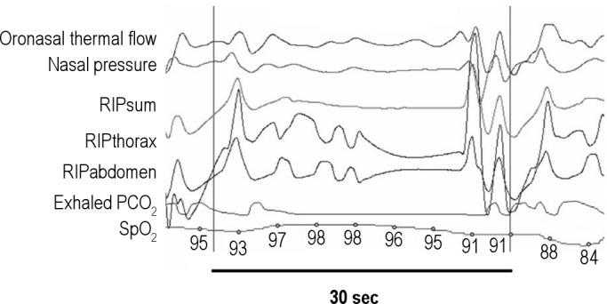
The vertical lines are 30 seconds apart.
Classification of Pediatric Apnea [Recommended] (Consensus)
Score a respiratory event as an obstructive apnea if it meets apnea criteria for at least the duration of 2 breaths during baseline breathing and is associated with the presence of respiratory effort throughout the entire period of absent airflow.
Score a respiratory event as a central apnea if it meets apnea criteria, is associated with absent inspiratory effort throughout the entire duration of the event, and at least one of the following is met:
The event lasts 20 seconds or longer.
The event lasts at least the duration of two breaths during baseline breathing and is associated with an arousal or ≥ 3% oxygen desaturation.
For infants younger than 1 year of age, the event lasts at least the duration of two breaths during baseline breathing and is associated with a decrease in heart rate to less than 50 beats per minute for at least 5 seconds or less than 60 beats per minute for 15 seconds.
Score a respiratory event as a mixed apnea if it meets apnea criteria for at least the duration of 2 breaths during baseline breathing and is associated with absent respiratory effort during one portion of the event and the presence of inspiratory effort in another portion, regardless of which portion comes first.
5.3. Hypopnea Rule for Pediatric Patients
The hypopnea definition for pediatrics in the 2007 scoring manual requires a 50% drop in flow with either ≥ 3% oxygen desaturation or arousal. This is very similar to the current recommendation of the task force for the hypopnea definition for adults except that the minimum duration is 2 breaths. The main issue was whether to use a 30% drop in flow as in the adult definition or retain the 50% drop in flow in the current pediatric hypopnea definition. Advantages of the 50% drop include the ability to compare results with some previously published papers. An advantage of using a 30% drop is that both pediatric and adult definitions would be similar. It was also felt that using a 50% drop would create a class of events with > 30% drop but less than a 50% drop in flow that if associated with an arousal or desaturation would not be scored (unless scored as a RERA). The task force consensus was to use 30% drop in flow; thus, the hypopnea rule is the same for children as for adults except for the minimum duration of 2 breaths (rather than 10 seconds).
Hypopnea Rule for Pediatric Patients [Recommended] (Consensus)
Score a respiratory event as a hypopnea if it meets all of the following criteria:
The peak signal excursions drop by ≥ 30% of pre-events baseline using nasal pressure (diagnostic study), PAP device flow (titration study) or an alternative hypopnea sensor (diagnostic study).
The duration of the ≥ 30% drop lasts for at least 2 breaths.
There is ≥ 3% desaturation from pre-event baseline or the event is associated with an arousal.
5.3.1. Classification of Pediatric Hypopnea
The recommended scoring rules for scoring pediatric hypopneas as either central or obstructive are the same as for adults [Recommended] (Consensus), and classifying hypopneas in pediatric patients should be considered as [Optional] (Consensus).
5.4. Respiratory Effort-Related Arousal Rule for Pediatric Patients
The task force revised the definition of RERA in pediatric patients to be similar to that recommended for adults except for a few differences. The minimum duration is 2 breaths, and snoring or an elevation in the end-tidal PCO2 is included as supporting the diagnosis of RERA. The pediatric RERA rule in the 2007 scoring manual includes an increase in the transcutaneous PCO2 included as a scoring criterion; however, the device response is too slow for RERA identification and the criterion was removed. As in adults, the task force felt that scoring RERAs should be [Optional] (Consensus).
RERA Rule for Pediatric Patients [Recommended] (Consensus)
If electing to score respiratory effort-related arousals, score a respiratory event as a RERA if there is a sequence of breaths lasting at least 2 breaths (or the duration of two breaths during baseline breathing) when the breathing sequence is characterized by increasing respiratory effort, flattening of the inspiratory portion of the nasal pressure or PAP device flow waveform, snoring, or an elevation in the end-tidal PCO2 leading to arousal from sleep when the sequence of breaths does not meet criteria for an apnea or hypopnea.
5.5. Hypoventilation Rule for Pediatric Patients
There are more normative data for the use of end-tidal PCO2 and transcutaneous PCO2 during sleep in children compared to adults.98–100 The existing scoring rule was felt to be consistent with currently available data. The task force agreed that the 2007 scoring manual scoring rule for hypoventilation in children should remain unchanged until further published data are available.
Hypoventilation Rule for Children [Recommended] (Consensus)
Score hypoventilation during sleep when > 25% of the total sleep time as measured by either the arterial PCO2 or surrogate is spent with a PCO2 > 50 mm Hg.
5.6. Periodic Breathing Rule for Pediatric Patients
The task force found no evidence suggesting that the current definition of periodic breathing be significantly changed. The task force recommends a periodic breathing respiratory definition that is similar to the 2007 scoring manual except that “greater than 3 episodes” is replaced by “greater than or equal to 3 episodes.” This was felt to be more inclusive.
Periodic Breathing Rule for Pediatric Patients [Recommended] (Consensus)
Score a respiratory event as periodic breathing if there are ≥ 3 episodes of central apnea lasting > 3 seconds separated by no more than 20 seconds of normal breathing.
6.0. SUMMARY
The definitions of respiratory events and recommendations concerning monitoring technology will continue to evolve as more knowledge is gained about the effect of using different definitions or technology on outcomes. Improved ability to predict patients who will improve symptomatically with treatment (especially in patients with “milder” obstructive sleep apnea) is clearly needed. It is hoped that this document is simply a starting point of a new process to provide a flexible and evolving set of respiratory definitions. The recommendations in this document are based predominantly on consensus. The task force attempted to carefully weigh the current evidence as well as to respond to concerns raised by the sleep community about the recommendations in the 2007 scoring manual. Many areas of uncertainty remain. No set of definitions can completely cover the wide variety of respiratory events encountered by clinicians. There is no substitute for clinical correlation of PSG findings with the clinical symptoms of the patient being evaluated.
DISCLOSURE STATEMENT
This was not an industry supported study. Drs. Berry and Redline have received a research grant from Dymedix, Inc. The other authors have indicated no financial conflicts of interest.
ACKNOWLEDGMENTS
The task force thanks Christine Stepanski, MS, and Richard Rosenberg, PhD, for their valuable assistance during the consensus process and the Board of Directors of the AASM for their guidance and support. The task force also thanks Drs. Berry and Marcus for supplying the figures.
REFERENCES
- 1.Iber C, Ancoli-Israel S, Chesson AL, Jr., Quan SF for the American Academy of Sleep Medicine. The AASM manual for the scoring of sleep and associated events: rules, terminology and technical specifications. 1st ed. Westchester, IL: American Academy of Sleep Medicine; 2007. [Google Scholar]
- 2.Ruehland WR, Rochford PD, O'Donoghue FJ, et al. The new AASM criteria for scoring hypopneas: impact on the apnea hypopnea index. Sleep. 2009;32:150–7. doi: 10.1093/sleep/32.2.150. [DOI] [PMC free article] [PubMed] [Google Scholar]
- 3.Accardo JA, Shults J, Leonard MB, Traylor J, Marcus CL. Differences in overnight polysomnography scores using the adult and pediatric criteria for respiratory events in adolescents. Sleep. 2010;33:1333–9. doi: 10.1093/sleep/33.10.1333. [DOI] [PMC free article] [PubMed] [Google Scholar]
- 4.Grigg-Damberger MM. The AASM scoring manual four years later. J Clin Sleep Med. 2012;8:323–32. doi: 10.5664/jcsm.1928. [DOI] [PMC free article] [PubMed] [Google Scholar]
- 5.Guilleminault C, Hagen CC, Huynh NT. Comparison of hypopnea definitions in lean patients with known obstructive sleep apnea hypopnea syndrome. Sleep Breath. 2009;13:341–7. doi: 10.1007/s11325-009-0253-7. [DOI] [PubMed] [Google Scholar]
- 6.Koo BB, Drummond C, Surovec S, Johnson N, Marvin SA, Redline S. Validation of a polyvinylidene fluoride impedance sensor for respiratory event classification during polysomnography. J Clin Sleep Med. 2011;7:479–85. doi: 10.5664/JCSM.1312. [DOI] [PMC free article] [PubMed] [Google Scholar]
- 7.Collop NA, Tracy SL, Kapur V, Mehra R, et al. Obstructive sleep apnea devices for out-of-center (OOC) testing: technology evaluation. J Clin Sleep Med. 2011;5:531–48. doi: 10.5664/JCSM.1328. [DOI] [PMC free article] [PubMed] [Google Scholar]
- 8.Redline S, Budhiraja R, Kapur V, et al. The scoring of respiratory events in sleep: reliability and validity. J Clin Sleep Med. 2007;3:169–200. [PubMed] [Google Scholar]
- 9.American Academy of Sleep Medicine Task Force. Sleep-related breathing disorders in adults: recommendations for syndrome definition and measurement techniques in clinical research. The Report of an American Academy of Sleep Medicine Task Force. Sleep. 1999;22:667–89. [PubMed] [Google Scholar]
- 10.American Academy of Sleep Medicine. International classification of sleep disorders: diagnostic and coding manual. 2nd ed. Westchester, IL: American Academy of Sleep Medicine; 2005. [Google Scholar]
- 11.Sackett DL, Strauss SE, Richardson WS, et al. Evidence-based medicine: how to practice and teach EBM. 2nd ed. Edinburgh, Scotland, UK: Churchill Livingstone; 2000. [Google Scholar]
- 12.Fitch K, Bernstein SJ, Aguilar MD, et al. The RAND/UCLA Appropriateness Method User's Manual. Santa Monica, CA: RAND; 2001. [Google Scholar]
- 13.Condos R, Norman RG, Krishnasamy I, et al. Flow limitation as a noninvasive assessment of residual upper-airway resistance during continuous positive airway pressure therapy of obstructive sleep apnea. Am J Respir Crit Care Med. 1994;150:475–80. doi: 10.1164/ajrccm.150.2.8049832. [DOI] [PubMed] [Google Scholar]
- 14.Kushida CA, Chediak A, Berry RB, et al. Clinical guidelines for the manual titration of positive airway pressure in patients with obstructive sleep apnea. J Clin Sleep Med. 2008;4:157–71. [PMC free article] [PubMed] [Google Scholar]
- 15.Berry RB, Chediak A, Brown LK, et al. Best clinical practices for the sleep center adjustment of noninvasive positive pressure ventilation (NPPV) in stable chronic alveolar hypoventilation syndromes. J Clin Sleep Med. 2010;6:491–509. [PMC free article] [PubMed] [Google Scholar]
- 16.Montserrat JM, Ballester E, Olivi H, et al. Time-course of stepwise CPAP titration: behavior of respiratory and neurological variables. Am J Respir Crit Care Med. 1995;152:1854–959. doi: 10.1164/ajrccm.152.6.8520746. [DOI] [PubMed] [Google Scholar]
- 17.Cohn MA, Rao AS, Broudy M, et al. The respiratory inductive plethysmograph: a new non-invasive monitor of respiration. Bull Eur Physiopathol Respir. 1982;18:643–8. [PubMed] [Google Scholar]
- 18.Tobin MJ, Cohn MA, Sackner MA. Breathing abnormalities during sleep. Arch Intern Med. 1983:1221–8. [PubMed] [Google Scholar]
- 19.Farré R, Montserrat JM, Navajas D. Noninvasive monitoring of respiratory mechanics during sleep. Eur Respir J. 2004;24:1052–60. doi: 10.1183/09031936.04.00072304. [DOI] [PubMed] [Google Scholar]
- 20.Kaplan V, Zhang JN, Russi EW, Bloch KE. Detection of inspiratory flow limitation during sleep by computer assisted respiratory inductive plethysmography. Eur Respir J. 2000;15:570–8. doi: 10.1034/j.1399-3003.2000.15.24.x. [DOI] [PubMed] [Google Scholar]
- 21.Cantineau JP, Escourrou P, Sartene R, Gaultier C, Goldman M. Accuracy of respiratory inductive plethysmography during wakefulness and sleep in patients with obstructive sleep apnea. Chest. 1992;102:1145–51. doi: 10.1378/chest.102.4.1145. [DOI] [PubMed] [Google Scholar]
- 22.Gould GA, Whyte KF, Rhind MA, et al. Sleep hypopnea syndrome. Am Rev Respir Dis. 1988;137:895–8. doi: 10.1164/ajrccm/137.4.895. [DOI] [PubMed] [Google Scholar]
- 23.Thurnheer R, Xie X, Bloch KE. Accuracy of nasal cannula pressure recordings for assessment of ventilation during sleep. Am J Respir Crit Care Med. 2001;146:1914–9. doi: 10.1164/ajrccm.164.10.2102104. [DOI] [PubMed] [Google Scholar]
- 24.Clark SA, Wilson CR, Satoh M, et al. Assessment of inspiratory flow limitation invasively and noninvasively during sleep. Am J Respir Crit Care Med. 1998;158:713–22. doi: 10.1164/ajrccm.158.3.9708056. [DOI] [PubMed] [Google Scholar]
- 25.Griffiths A, Maul J, Wilson A, Stick S. Improved detection of obstructive events in childhood sleep apnoea with the use of the nasal cannula and the differentiated sum signal. J Sleep Res. 2005;14:431–6. doi: 10.1111/j.1365-2869.2005.00474.x. [DOI] [PubMed] [Google Scholar]
- 26.Sackner MA, Watson H, Belsito AS, et al. Calibration of respiratory inductive plethysmograph during natural breathing. J Appl Physiol. 1989;66:410–20. doi: 10.1152/jappl.1989.66.1.410. [DOI] [PubMed] [Google Scholar]
- 27.Masa JF, Corral J, Martin MJ, et al. Assessment of thoracoabdominal bands to detect respiratory effort-related arousals. Eur Resp J. 2003;33:661–7. doi: 10.1183/09031936.03.00010903. [DOI] [PubMed] [Google Scholar]
- 28.Loube DI, Andrada T, Howard RS. Accuracy of respiratory inductive plethysmography for the diagnosis of upper airway resistance syndrome. Chest. 1999;115:1333–7. doi: 10.1378/chest.115.5.1333. [DOI] [PubMed] [Google Scholar]
- 29.Hosselet JJ, Norman RG, Ayappa I, Rapoport DM. Detection of flow limitation with a nasal cannula/pressure transducer system. Am J Respir Crit Care Med. 1998;157:1461–7. doi: 10.1164/ajrccm.157.5.9708008. [DOI] [PubMed] [Google Scholar]
- 30.Norman RG, Ahmed MM, Walsleben JA, Rapoport DM. Detection of respiratory events during NPSG: nasal cannula/pressure sensor versus thermistor. Sleep. 1997;20:1175–84. [PubMed] [Google Scholar]
- 31.Ayappa I, Norman RG, Krieger AC, et al. Non-invasive detection of respiratory effort-related arousals (RERAs) by a nasal cannula/pressure transducer system. Sleep. 2000;23:763–71. doi: 10.1093/sleep/23.6.763. [DOI] [PubMed] [Google Scholar]
- 32.Hernandez L, Ballester E, Farre R, et al. Performance of nasal prongs in sleep studies. Chest. 2001;119:442–50. doi: 10.1378/chest.119.2.442. [DOI] [PubMed] [Google Scholar]
- 33.Farré R, Montserrat JM, Rotger M, et al. Accuracy of thermistors and thermocouples as flow-measuring devices for detecting hypopneas. Eur Respir J. 1998;11:179–82. doi: 10.1183/09031936.98.11010179. [DOI] [PubMed] [Google Scholar]
- 34.Berry RB, Koch GL, Trautz S, Wagner MH. Comparison of respiratory event detection by a polyvinylidene fluoride film airflow sensor and a pneumotachograph in sleep apnea patients. Chest. 2005;128:1331–8. doi: 10.1378/chest.128.3.1331. [DOI] [PubMed] [Google Scholar]
- 35.Nakano H, Tanigawa T, Ohnishi Y. Validation of a single-channel airflow monitor for screening of sleep-disordered breathing. Eur Respir J. 2008;32:1060–7. doi: 10.1183/09031936.00130907. [DOI] [PubMed] [Google Scholar]
- 36.Farré R, Rigau J, Montserrat JM, et al. Relevance of linearizing nasal prongs for assessing hypopneas and flow limitation during sleep. Am J Respir Crit Care Med. 2001;163:494–7. doi: 10.1164/ajrccm.163.2.2006058. [DOI] [PubMed] [Google Scholar]
- 37.Whyte KF, Gugger M, Gould GA, et al. Accuracy of respiratory inductance plethysmograph in measuring tidal volume during sleep. J Appl Physiol. 1991;71:1866–71. doi: 10.1152/jappl.1991.71.5.1866. [DOI] [PubMed] [Google Scholar]
- 38.Heitman SJ, Atkar RS, Hajduk EA, et al. Validation of nasal pressure for the identification of apneas/hypopneas during sleep. Am J Respir Crit Care Med. 2002;166:386–91. doi: 10.1164/rccm.2105085. [DOI] [PubMed] [Google Scholar]
- 39.Weese-Mayer DE, Corwin MJ, Peucker MR, et al. Comparison of apnea identified by respiratory inductance plethysmography with that detected by end-tidal CO(2) or thermistor. The CHIME Study Group. Am J Respir Crit Care Med. 2000;162(2 Pt 1):471–80. doi: 10.1164/ajrccm.162.2.9904029. [DOI] [PubMed] [Google Scholar]
- 40.West P, Kryger MH. Sleep and respiration: Terminology and methodology. Clin Chest Med. 1985;6:706. [PubMed] [Google Scholar]
- 41.Berg S, Haight JSJ, Yap V, et al. Comparison of direct and indirect measurement of respiratory airflow: implications for hypopneas. Sleep. 1997;20:60–4. doi: 10.1093/sleep/20.1.60. [DOI] [PubMed] [Google Scholar]
- 42.Kushida CA, Giacomini A, Lee MK, Guilleminault C, et al. Technical protocol for the use of esophageal manometry in the diagnosis of sleep-related breathing disorders. Sleep Med. 2002;3:163–73. doi: 10.1016/s1389-9457(01)00143-5. [DOI] [PubMed] [Google Scholar]
- 43.Boudewyns A, Willemen M, Wagemans M, De Cock W, Van de Heyning P, De Backer W. Assessment of respiratory effort by means of strain gauges and esophageal pressure swings: a comparative study. Sleep. 1997;20:168–70. doi: 10.1093/sleep/20.2.168. [DOI] [PubMed] [Google Scholar]
- 44.Stats BA, Bonekat W, Harris CD, Offord KP. Chest wall motion in sleep apnea. Am Rev Respir Dis. 1984;130:59–63. doi: 10.1164/arrd.1984.130.1.59. [DOI] [PubMed] [Google Scholar]
- 45.Adams JA, Zabaleta IA, Stroh D, Sackner MA. Measurement of breath amplitudes: comparison of three noninvasive respiratory monitors to integrated pneumotachograph. Pediatr Pulmonol. 1993;16:254–8. doi: 10.1002/ppul.1950160408. [DOI] [PubMed] [Google Scholar]
- 46.Sackner MA, Kreiger BP. Non-invasive respiratory monitoring. In: Scharf SM, Cassidy SS, editors. Heart-lung interactions in health and disease. New York: Marcel Dekker; 1989. pp. 663–805. [Google Scholar]
- 47.Stoohs RA, Blum HC, Knaack L, Butsch-von-der-Heydt B, Guilleminault C. Comparison of pleural pressure and transcutaneous diaphragmatic electromyogram in obstructive sleep apnea syndrome. Sleep. 2005;28:321–9. [PubMed] [Google Scholar]
- 48.Stoohs RA, Blum HC, Kanack L, Guillenault C. Non-invasive estimation of esophageal pressure based on intercostal EMG. Proceedings of the 26th Annual International Conference of the IEE EMBSS; Sept 1-5, 2004; San Francisco, CA. pp. 3867–9. [DOI] [PubMed] [Google Scholar]
- 49.Toffaletti J, Zijlstra WG. Misconceptions in reporting oxygen saturation. Anesth Anal. 2007;105:S5–9. doi: 10.1213/01.ane.0000278741.29274.e1. [DOI] [PubMed] [Google Scholar]
- 50.Zavorsky GS, Cao J, Mayo NE, Gabbay R, Murias JM. Arterial versus capillary blood gases: a meta-analysis. Respir Physiol Neurobiol. 2007;155:268–79. doi: 10.1016/j.resp.2006.07.002. [DOI] [PubMed] [Google Scholar]
- 51.Beck SE, Marcus CL. Pediatric polysomnography. Sleep Med Clin. 2009;4:393–406. doi: 10.1016/j.jsmc.2009.04.007. [DOI] [PMC free article] [PubMed] [Google Scholar]
- 52.D'Mello J, Butani M. Capnography. Indian J Anaesth. 2002;46:269–78. [Google Scholar]
- 53.Sanders MH, Kern NB, Costantino JP, et al. Accuracy of end-tidal and transcutaneous PCO2 monitoring during sleep. Chest. 1994;106:472–83. doi: 10.1378/chest.106.2.472. [DOI] [PubMed] [Google Scholar]
- 54.Eberhard P. The design, use, and results of transcutaneous carbon dioxide analysis: current and future directions. Anesth Analg. 2007;195:S48–S52. doi: 10.1213/01.ane.0000278642.16117.f8. [DOI] [PubMed] [Google Scholar]
- 55.Storre JH, Steurer B, Kabitz HJ, Dreher M, Windisch W. Transcutaneous PCO2 monitoring during initiation of noninvasive ventilation. Chest. 2007;132:1810–6. doi: 10.1378/chest.07-1173. [DOI] [PubMed] [Google Scholar]
- 56.Kasuya Y, Akça O, Sessler DI, Ozaki M, Komatsu R. Accuracy of postoperative end-tidal Pco2 measurements with mainstream and sidestream capnography in non-obese patients and in obese patients with and without obstructive sleep apnea. Anesthesiology. 2009;111:609–15. doi: 10.1097/ALN.0b013e3181b060b6. [DOI] [PubMed] [Google Scholar]
- 57.Kim SM, Park KS, Nam H, et al. Capnography for assessing nocturnal hypoventilation and predicting compliance with subsequent noninvasive ventilation in patients with ALS. PLoS One. 2011;6:e17893. doi: 10.1371/journal.pone.0017893. [DOI] [PMC free article] [PubMed] [Google Scholar]
- 58.Maniscalco M, Zedda A, Faraone S, Carratu P, Sofia M. Evaluation of a transcutaneous carbon dioxide monitor in severe obesity. Intensive Care Med. 2008;34:1340–4. doi: 10.1007/s00134-008-1078-8. [DOI] [PubMed] [Google Scholar]
- 59.Storre JH, Magnet F, Dreher M, et al. Transcutaneous monitoring as a replacement for arterial pCO2 monitoring during nocturnal non-invasive ventilation. Respir Med. 2011;105:143–50. doi: 10.1016/j.rmed.2010.10.007. [DOI] [PubMed] [Google Scholar]
- 60.Paiva R, Krivec U, Aubertin G, Cohen E, Clément A, Fauroux B. Carbon dioxide monitoring during long-term noninvasive respiratory support in children. Intensive Care Med. 2009;35:1068–74. doi: 10.1007/s00134-009-1408-5. [DOI] [PubMed] [Google Scholar]
- 61.Senn O, Clarenbach CF, Kaplan V, et al. Monitoring carbon dioxide tension and arterial oxygen saturation by a single earlobe sensor in patients with critical illness or sleep apnea. Chest. 2005;128:1291–6. doi: 10.1378/chest.128.3.1291. [DOI] [PubMed] [Google Scholar]
- 62.Kirk VG, Batuyong ED, Bohn SG. Transcutaneous carbon dioxide monitoring and capnography during pediatric polysomnography. Sleep. 2006;29:1601–8. doi: 10.1093/sleep/29.12.1601. [DOI] [PubMed] [Google Scholar]
- 63.Hirabayashi M, Fujiwara C, Ohtani N, Kagawa S, Kamide M. Transcutaneous PCO2 monitors are more accurate than end-tidal PCO2 monitors. J Anesth. 2009;23:198–202. doi: 10.1007/s00540-008-0734-z. [DOI] [PubMed] [Google Scholar]
- 64.Casati A, Squicciarini G, Malagutti G, et al. Transcutaneous monitoring of partial pressure of carbon dioxide in the elderly patient: a prospective, clinical comparison with end-tidal monitoring. J Clin Anesth. 2006;18:436–80. doi: 10.1016/j.jclinane.2006.02.007. [DOI] [PubMed] [Google Scholar]
- 65.Redline S, Sanders M. Hypopnea, a floating metric: implications for prevalence, morbidity estimates, and case finding. Sleep. 1997;20:1209–17. doi: 10.1093/sleep/20.12.1209. [DOI] [PubMed] [Google Scholar]
- 66.Redline S, Kapur VK, Sanders MH, et al. Effects of varying approaches for identifying respiratory disturbances on sleep apnea assessment. Am J Respir Crit Care Med. 2000;161(2 Pt 1):369–74. doi: 10.1164/ajrccm.161.2.9904031. [DOI] [PubMed] [Google Scholar]
- 67.Redline S, Min NI, Shahar E, Rapoport D, O'Connor G. Polysomnographic predictors of blood pressure and hypertension: is one index best? Sleep. 2005;28:1122–30. doi: 10.1093/sleep/28.9.1122. [DOI] [PubMed] [Google Scholar]
- 68.Manser RL, Rochford P, Pierce RJ, Byrnes GB, Campbell DA. Impact of different criteria for defining hypopneas in the apnea-hypopnea index. Chest. 2001;120:909–14. doi: 10.1378/chest.120.3.909. [DOI] [PubMed] [Google Scholar]
- 69.Block AJ, Boysen PG, Wynne JW, et al. Sleep apnea, hypopnea and oxygen desaturation in normal subjects. N Engl J Med. 1979;300:513–7. doi: 10.1056/NEJM197903083001001. [DOI] [PubMed] [Google Scholar]
- 70.Mehra R, Benjamin EJ, Shahar E, et al. Association of nocturnal arrhythmias with sleep-disordered breathing: The Sleep Heart Health Study. Am J Respir Crit Care Med. 2006;173:910–6. doi: 10.1164/rccm.200509-1442OC. [DOI] [PMC free article] [PubMed] [Google Scholar]
- 71.Punjabi NM, Newman AB, Young TB, Resnick HE, Sanders MH. Sleep-disordered breathing and cardiovascular disease: an outcome-based definition of hypopneas. Am J Respir Crit Care Med. 2008;177:1150–5. doi: 10.1164/rccm.200712-1884OC. [DOI] [PMC free article] [PubMed] [Google Scholar]
- 72.Mehra R, Stone KL, Varosy PD, et al. Nocturnal arrhythmias across a spectrum of obstructive and central sleep-disordered breathing in older men: outcomes of sleep disorders in older men (MrOS sleep) study. Arch Intern Med. 2009;169:1147–55. doi: 10.1001/archinternmed.2009.138. [DOI] [PMC free article] [PubMed] [Google Scholar]
- 73.Stamatakis K, Sanders MH, Caffo B, et al. Fasting glycemia in sleep disordered breathing: lowering the threshold on oxyhemoglobin desaturation. Sleep. 2008;31:1018–24. [PMC free article] [PubMed] [Google Scholar]
- 74.Redline S, Yenokyan G, Gottlieb DJ. Obstructive sleep apnea-hypopnea and incident stroke: the sleep heart health study. Am J Respir Crit Care Med. 2010;182:269–77. doi: 10.1164/rccm.200911-1746OC. [DOI] [PMC free article] [PubMed] [Google Scholar]
- 75.Whitney CW, Gottlieb DJ, Redline S, et al. Reliability of scoring respiratory disturbance indices and sleep staging. Sleep. 1998;21:749–57. doi: 10.1093/sleep/21.7.749. [DOI] [PubMed] [Google Scholar]
- 76.Guilleminault C, Stoohs R, Clerk A, Cetel M, Maistros P. A cause of excessive daytime sleepiness. The upper airway resistance syndrome. Chest. 1993;104:781–7. doi: 10.1378/chest.104.3.781. [DOI] [PubMed] [Google Scholar]
- 77.Bonnet MH, Doghramji K, Roehrs T, et al. The scoring of arousal in sleep: reliability, validity, and alternatives. J Clin Sleep Med. 2007;3:133–45. [PubMed] [Google Scholar]
- 78.Cracowski C, Pepin JL, Wuyam B, Levy P. Characterization of obstructive nonapneic respiratory events in moderate sleep apnea syndrome. Am J Respir Crit Care Med. 2001;164:944–8. doi: 10.1164/ajrccm.164.6.2002116. [DOI] [PubMed] [Google Scholar]
- 79.Masa JF, Corral J, Teran J, et al. Apnoeic and obstructive nonapnoeic sleep respiratory events. Eur Respir J. 2009;34:156–61. doi: 10.1183/09031936.00160208. [DOI] [PubMed] [Google Scholar]
- 80.Loredo JS, Ziegler MG, Ancoli-Israel S, et al. Relationship of arousals from sleep to sympathetic nervous system activity and BP in obstructive sleep apnea. Chest. 1999;116:655–9. doi: 10.1378/chest.116.3.655. [DOI] [PubMed] [Google Scholar]
- 81.Somers VK, Dyken ME, Mark AL, et al. Sympathetic-nerve activity during sleep in normal subjects. N Engl J Med. 1993;328:303–7. doi: 10.1056/NEJM199302043280502. [DOI] [PubMed] [Google Scholar]
- 82.Sulit L, Storfer-Isser A, Kirchner HL, Redline S. Differences in polysomnography predictors for hypertension and impaired glucose tolerance. Sleep. 2006;29:777–83. doi: 10.1093/sleep/29.6.777. [DOI] [PubMed] [Google Scholar]
- 83.Ding J, Nieto FJ, Beauchamp NJ, Jr., et al. Sleep-disordered breathing and white matter disease in the brainstem in older adults. Sleep. 2004;27:474–9. doi: 10.1093/sleep/27.3.474. [DOI] [PubMed] [Google Scholar]
- 84.Gooneratne NS, Richards KC, Joffe M, et al. Sleep disordered breathing with excessive daytime sleepiness is a risk factor for mortality in older adults. Sleep. 2011;34:435–42. doi: 10.1093/sleep/34.4.435. [DOI] [PMC free article] [PubMed] [Google Scholar]
- 85.Blackwell T, Yaffe K, Ancoli-Israel S, Redline S, et al. Associations between sleep architecture and sleep-disordered breathing and cognition in older community-dwelling men: the Osteoporotic Fractures in Men Sleep Study. J Am Geriatr Soc. 2011;59:2217–25. doi: 10.1111/j.1532-5415.2011.03731.x. [DOI] [PMC free article] [PubMed] [Google Scholar]
- 86.Bradley TD, Martinez D, Rutherford R, et al. Physiological determinants of nocturnal arterial oxygenation in patients with obstructive sleep apnea. J Appl Physiol. 1985;59:1364–8. doi: 10.1152/jappl.1985.59.5.1364. [DOI] [PubMed] [Google Scholar]
- 87.Peppard PE, Ward NR, Morrell MJ. The impact of obesity on oxygen desaturation during sleep disordered breathing. Am J Respir Crit Care Med. 2009;180:788–93. doi: 10.1164/rccm.200905-0773OC. [DOI] [PMC free article] [PubMed] [Google Scholar]
- 88.Centers for Medicare – Medicaid Services, “LCD for Respiratory Assist Devices (L11504, L5023, L11493),” U.S. Department of Health and Human Services, (revision effective date 2/4/2011) [Google Scholar]
- 89.Bradley TD, Logan AG, Kimoff RJ, et al. Continuous positive airway pressure for central sleep apnea and heart failure. N Engl J Med. 2005;353:2025–33. doi: 10.1056/NEJMoa051001. [DOI] [PubMed] [Google Scholar]
- 90.Javaheri S. Effects of continuous positive airway pressure on sleep apnea and ventricular irritability in patients with heart failure. Circulation. 2000;101:392–7. doi: 10.1161/01.cir.101.4.392. [DOI] [PubMed] [Google Scholar]
- 91.Sin DD, Logan AG, Fitzgerald FS, Liu PP, Bradley TD. Effects of continuous positive airway pressure on cardiovascular outcomes in heart failure patients with and without Cheyne-Stokes respiration. Circulation. 2000;102:61–6. doi: 10.1161/01.cir.102.1.61. [DOI] [PubMed] [Google Scholar]
- 92.Lanfranchi PA, Braghiroli A, Bosimini E, et al. Prognostic value of nocturnal Cheyne-Stokes respiration in chronic heart failure. Circulation. 1999;99:1435–40. doi: 10.1161/01.cir.99.11.1435. [DOI] [PubMed] [Google Scholar]
- 93.Ryan CM, Floras JS, Logan AG, et al. Shift in sleep apnoea type in heart failure patients in the CANPAP trial. Eur Respir J. 2010;35:592–7. doi: 10.1183/09031936.00070509. [DOI] [PubMed] [Google Scholar]
- 94.Kushida CA, Littner MR, Morgenthaler TI, et al. Practice parameters for polysomnography and related procedures: an update for 2005. Sleep. 2005;28:499–521. doi: 10.1093/sleep/28.4.499. [DOI] [PubMed] [Google Scholar]
- 95.Loube DI, Gay PC, Strohl KP, Pack AI, White DP, Collop NA. Indications for positive airway pressure treatment of adult obstructive sleep apnea patients: a consensus statement. Chest. 1999;115:863–6. doi: 10.1378/chest.115.3.863. [DOI] [PubMed] [Google Scholar]
- 96.Birchfield RI, Sieker HO, Heyman A. Alterations in blood gases during natural sleep and narcolepsy; a correlation with the electroencephalographic stages of sleep. Neurology. 1958;8:107–12. doi: 10.1212/wnl.8.2.107. [DOI] [PubMed] [Google Scholar]
- 97.Bristow JD, Honour AJ, Pickering TG, Sleight P. Cardiovascular and respiratory changes during sleep in normal and hypertensive subjects. Cardiovasc Res. 1969;3:476–85. doi: 10.1093/cvr/3.4.476. [DOI] [PubMed] [Google Scholar]
- 98.Marcus CL, Omlin KJ, Basinki DJ, et al. Normal polysomnographic values for children and adolescents. Am Rev Respir Dis. 1992;146(5 Pt 1):1235–9. doi: 10.1164/ajrccm/146.5_Pt_1.1235. [DOI] [PubMed] [Google Scholar]
- 99.Uliel S, Tauman R, Greenfeld M, Sivan Y. Normal polysomnographic respiratory values in children and adolescents. Chest. 2004;125:872–8. doi: 10.1378/chest.125.3.872. [DOI] [PubMed] [Google Scholar]
- 100.Montgomery-Downs HE, O'Brien LM, Gulliver TE, Gozal D. Polysomnographic characteristics in normal preschool and early school-aged children. Pediatrics. 2006;117:741–53. doi: 10.1542/peds.2005-1067. [DOI] [PubMed] [Google Scholar]
- 101.Midgren B, Hansson L. Changes in transcutaneous PCO2 with sleep in normal subjects and in patients with chronic respiratory diseases. Eur J Respir Dis. 1987;71:388–94. [PubMed] [Google Scholar]
- 102.Morrell MJ, Harty HR, Adams L, et al. Changes in total pulmonary resistance and PCO2 between wakefulness and sleep in normal human subjects. J Appl Physiol. 1995;78:1339–49. doi: 10.1152/jappl.1995.78.4.1339. [DOI] [PubMed] [Google Scholar]
- 103.Chin K, Hirai M, Kuriyama T, et al. Changes in the arterial PCO2 during a single night's sleep in patients with obstructive sleep apnea. Intern Med. 1997;36:454–60. doi: 10.2169/internalmedicine.36.454. [DOI] [PubMed] [Google Scholar]
- 104.Hanly PJ, Zuberi-Khokhar NS. Increased mortality associated with Cheyne-Stokes respiration in patients with congestive heart failure. Am J Respir Crit Care Med. 1996;153:272–6. doi: 10.1164/ajrccm.153.1.8542128. [DOI] [PubMed] [Google Scholar]
- 105.Hall MJ, Xie A, Rutherford R, Ando S, Floras JS, Bradley TD. Cycle length of periodic breathing in patients with and without heart failure. Am J Respir Crit Care Med. 1996;154:376–81. doi: 10.1164/ajrccm.154.2.8756809. [DOI] [PubMed] [Google Scholar]
- 106.Wedewardt J, Bitter T, Prinz C, Faber L, Horstkotte D, Oldenburg O. Cheyne-Stokes respiration in heart failure: cycle length is dependent on left ventricular ejection fraction. Sleep Med. 2010;11:137–42. doi: 10.1016/j.sleep.2009.09.004. [DOI] [PubMed] [Google Scholar]
- 107.Siccoli MM, Valko PO, Hermann DM, Bassetti CL. Central periodic breathing during sleep in 74 patients with acute ischemic stroke-neurogenic and cardiogenic factors. J Neurol. 2008;255:1687–92. doi: 10.1007/s00415-008-0981-9. [DOI] [PubMed] [Google Scholar]
- 108.Bitter T, Faber L, Hering D, Langer C, Horstkotte D, Oldenburg O. Sleep-disordered breathing in heart failure with normal left ventricular ejection fraction. Eur J Heart Fail. 2009;11:602–8. doi: 10.1093/eurjhf/hfp057. [DOI] [PubMed] [Google Scholar]
- 109.Tkacova R, Niroumand M, Lorenzi-Filho G, Bradley TD. Overnight shift from obstructive to central apneas in patients with heart failure: role of PCO2 and circulatory delay. Circulation. 2001;103:238–43. doi: 10.1161/01.cir.103.2.238. [DOI] [PubMed] [Google Scholar]
- 110.Mared L, Cline C, Erhardt L, et al. Cheyne-Stokes respiration in patients hospitalised for heart failure. Respir Res. 2004 Sep 20;5:14. doi: 10.1186/1465-9921-5-14. [DOI] [PMC free article] [PubMed] [Google Scholar]
- 111.Amir O, Reisfeld D, Sberro H, et al. Implications of Cheyne-Stokes breathing in advanced systolic heart failure. Clin Cardiol. 2010;33:E8–12. doi: 10.1002/clc.20521. [DOI] [PMC free article] [PubMed] [Google Scholar]
- 112.Tapia IE, Karamessinis L, Bandla P, et al. Polysomnographic values in children undergoing puberty: pediatric vs. adult respiratory rules in adolescents. Sleep. 2008;31:1737–44. doi: 10.1093/sleep/31.12.1737. [DOI] [PMC free article] [PubMed] [Google Scholar]
- 113.Ramanathan R, Corwin MJ, Hunt CE, et al. Cardiorespiratory events recorded on home monitors: comparison of healthy infants with those at increased risk for SIDS. JAMA. 2001;285:2199–207. doi: 10.1001/jama.285.17.2199. [DOI] [PubMed] [Google Scholar]
- 114.Kelly DH, Stellwagen LM, Kaitz E, Shannon DC. Apnea and periodic breathing in normal full term infants during the first twelve months. Pediatr Pulmonol. 1985;1:215–9. doi: 10.1002/ppul.1950010409. [DOI] [PubMed] [Google Scholar]


