Abstract
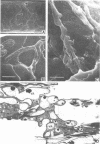
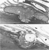
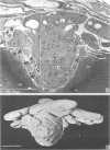
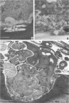
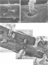
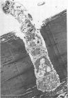
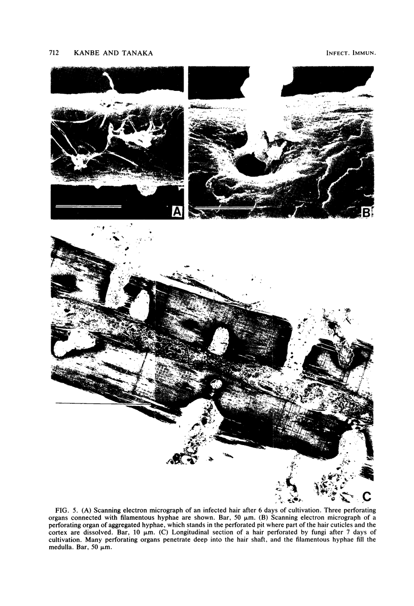
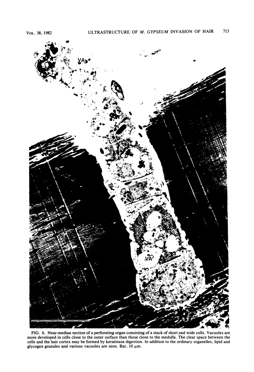
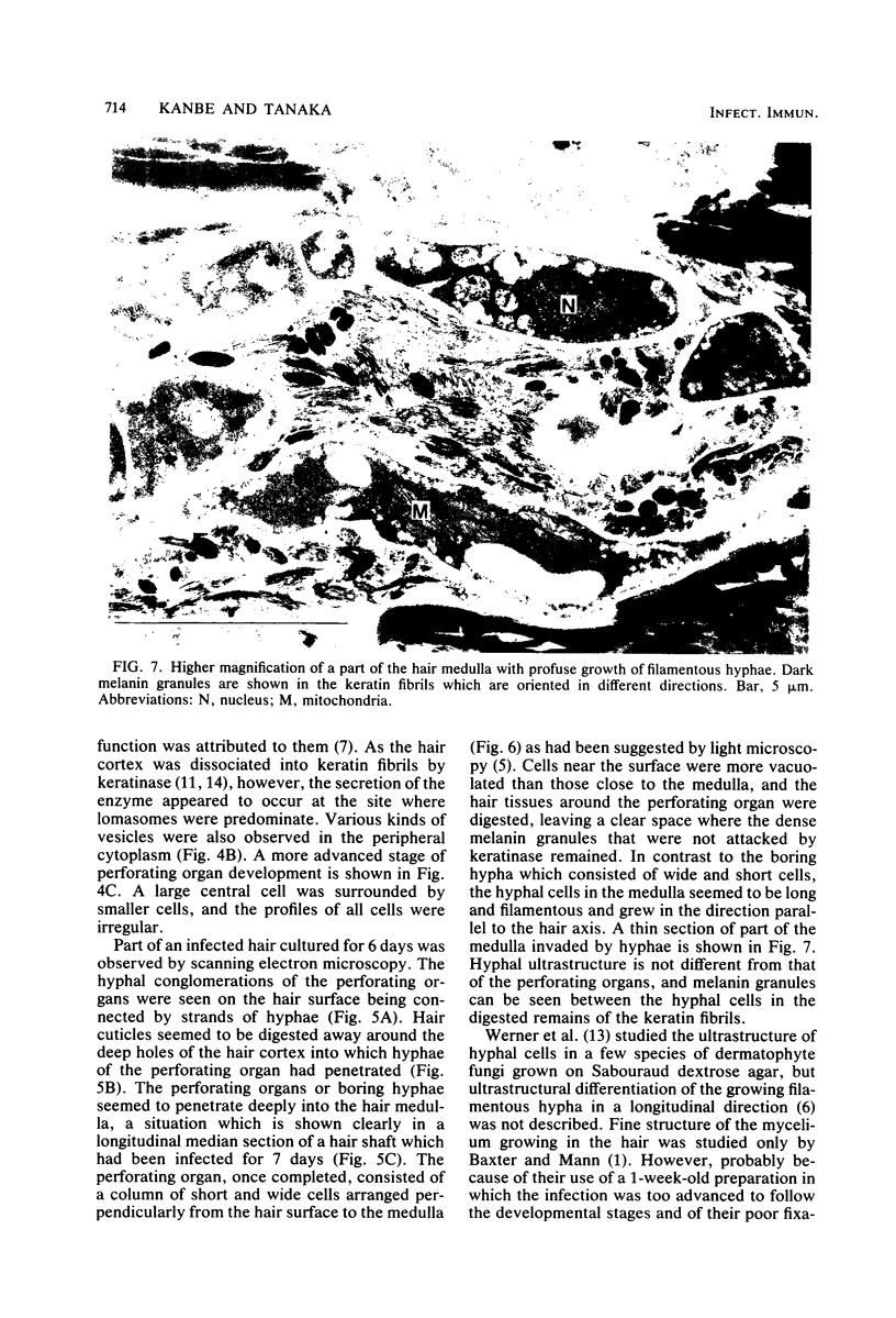
The pattern of invasion of human hair in vitro by the dermatophyte Microsporum gypseum was studied by transmission and scanning electron microscopy. Mycelia that invaded the hair cortex through the edge of cuticles showed a flattened "frond" growth in contrast to the filamentous form seen on ordinary laboratory media. The frond cells were characterized by the presence of vesicles formed by invaginations of plasmalemma, and lomasomes were prominent in the region adjacent to the hard keratinized tissue of the hair cortex being degraded as well. The initial perforating organ, which originated from the frond mycelium, appeared as an enlarged spherical cell which integrated with the laterally branched hyphae, as revealed by analysis of a three-dimensional model reconstructed from a series of sections. The fully developed perforating organ consisted of a column of wide and short cells which penetrated perpendicularly through the hair cortex. Through the medulla the filamentous hyphae had grown profusely in a longitudinal direction. Our studies confirm earlier light microscope observations and provide new ultrastructural details on the development of the eroding frond and the perforating organ.
Full text
PDF
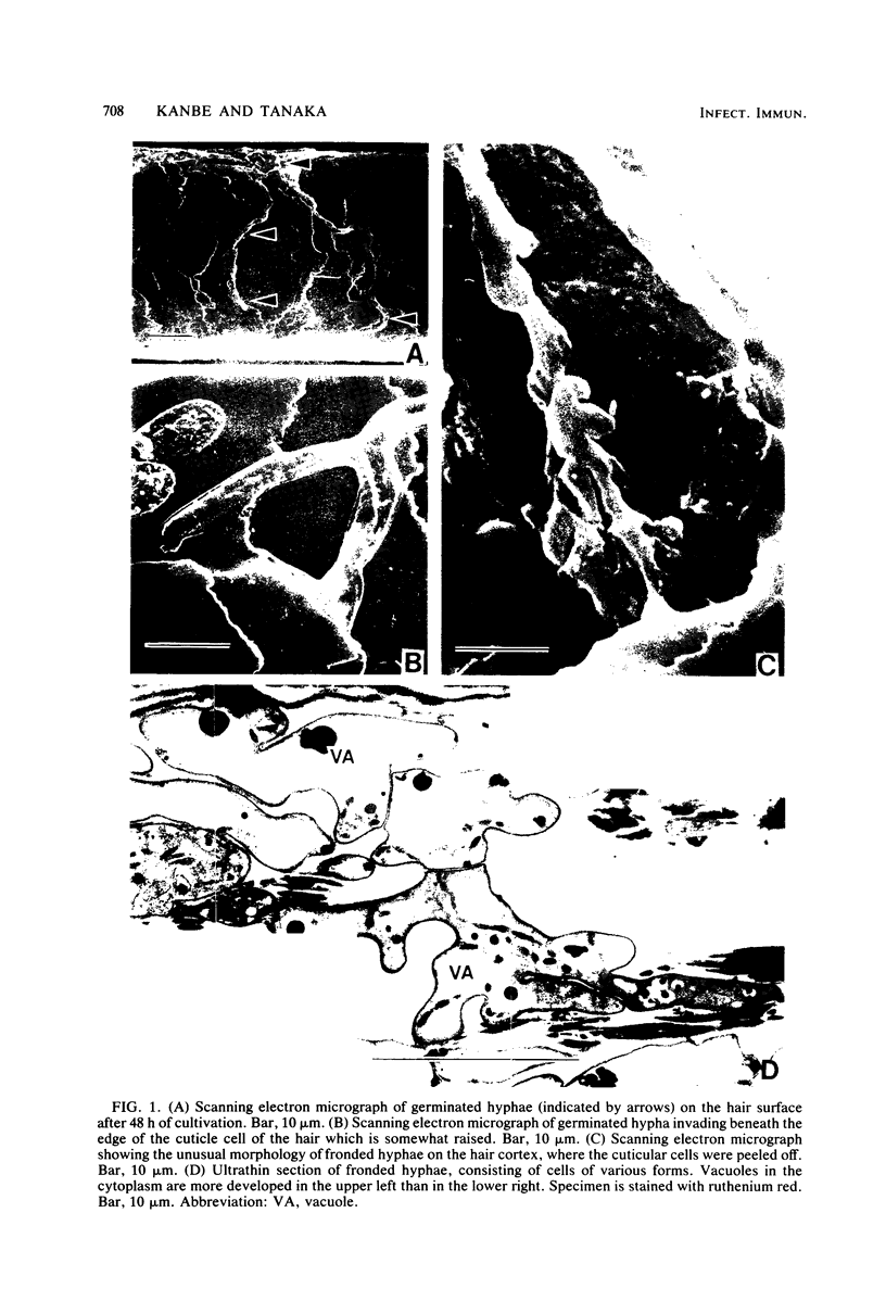
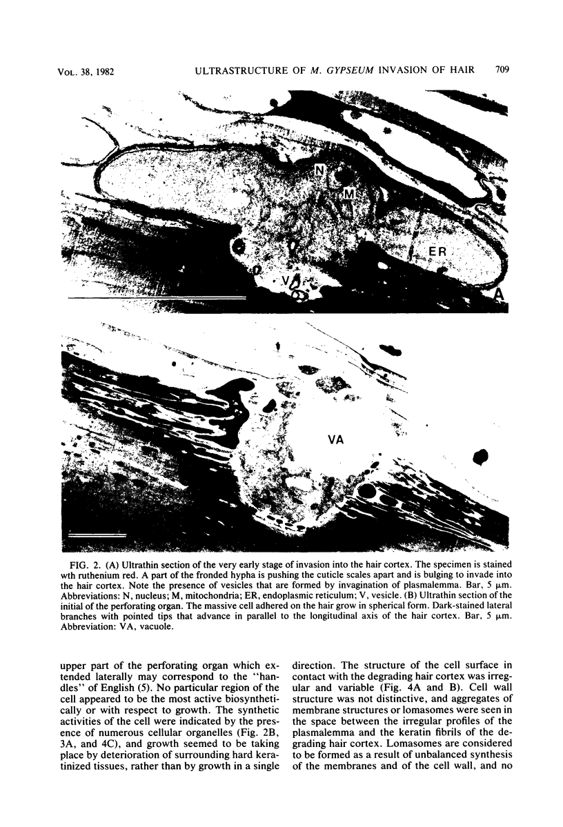
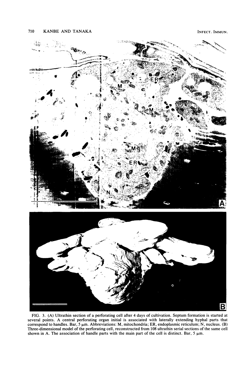
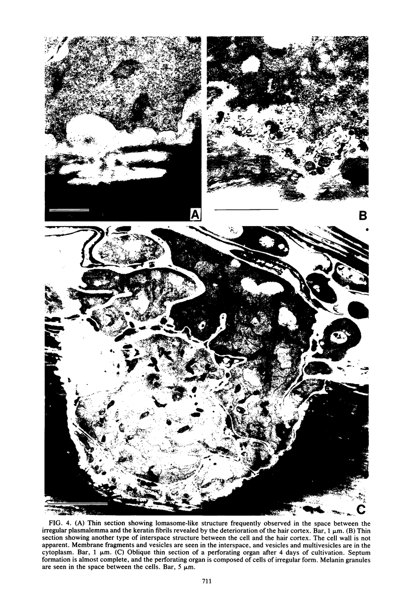
Images in this article
Selected References
These references are in PubMed. This may not be the complete list of references from this article.
- Baxter M., Mann P. R. Electron microscopic studies of the invasion of human hair in vitro by three keratinophilic fungi. Sabouraudia. 1969 Feb;7(1):33–37. [PubMed] [Google Scholar]
- English M. P. The developmental morphology of the perforating organs and eroding mycelium of dermatophytes. Sabouraudia. 1968 Jun;6(3):218–227. doi: 10.1080/00362176885190421. [DOI] [PubMed] [Google Scholar]
- Hutton R. D., Kerbs S., Yee K. Scanning electron microscopy of experimental Trichophyton mentagrophytes infections in guinea pig skin. Infect Immun. 1978 Jul;21(1):247–253. doi: 10.1128/iai.21.1.247-253.1978. [DOI] [PMC free article] [PubMed] [Google Scholar]
- Pellegrini M. Three-dimensional reconstruction of organelles in Euglena gracilis Z. I. Qualitative and quantitative changes of chloroplasts and mitochondrial reticulum in synchronous photoautotrophic culture. J Cell Sci. 1980 Jun;43:137–166. doi: 10.1242/jcs.43.1.137. [DOI] [PubMed] [Google Scholar]
- RAUBITSCHEK F., EVRON R. EXPERIMENTAL INVASION OF HAIR BY DERMATOPHYTES. Arch Dermatol. 1963 Dec;88:837–845. doi: 10.1001/archderm.1963.01590240161028. [DOI] [PubMed] [Google Scholar]
- Takiuchi I., Higuchi D. [Isolation, purification and biochemical properties of keratinase elaborated from Microsporum gypseum (author's transl)]. Nihon Hifuka Gakkai Zasshi. 1977 Apr;87(5):305–309. [PubMed] [Google Scholar]
- Tosti A., Villardita S., Fazzini M. L., Scalici R. Contribution to the knowledge of dermatophytic invasion of hair. An investigation with the scanning electron microscope. J Invest Dermatol. 1970 Aug;55(2):123–134. doi: 10.1111/1523-1747.ep12291637. [DOI] [PubMed] [Google Scholar]
- Werner H. J., Catsulis C., Jolly H. W., Jr, Carpenter C. L., Jr Electron microscope observations of the fine structure of Microsporum gypseum. J Invest Dermatol. 1967 May;48(5):481–484. doi: 10.1038/jid.1967.74. [DOI] [PubMed] [Google Scholar]
- Yu R. J., Harmon S. R., Blank F. Hair digestion by a keratinase of Trichophyton mentagrophytes. J Invest Dermatol. 1969 Aug;53(2):166–171. [PubMed] [Google Scholar]


