Abstract
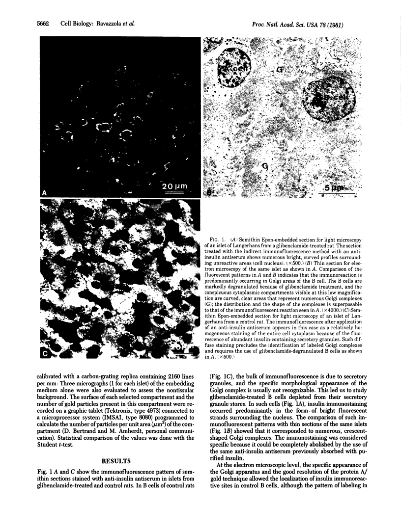
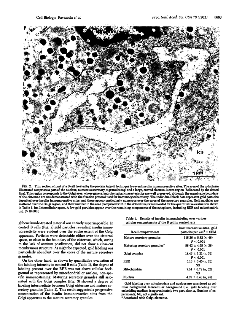
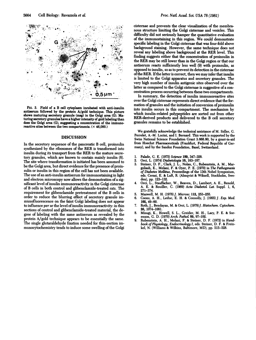
Insulin immunoreactive sites were localized in the Golgi apparatus of pancreatic B cells by light and electron microscopy. Identification of the Golgi apparatus by immunofluorescence required the prior degranulation of B cells with glibenclamide to reduce the insulin immunostaining due to secretory granules. In such cells, insulin immunofluorescence revealed brightly stained, crescent-shaped strands with form and location super-imposable on that of Golgi complexes seen in thin sections of the same cells. With the electron microscope, the insulin immunoreactive sites revealed by the protein A/gold technique were localized in the cisternae and vesicles of the Golgi apparatus of glibenclamide-treated and control B cells and over maturing and mature secretory granules. The quantitative evaluation of the intensity of the insulin immunoreactive sites in the Golgi apparatus revealed a density of sites 4 times more than cellular background values. The demonstration of insulin immunoreactivity in the Golgi apparatus provides direct evidence for the involvement of this compartment in the transport and maturation of proinsulin into insulin.
Full text
PDF
Images in this article
Selected References
These references are in PubMed. This may not be the complete list of references from this article.
- COONS A. H., LEDUC E. H., CONNOLLY J. M. Studies on antibody production. I. A method for the histochemical demonstration of specific antibody and its application to a study of the hyperimmune rabbit. J Exp Med. 1955 Jul 1;102(1):49–60. doi: 10.1084/jem.102.1.49. [DOI] [PMC free article] [PubMed] [Google Scholar]
- Maxwell M. H. Two rapid and simple methods used for the removal of resins from 1.0 micron thick epoxy sections. J Microsc. 1978 Mar;112(2):253–255. doi: 10.1111/j.1365-2818.1978.tb01174.x. [DOI] [PubMed] [Google Scholar]
- Misugi K., Howell S. L., Greider M. H., Lacy P. E., Sorenson G. D. The pancreatic beta cell. Demonstration with peroxidase-labeled antibody technique. Arch Pathol. 1970 Feb;89(2):97–102. [PubMed] [Google Scholar]
- Orci L. A portrait of the pancreatic B-cell. The Minkowski Award Lecture delivered on July 19, 1973, during the 8th Congress of the International Diabetes Federation, held in Brussels, Belgium. Diabetologia. 1974 Jun;10(3):163–187. doi: 10.1007/BF00423031. [DOI] [PubMed] [Google Scholar]
- Orci L., Stauffacher W., Renold A. E., Beaven D., Lambert A., Rouiller C. [Ultrastructural events associated with the action of tolbutamide and glybenclamide on pancreatic B-cells in vivo and in vitro]. Acta Diabetol Lat. 1969 Sep;6 (Suppl 1):271–374. [PubMed] [Google Scholar]
- Palade G. Intracellular aspects of the process of protein synthesis. Science. 1975 Aug 1;189(4200):347–358. doi: 10.1126/science.1096303. [DOI] [PubMed] [Google Scholar]
- Roth J., Bendayan M., Orci L. Ultrastructural localization of intracellular antigens by the use of protein A-gold complex. J Histochem Cytochem. 1978 Dec;26(12):1074–1081. doi: 10.1177/26.12.366014. [DOI] [PubMed] [Google Scholar]


