Abstract
Bunyamwera virus (BUNV) is the prototype virus for both the genus Orthobunyavirus and the family Bunyaviridae. BUNV has a tripartite, negative-sense RNA genome. The coding region of each segment is flanked by untranslated regions (UTRs) that are partially complementary. The UTRs play an important role in the virus life cycle by promoting transcription, replication, and encapsidation of the viral genome. Using reverse genetics, we generated recombinant viruses that contained deletions within the 3′ and/or 5′ UTRs of the L or M segments to determine the minimal UTRs competent for virus viability. We then generated viruses carrying deleted UTRs in all three segments. These viruses were grossly attenuated in tissue culture, being significantly impaired in their ability to produce plaques in BHK cells, and had a reduced capacity to cause host cell protein shutoff. After serial passage in tissue culture, some viruses partially recovered fitness, generating higher titers and producing larger plaques. We determined the complete nucleotide sequence for each virus. The deleted UTR sequences were maintained, and no amino acid changes were observed in the nonstructural proteins (NSs and NSm), the nucleocapsid protein (N), or the Gn glycoprotein. One virus had a single amino acid substitution in Gc. Three viruses contained amino acid changes in the viral polymerase that mostly occurred in the C-terminal domain of the L protein. Although the role of this domain remains unknown, we suggest that those changes might be involved in the evolution of the polymerase to recognize the deleted UTRs more efficiently.
INTRODUCTION
The genus Orthobunyavirus and the family Bunyaviridae take their name from Bunyamwera virus (BUNV), the prototype bunyavirus, which was isolated from mosquitoes of Aedes species in the Semliki Forest in Uganda (25). The BUNV genome is composed of three single-stranded RNA segments of negative polarity, named large (L), medium (M), and small (S), which encode four structural proteins. The L segment codes for the RNA-dependent RNA polymerase or L protein; the M segment codes for the two envelope glycoproteins, Gn and Gc; and the S segment codes for the nucleocapsid protein (N). Nonstructural proteins are encoded on the M and S segment and are named NSm and NSs, respectively (reviewed in reference 10). The NSm protein was shown to be involved in virion assembly (24), while NSs plays a role in counteracting the host cell innate immune response through a general block in transcription and translation (4, 16, 26, 27). Each genome segment is encapsidated by numerous copies of the N protein and is associated with a few copies of the L protein to form ribonucleoprotein (RNP) complexes that are the functional templates for both transcription and replication.
The coding regions are flanked by untranslated regions (UTRs), whose size and sequence vary greatly between segments, but as a general rule, the 3′ UTR is shorter than the 5′ UTR. BUNV 3′ and 5′ UTRs on the L and M segments are about 50 nucleotides (nt) and 100 nt, respectively, while the S segment UTRs are longer, 85 nt and 174 nt, respectively. Similar to other orthobunyaviruses, the terminal 11 nt of each segment are identical and invertedly complementary, with the exception of a G-U mismatch at positions 9 and −9 at each end. The complementarity of the UTRs allows base pairing of the termini and formation of a panhandle structure characteristic of segmented negative-sense RNA viruses (12, 19). Exact complementarity is extended in a segment-specific manner for 4 nt on the L and S segment and 3 nt on the M segment, and imperfect complementarity is observed within each segment until about nt position 30 (11). Thereafter, the UTRs are highly variable between segments and consist of largely nonpaired sequences.
The UTRs are multifunctional and contain signals that are necessary and sufficient to drive viral RNA and protein synthesis, as well as nucleocapsid formation and virion assembly. Some functions have been mapped within the BUNV S segment UTRs and localize mainly to the extremities: (i) the core promoter for transcription and replication is encompassed in the extreme 16 nt of the 3′ and 5′ genomic termini (2); (ii) the N protein was shown to interact preferentially with the 5′-terminal 32 nt (13, 20); (iii) nt positions 20 to 33 of the 3′ and 5′ UTRs were identified as important for genome packaging in a minigenome assay (14). However, very little information is available concerning the role of the internal regions of the UTRs. BUNV carries longer UTRs than do most segmented, negative-strand RNA viruses such as influenza viruses (15), arenaviruses (8), and even other orthobunyaviruses. Previously, Lowen and Elliott (17) used a rational approach to reduce the lengths of BUNV S segment UTRs outwards from the coding region toward the termini and showed that the internal parts of the UTR were nonessential for virus viability but played an important role during virus replication, as viruses carrying such deletions showed restricted growth in cell culture.
In this study, we extended this approach to investigate the other UTRs. First, we rescued viruses carrying deletions in either their L or M segment UTRs to determine the minimal L and M UTRs required for virus growth. Then, we determined the minimal sequences that could support virus replication when deleted UTRs were present on all three segments. Rescued viruses were grossly attenuated in tissue culture but proved able to regain some level of fitness through serial passage. When we investigated further the mechanism behind the regain of fitness, we observed the accumulation of amino acid mutations within the C-terminal part of the viral polymerase protein in some viruses while other coding sequences and remaining UTRs were stable through serial passage.
MATERIALS AND METHODS
Cells and viruses.
BHK-21 cells were grown in Glasgow's minimal essential medium (GMEM) supplemented with 10% tryptose phosphate broth (TPB) and 10% newborn calf serum (NCS). BSR-T7/5 cells, which stably express T7 RNA polymerase (7), were provided by K.-K. Conzelmann (Max-von-Pettenkofer Institut, Munich, Germany) and were grown in GMEM supplemented with 10% TPB, 10% fetal calf serum (FCS), and 1 mg/ml G418. All cell lines were grown at 37°C with 5% CO2.
Where possible, recombinant viruses were purified by plaque formation on BHK-21 cells; working stocks were grown at 33°C in BHK-21 cells in medium supplemented with 2% NCS, and supernatants were harvested when a marked cytopathic effect (CPE) was observed. For recombinant viruses that did not show CPE, working stocks were grown at 33°C in BHK-21 cells and supernatants were harvested 5 days postinfection.
Plasmids.
pT7riboBUNL(+), pT7riboBUNM(+), and pT7riboBUNS(+) have been described previously (5); briefly, each plasmid contains a full-length, antigenome-sense BUNV segment flanked immediately upstream by the T7 promoter and immediately downstream by the hepatitis δ ribozyme, followed by the T7 terminator sequence. Deletions in the L and M UTRs were introduced by excision PCR using primer pairs flanking the region to be deleted in outward orientation using pT7riboBUNL(+) or pT7riboBUNM(+) as the template. Primer sequences and PCR conditions are available from us on request. All constructs were verified by sequence analysis.
Generation of recombinant viruses from cDNAs.
Recombinant viruses were produced using the three-plasmid rescue system (18). Subconfluent BSR-T7/5 cells (4 × 10⁵ cells in a 60-mm-diameter dish) were transfected with 1 μg each of pT7riboBUNL(+), pT7riboBUNM(+), and pT7riboBUNS(+), or the appropriate mutant construct(s), by using Lipofectamine 2000 (Invitrogen) according to the manufacturer's instructions. Supernatants were harvested when marked CPE was observed or every 5 to 6 days up to 21 days posttransfection. Rescue outcome was assessed by either plaque assay or immunostaining. The genome segments of the recovered viruses were amplified by reverse transcription-PCR (RT-PCR), and their nucleotide sequences were determined to confirm the presence of the expected mutations.
Virus titration by plaque assays or immunostaining.
Monolayers of BHK-21 cells were infected with serial dilutions of virus and incubated under an overlay consisting of GMEM supplemented with 2% NCS and 0.6% Avicel (FMC) at 33°C for up to 5 days. Cell monolayers were fixed with 4% formaldehyde, and plaques were visualized by Giemsa staining. For viruses that did not form plaques, after fixation, cell monolayers were permeabilized with 0.5% Triton X-100 in phosphate-buffered saline (PBS) for 30 min. The cells were incubated with blocking buffer (PBS containing 5% skimmed milk) for 30 min before being reacted with a monospecific anti-BUNV N protein antibody, followed by a peroxidase-labeled secondary antibody. Foci were visualized using TrueBlue peroxidase substrate (InSight Biotechnology).
Serial passage.
Recombinant viruses were serially passaged in BHK-21 cells. To produce passage 1 (p1), cells were infected at a low multiplicity of infection (MOI) (approximately 0.001 PFU/cell or focus-forming unit [FFU]/cell) with the p0 stock and incubated at 33°C in medium supplemented with 2% NCS. The supernatant was harvested when CPE appeared or at 7 days postinfection. The same procedure was repeated up to passage 10 (p10).
Metabolic labeling of viral proteins.
BHK-21 cells were infected as described above, and at different times postinfection, cells were labeled with 35 μCi per well of Tran35S-label (MPbio) for 2 h in methionine-free Dulbecco's modified Eagle's medium. Cell lysates were prepared by addition of 300 μl lysis buffer (100 mM Tris-HCl [pH 6.8], 4% SDS, 20% glycerol, 200 mM dithiothreitol, 0.2% bromophenol blue, and 25 U/ml benzonase [Novagen]). Proteins were separated by SDS-PAGE on a 4 to 12% gel (Invitrogen). After fixation and drying, the gel was exposed to X-ray film.
RESULTS
Rescue of recombinant BUNV with minimal L or M segment UTRs.
Plasmids expressing L or M segment RNAs with deletions in the UTRs were transfected into BSR-T7/5 cells together with plasmids expressing two wild-type (wt) segments. For the L segment, we designed 16 constructs with deletions within the 3′, 5′, or both UTRs, and 12 led to successful rescue of virus. The complete 3′ and 5′ UTRs are 108 nt and 85 nt, respectively. The minimal UTRs for the L segment generating an infectious virus were 39 nt left in the 3′ UTR and 38 nt left in the 5′ UTR (Fig. 1, top). The full-length M segment UTRs are 100 nt (3′) and 56 nt (5′), and of 9 constructs containing deletions within the UTRs, only 2 led to successful rescue. One contained a deletion in the 3′ UTR, and the other contained a deletion in the 5′ UTR. It was not possible to rescue a virus containing deletions in both UTRs. Therefore, the minimal 3′ UTR was 33 nt and the minimal 5′ UTR was 40 nt (Fig. 1, bottom). All negative virus rescues were confirmed by performing at least one other rescue attempt under conditions where the wt virus was successfully rescued and generated at least 107 PFU The modified segment of each recovered virus was amplified by reverse transcription-PCR (RT-PCR), and nucleotide sequence determination confirmed the presence of the expected deletion.
Fig 1.
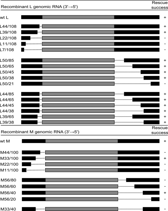
Schematic of the deletions introduced in the UTRs of the L segment (upper part) and M segment (lower part). Black bars on each side represent the UTRs; gray bars in the middle represent the coding region. Only the UTRs are drawn to scale. The names of the recombinant segments follow the pattern Xa/b, where X is the segment concerned, a is the length of the 3′ UTR, and b is the length of the 5′ UTR in nucleotides. + denotes recovery of a recombinant virus and − denotes no recovery, when virus rescue was performed with two wt segments.
After propagation in BHK-21 cells, the resulting virus stock was used to assess the plaque phenotype of the rescued viruses. Mutant viruses appeared to display a great diversity in their plaque size; however, they were always smaller than those formed by wtBUNV (Fig. 2A). For the L segment, the size of the plaque appeared to correlate with the size of the deletion, except for rBUNL39/65. The virus with the minimal L segment UTRs, rBUNL39/38, showed the smallest plaque phenotype. Both viruses with deletions in either of their M UTRs produced plaques that were smaller than those of the wtBUNV. The mutant viruses also appeared to grow more slowly than did wtBUNV (Fig. 2B). Differences were most striking at 24 h postinfection, where most mutant viruses gave titers that were at least 10-fold lower than those of wtBUNV and up to 10,000-fold lower for rBUNL38/39. However, by 48 h postinfection all viruses except rBUNL39/38 produced yields that were within 10-fold of those of the wt virus.
Fig 2.
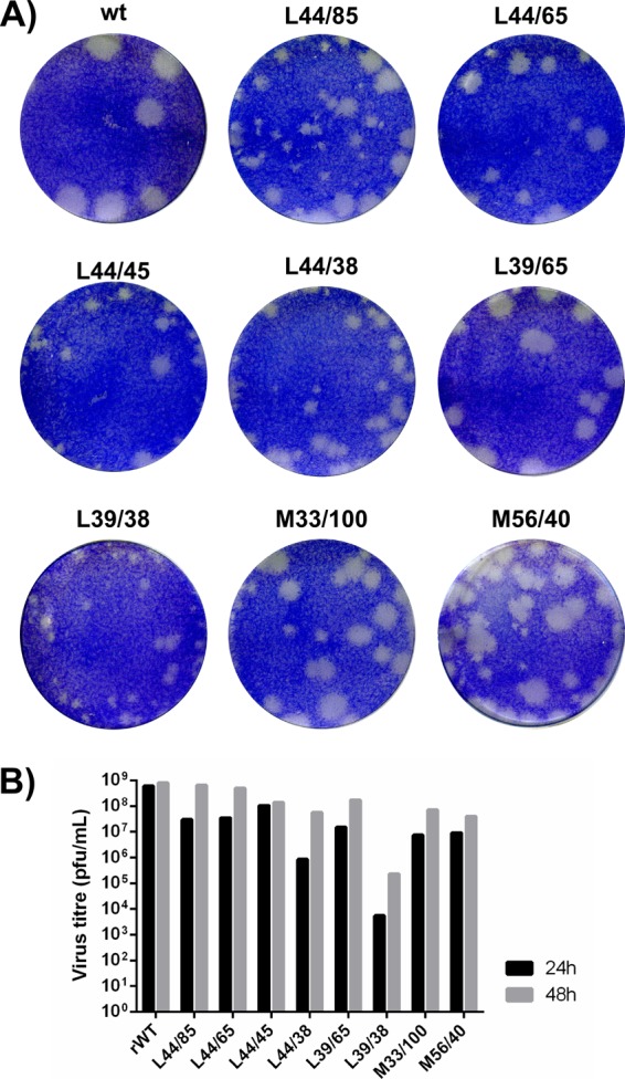
Growth properties of recombinant viruses compared to those of wtBUNV. (A) Plaque phenotypes. BHK-21 cell monolayers were fixed 4 days postinfection with 4% formaldehyde and stained with Giemsa solution. The name above each well indicates the mutant segment in a wt background. (B) Virus yields. BHK-21 cells were infected at an MOI of 0.5 PFU/cell. Virus titers in the supernatant were determined at 24 h and 48 h postinfection by titration in BHK-21 cells.
Rescue of recombinant BUNV with minimal UTRs in all three segments.
In an attempt to rescue viruses with the minimal UTRs, defined above for the L and M segments, and previously by Lowen and Elliott (17) for the S segment, BSR-T7/5 cells were transfected with plasmids representing the two possible combinations: L39/45 + M56/40 + S29/112 or L39/45 + M33/100 + S29/112 (Table 1). We speculated that rescued viruses might be grossly attenuated, and thus, the supernatant from transfected cells was assayed by an immunostaining procedure (see Materials and Methods). However, despite repeated attempts, no evidence for infectious virus could be detected, leading to the conclusion that viruses with minimal UTRs (as defined for individual segments) on all three segments were not viable.
TABLE 1.
Combinations of three deleted segments attempted in rescue experiments
| Expt | Presence or absence of plasmid (abbreviated name): |
Rescue outcomea | |||||||
|---|---|---|---|---|---|---|---|---|---|
| L44/85 | L44/65 (L1) | L44/45 (L2) | L39/65 (L3) | M56/40 (M1) | M33/100 (M2) | S85/112 (S1) | S62/112 (S2) | ||
| 1 | + | − | − | − | + | − | + | − | − |
| 2 | + | − | − | − | + | − | − | + | − |
| 3 | + | − | − | − | − | + | + | − | − |
| 4 | + | − | − | − | − | + | − | + | − |
| 5 | − | + | − | − | + | − | + | − | + |
| 6 | − | + | − | − | + | − | − | + | − |
| 7 | − | + | − | − | − | + | + | − | − |
| 8 | − | + | − | − | − | + | − | + | + |
| 9 | − | − | + | − | + | − | + | − | + |
| 10 | − | − | + | − | + | − | − | + | − |
| 11 | − | − | + | − | − | + | + | − | − |
| 12 | − | − | + | − | − | + | − | + | − |
| 13 | − | − | − | + | + | − | + | − | − |
| 14 | − | − | − | + | + | − | − | + | + |
| 15 | − | − | − | + | − | + | + | − | + |
| 16 | − | − | − | + | − | + | − | + | + |
+, virus rescued; −, no virus recovered.
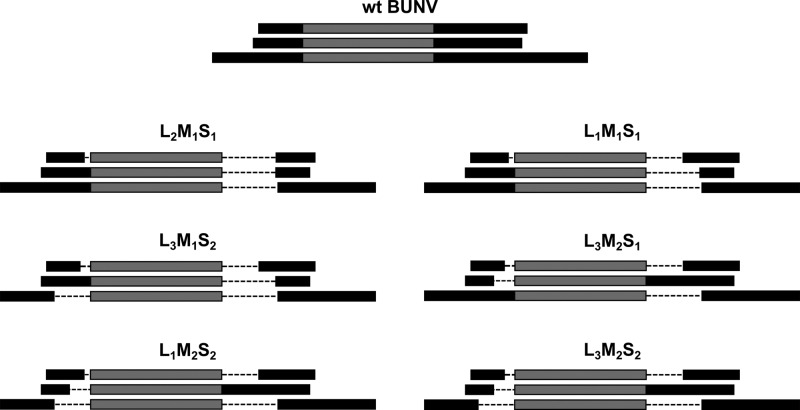
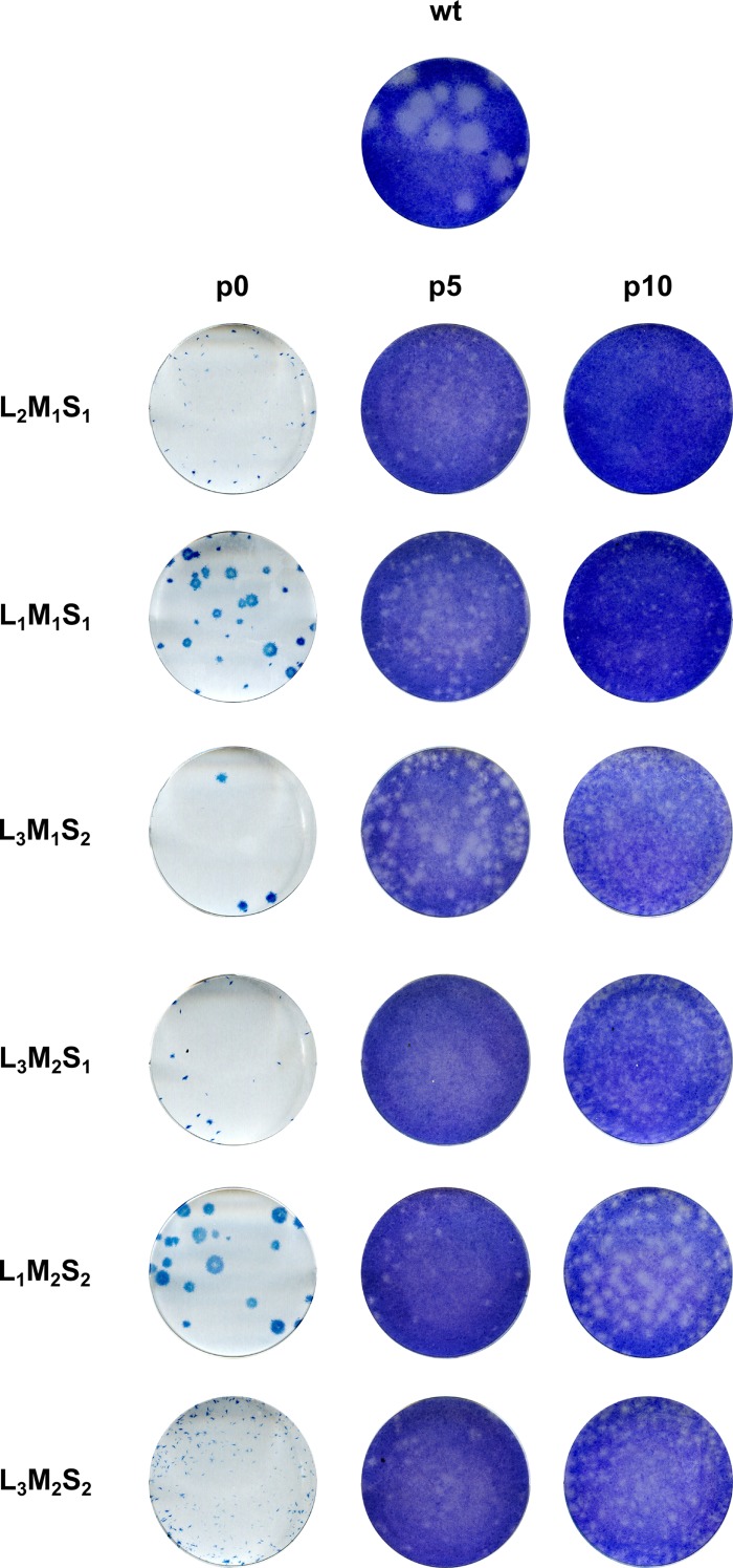
As it was not possible to rescue a virus containing all the minimal UTRs, we attempted virus rescue with different combinations of plasmids expressing mutant segments. First, we performed rescue experiments where only two segments contained deleted UTRs while the third segment had wt UTRs. Through this screening, we found that L segments L44/38 and L39/38 did not allow efficient virus rescue with any other deleted segments. This screening also showed that S segments with UTRs shorter than S62/112 did not allow efficient virus rescue with other segments carrying deletions in their UTRs. Both deleted M segment plasmids, however, could be used in combination with deleted L or S segments. Next, we attempted virus rescue with 16 different combinations of mutated plasmids, and six led to the recovery of viable viruses (Table 1; Fig. 3). The viruses were detected only by immunostaining of infectious foci, and none of the mutant viruses formed obvious plaques in BHK cells according to our standard protocol (Fig. 4). A negative rescue outcome was confirmed by carrying out at least three rescue experiments where rescue of wtBUNV was successful each time. Interestingly, we were unable to recover a virus with L44/85 UTRs in combination with shorter M and S segments whereas we were successful with the construct expressing shorter L segment UTRs, L39/65. Viruses with shorter L and S segment UTRs were recovered with either of the two deleted M segments, M33/100 and M56/40. Of the six rescued viruses, three had deletions in 4 out of the 6 UTRs, and three viruses had deletions in 5 UTRs. Each of the segments leading to virus rescue was given an abbreviated name for ease of use as shown in Table 1. The virus with the overall smallest genome, L39/65 + M56/40 + S62/112, is thus called L3M1S2; its total genome length is 12,085 nt, compared to 12,294 nt contained by wtBUNV.
Fig 3.
Schematic representing the genome segments present in minimal viruses. The coding regions, not to scale, are in gray. The UTRs, to scale, are in black. For each set of three, the top segment represents the L genome, the middle segment represents the M genome, and the bottom segment represents the S genome, with the 3′ UTR on the right of the coding sequence and the 5′ UTR on the left. The abbreviated segment designations are given in Table 1.
Fig 4.
Plaque phenotypes of mutant viruses at p0, p5, and p10 in BHK-21 cells. BHK-21 cells were infected with wtBUNV or different minimal genome recombinant BUNVs. Cells were fixed at 3 days postinfection in 4% formaldehyde and immunostained for N protein (p0) or stained with Giemsa's solution (p5 and p10).
Mutant viruses recover fitness through serial passage.
The supernatants from transfected cells were used to prepare elite stocks of each virus in BHK-21 cells and were designated p0. Using the immunostaining protocol, titers of p0 stocks were in the range of 104 to 105 FFU/ml, whereas recombinant wtBUNV produced at the same time reached titers above 107 PFU/ml. Two types of foci were observed for the mutant viruses: very small (L2M1S1, L3M2S1, and L3M2S2) or larger, round, and well-defined (L1M1S1, L3M1S2, and L1M2S2) (Fig. 4). The viruses were serially passaged in BHK cells, using the same volume of supernatant from each passage to infect the subsequent flask of cells, and evidence for regain of fitness during the process was investigated. Thus, the ability of each virus to form plaques in BHK-21 cells was assessed from passage 5 (p5) and 10 (p10) stocks (Fig. 4). While wtBUNV displayed very similar plaque sizes at every passage, we observed a positive effect of passage on the ability of the mutant viruses to form plaques. All p5 mutant viruses had apparently acquired the ability to form plaques but to different extents. For L2M1S1, L3M2S1, and L3M2S2, plaques were small, fuzzy, and difficult to visualize while plaques produced by L1M1S1, L3M1S2, and L1M2S2 were small, clear, and distinct. These phenotypes remained the same at p10.
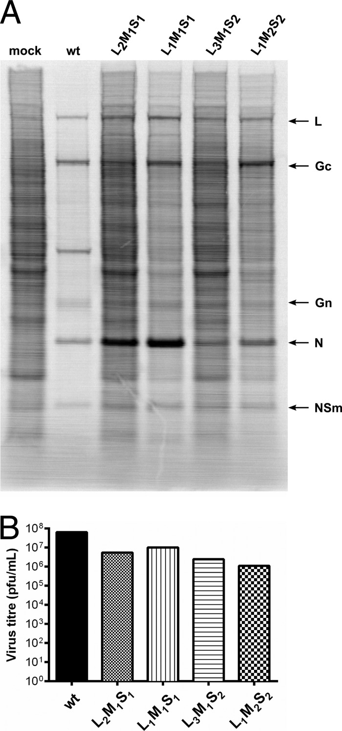
After 10 passages, the titers of three mutants had not increased above the titers of the p0 stocks: L2M1S1 remained at around 105 PFU/ml, whereas the titers of L3M2S1 and L3M2S2 did not exceed 104 PFU/ml, making further characterization difficult. In contrast, the titers of L1M1S1, L3M1S2, and L1M2S2 increased by at least 100-fold by p10. Viral protein synthesis and the ability to induce host cell protein shutoff were assessed for recombinant viruses L2M1S1, L1M1S1, L3M1S2, and L1M2S2 in BHK cells infected at an MOI of 5; wtBUNV was included for comparison (Fig. 5). wtBUNV induced significant shutoff of host cell protein synthesis by 24 h postinfection compared to that in mock-infected cells, and while synthesis of viral protein was clearly visible, it had already begun to decrease compared to earlier time points (not shown). Viruses L2M1S1 and L3M1S2 did not cause host cell protein shutoff compared to uninfected cells, while viruses L1M1S1 and L1M2S2 induced moderate shutoff (Fig. 5A). Interestingly, the level of viral N (and presumably NSs) protein did not correlate with the level of observed host cell protein shutoff. Indeed, levels of N protein were higher in L1M1S1- and L2M1S1- than in L3M1S2- and L1M2S2-infected cells, whereas levels of other viral proteins appeared more similar between mutant viruses. Virus titers in the supernatants from these cells at 24 h postinfection were determined by plaque assay in BHK-21 cells (Fig. 5B). The mutant viruses achieved titers between 106 and 107 PFU/ml, whereas wtBUNV titers approached 108 PFU/ml.
Fig 5.
Protein synthesis and associated virus yields in BHK-21 cells. BHK-21 cells were infected at an MOI of 5 PFU/cell. (A) Proteins were labeled at 24 h postinfection with [35S]methionine over 2 h. The same volume of lysate was loaded in each lane. Positions of viral proteins are indicated on the right. (B) The supernatants were harvested at 24 h postinfection and titrated in BHK-21 cells.
Complete nucleotide sequence analysis after serial passage.
The complete genomic sequences of the six mutant viruses were determined. No changes in the UTR sequences were found in any virus, confirming that the introduced deletions were stable for up to 10 passages. No nucleotide changes were observed in the S segment coding region. For the M segment coding region, only virus L1M2S2 showed a change, a single nucleotide substitution at position 3070 (G→T), leading to an amino acid (E→K) change at position 925 of the glycoprotein precursor, corresponding to position 448 in the Gc glycoprotein. The L segment coding region in all mutant viruses, with the exception of L3M2S2, contained mutations. Viruses L2M1S1 and L3M2S1 had 1 nucleotide substitution each, but these did not result in an amino acid change. On the other hand, viruses L1M1S1, L3M1S2, and L1M2S2 all had several nucleotide substitutions, some of which resulted in amino acid changes; these are listed in Table 2. Notably, these three viruses all showed an increase in fitness, as evidenced by improved titers, following serial passage. In most cases, the amino acids retained similar biochemical properties. For instance, in virus L1M1S1, at position 1118 R was replaced by K, and at position 1945 V was replaced by I. However, in virus L3M1S2, the alteration at position 1619 was from E to Q, and in virus L1M2S2, the second mutation at position 1595 was from H to P.
TABLE 2.
List of nucleotide and amino acid substitutions in the L sequence of mutant virusesa
| Virus | Nucleotide change |
Amino acid change |
||
|---|---|---|---|---|
| Position | Substitution | Position | Substitution | |
| L1M1S1 | 3375 | C→T | 1108 | None |
| 3403 | G→A | 1118 | R→K | |
| 5883 | G→A | 1945 | V→I | |
| L2M1S1 | 6383 | C→T | 2111 | None |
| L3M1S2 | 3545 | C→T | 1165 | None |
| 4905 | G→C | 1619 | E→Q | |
| 5449 | G→A | 1800 | R→K | |
| 5468 | G→A | 1806 | None | |
| 6340 | C→T | 2097 | A→V | |
| L3M2S1 | 6014 | G→A | 1988 | None |
| L1M2S2 | 414 | A→G | 122 | I→V |
| 3557 | G→C | 1169 | None | |
| 4834 | A→C | 1595 | H→P | |
Nucleotide substitutions and amino acid changes were positioned according to the wtBUNV L sequence.
DISCUSSION
The untranslated sequences at the ends of bunyavirus genome segments perform critical functions in the viral life cycle (10). The very terminal sequences are conserved between the three genome segments for viruses within each genus but are different between genera, and the consensus terminal sequences are used as a criterion in classification (21). The sequences between the conserved termini and the coding regions generally display considerable variability. However, with regard to members of the Orthobunyavirus genus, conserved CA- and GU-rich motifs are found within the variable 5′ UTR of the S segment (9). Considering that some viruses within the genus Orthobunyavirus possess very short UTRs (e.g., those in the Simbu serogroup such as Akabane or Oropouche viruses [10, 17]), it begs the question of the function of these “additional” sequences and whether the UTR sequences in BUNV can be reduced while maintaining virus viability. Previously, it had been shown that the S segment UTRs could be shortened from 85 nt to 29 nt at the 3′ end and from 174 nt to 112 nt at the 5′ end (17). The purpose of this study was first to delineate the minimal L or M segment UTRs that would support virus growth in the presence of complete UTRs on the two other segments and then to define the minimal UTR combinations for all three segments, i.e., to create a recombinant BUNV with the shortest genome.
We were able to recover a virus in which the UTRs of the L segment were shortened from 108 nt to 39 nt at the 3′ end and from 50 nt to 38 nt at the 5′ end, BUNL39/38. Recovering a virus with deletions within the M UTRs proved more difficult. It was possible only to reduce one or the other UTR but not both at the same time. The minimal UTRs for the M segment were either 33 nt (instead of 100 nt) at the 3′ end or 40 nt (rather than 56 nt) at the 5′ end, BUNM33/100 and BUNM56/40, respectively. Despite the fact that nonconserved sequences were dispensable, they contributed to viral growth, and as a general rule, the shorter the UTRs, the slower the virus grew. The replication cycles of the mutant viruses carrying shortened L or M segment UTRs appeared slower than that of wtBUNV, resulting in the formation of smaller plaques. These observations correlate with what has been observed when introducing deletions within the S segment (17).
Virus growth is a complex process during which UTRs play a critical role. Indeed, UTRs are involved in replication, transcription, encapsidation, and packaging of the viral genome. Using minigenome assays, important packaging elements have been mapped between nucleotides 20 and 33 in the 3′ and 5′ UTRs (14). In our study, the terminal 33 nt of both the 3′ and 5′ UTRs were present in all constructs, and thus, we predicted that segments with deleted UTRs would be packaged with an efficiency similar to that of wt segments. Also, using a minigenome assay, two termination signals have been identified in the BUNV S segment (1), one playing a major role at nt 865 to 870 (3′-GUCGAC-5′) and a second, 32 nt downstream, at nt 898 to 902 (3′-UGUCG-5′). However, through the creation of appropriate recombinant viruses, it was recently shown that only the first motif signals transcription termination in the context of virus infection (3). When the S segment termination signal was deleted, runoff RNA transcripts were detected in recombinant BUNV-infected cells (3). It would be of interest to determine the situation with the viruses carrying deletions in the M and L segment UTRs. Transcription termination signals have not been identified for the L and M segments, and it seems that their 5′ UTRs were less sensitive to deletion. This suggests that mRNA termination signals might be located close to the genomic 5′ end. Deletion of just the termination signal, however, did not attenuate virus replication; attenuation was seen when larger regions of the 5′ UTR were excised (3, 17). This implies that some other function associated with the S segment 5′ UTR is more important for efficient virus replication, and presumably this applies also for the M and L segment UTRs.
Considering that deleted UTRs in only one segment led to virus attenuation, it was thought that if a virus containing three segments with deletions could be rescued, it would be attenuated even further. This proved to be the case. First, we were unable to recover a virus carrying the minimal UTRs, as defined when only one segment was mutated, on all segments. It appeared that when the number of segments carrying deleted UTRs increased, the size of each UTR had also to be increased. After screening rescue attempts with numerous combinations of plasmids expressing genomic RNAs with deleted UTRs, a few recombinant viruses with deletions in 4 or 5 UTRs were recovered, but the viruses were extremely attenuated. Indeed, the only reliable way to detect such viruses was by immunostaining of viral foci with an anti-N protein antiserum, as the viruses were markedly impaired in their ability to form plaques. Furthermore, the titers of initial virus stocks were much lower than those of recombinant wtBUNV rescued in parallel. However, after 10 passages, three mutant viruses showed increased titers, though they remained attenuated compared to wtBUNV, forming smaller plaques, and displaying a delay in the induction of host cell protein shutoff. When looking at host cell protein shutoff, it seems that the level of NSs was not the only factor regulating inhibition of protein synthesis. For instance, viruses L1M1S1 and L2M1S1 both carried the same S segment, expressed similar amounts of N protein, and presumably expressed similar amounts of NSs, but L1M1S1 induced a more pronounced protein shutoff. The same applies to viruses L3M1S2 and L1M2S2, with L1M2S2 inducing more protein shutoff than did L3M1S2. The M segment did not seem to be involved in protein shutoff, as L1M1S1, L2M1S1, and L3M1S2 carried the same M1 segment but displayed different levels of protein shutoff, suggesting that the L segment might be involved. The two viruses displaying the more pronounced protein shutoff carried the same mutated segment, L1, whereas viruses for which protein shutoff was poor carried either the L2 or the L3 segment. Whether this reflects differences in the expression levels of L protein, and thus differences in cap-snatching activity, unfortunately cannot be easily measured.
Serial passage can be compared to forced evolution, and as the viruses replicate, they will accumulate point mutations in their genomes. In order to understand the mechanism behind the regain of fitness, the entire nucleotide sequences of a number of viral genomes were determined to assess the extent of mutation. A previous in vitro study using a minigenome system showed that BUNV was able to repair both insertions and deletions in its UTRs (28). Therefore, the first concern was to ensure that the UTRs were unchanged and the deletions were still present after 10 passages. For the viruses examined, no repair of the deleted UTRs was observed, and moreover, no nucleotide changes were detected within the remaining UTR sequences. Hence, the mutant viruses did not regain fitness through modification of their UTRs. We next evaluated the coding sequences. Proteins encoded by the S segment play a role in virus replication: the N protein is involved in genome encapsidation (20) and NSs plays a role in counteracting the host cell interferon response and inhibiting host cell protein synthesis (6, 16). NSs has also been shown to have an impact on viral polymerase activity in a minigenome system (29). Despite N and NSs potentially having important roles in virus fitness, not a single nucleotide change was detected in the S open reading frame (ORF) of any mutant virus after serial passage. The M segment codes for the two glycoproteins Gn and Gc, involved in entry mechanisms, and NSm, which is required for assembly and morphogenesis (24). One nucleotide mutation was detected in the M segment of the virus L1M2S2, at position 925 in the glycoprotein precursor. As a recombinant virus with the NH2 terminus of Gc deleted (corresponding to residue 930 in the precursor) is viable, though attenuated, this indicates that residue 925 is not in a critical region for viral replication (23). It was on the L segment, coding for the polymerase protein, that sequencing revealed the generation of most mutations. Every mutant virus except L3M2S2 contained at least one nucleotide change, while the L segment of wtBUNV did not accumulate any nucleotide mutations after 10 passages. Nevertheless, not every nucleotide substitution led to an amino acid change. Viruses L2M1S1 and L3M2S1 had only silent nucleotide changes, whereas the three viruses that showed an increase in titer over passage had nucleotide changes that led to amino acid substitutions (Table 2). It is noteworthy that these three mutants (L1M1S1, L3M1S2, and L1M2S2) produced larger foci after rescue whereas those viruses that did not regain fitness over 10 passages (as defined by increase in titer) initially displayed small foci (Fig. 4). The majority of amino acid changes were located in the C-terminal part of the L protein. The function of this part of the BUNV polymerase is currently unknown, though Reguera et al. (22) suggest that it may be functionally similar to the PB2 protein of influenza virus, which has cap-binding activity. It would be of interest to investigate the impact of the mutations on BUNV polymerase activity and to determine if they influence the levels of transcription/replication of template RNAs containing wt or mutant UTRs.
In summary, we have shown that the nonconserved sequences of BUNV UTRs on the L and M segments are nonessential for viral growth, extending previous studies on nonconserved sequences in the S segment UTRs. However, the sequences are important for promoting optimal virus replication, perhaps by ensuring the correct balance between transcription and replication. Interestingly, when the UTRs are altered and viruses are serially passaged, mutations appeared in the polymerase. Those mutations are potentially linked to the observed regain of fitness.
ACKNOWLEDGMENTS
We thank Angela McLees for expert technical assistance.
B.M.-S. was supported by a postgraduate studentship from the BBSRC, and work in R.M.E.'s group is funded by the BBSRC, MRC, and Wellcome Trust.
Footnotes
Published ahead of print 3 October 2012
REFERENCES
- 1. Barr JN, Rodgers JW, Wertz GW. 2006. Identification of the Bunyamwera bunyavirus transcription termination signal. J. Gen. Virol. 87:189–198 [DOI] [PubMed] [Google Scholar]
- 2. Barr JN, Wertz GW. 2005. Role of the conserved nucleotide mismatch within 3′- and 5′-terminal regions of Bunyamwera virus in signaling transcription. J. Virol. 79:3586–3594 [DOI] [PMC free article] [PubMed] [Google Scholar]
- 3. Blakqori G, Lowen AC, Elliott RM. 2012. The small genome segment of Bunyamwera orthobunyavirus harbours a single transcription-termination signal. J. Gen. Virol. 93:1449–1455 [DOI] [PMC free article] [PubMed] [Google Scholar]
- 4. Blakqori G, van Knippenberg I, Elliott RM. 2009. Bunyamwera orthobunyavirus S-segment untranslated regions mediate poly(A) tail-independent translation. J. Virol. 83:3637–3646 [DOI] [PMC free article] [PubMed] [Google Scholar]
- 5. Bridgen A, Elliott RM. 1996. Rescue of a segmented negative-strand RNA virus entirely from cloned complementary DNAs. Proc. Natl. Acad. Sci. U. S. A. 93:15400–15404 [DOI] [PMC free article] [PubMed] [Google Scholar]
- 6. Bridgen A, Weber F, Fazakerley JK, Elliott RM. 2001. Bunyamwera bunyavirus nonstructural protein NSs is a nonessential gene product that contributes to viral pathogenesis. Proc. Natl. Acad. Sci. U. S. A. 98:664–669 [DOI] [PMC free article] [PubMed] [Google Scholar]
- 7. Buchholz UJ, Finke S, Conzelmann KK. 1999. Generation of bovine respiratory syncytial virus (BRSV) from cDNA: BRSV NS2 is not essential for virus replication in tissue culture, and the human RSV leader region acts as a functional BRSV genome promoter. J. Virol. 73:251–259 [DOI] [PMC free article] [PubMed] [Google Scholar]
- 8. Buchmeier JM, Bowen MD, Peters CJ. 2001. Arenaviridae: the virus and their replication, p 1635–1668 In Knipe DM, et al. (ed), Fields virology, vol 1 Lippincott Williams & Wilkins, Philadelphia, PA [Google Scholar]
- 9. Dunn EF, Pritlove DC, Elliott RM. 1994. The S RNA genome segments of Batai, Cache Valley, Guaroa, Kairi, Lumbo, Main Drain and Northway bunyaviruses: sequence determination and analysis. J. Gen. Virol. 75:597–608 [DOI] [PubMed] [Google Scholar]
- 10. Elliott RM, Blakqori G. 2011. Molecular biology of orthobunyaviruses, p 1–39 In Plyusnin A, Elliott RM. (ed), Bunyaviridae. Molecular and cellular biology. Caister Academic Press, Norwich, Norfolk, United Kingdom [Google Scholar]
- 11. Elliott RM, Schmaljohn CS, Collett MS. 1991. Bunyaviridae genome structure and gene expression. Curr. Top. Microbiol. Immunol. 169:91–141 [DOI] [PubMed] [Google Scholar]
- 12. Hewlett MJ, Pettersson RF, Baltimore D. 1977. Circular forms of Uukuniemi virion RNA: an electron microscopic study. J. Virol. 21:1085–1093 [DOI] [PMC free article] [PubMed] [Google Scholar]
- 13. Kohl A, Dunn EF, Lowen AC, Elliott RM. 2004. Complementarity, sequence and structural elements within the 3′ and 5′ non-coding regions of the Bunyamwera orthobunyavirus S segment determine promoter strength. J. Gen. Virol. 85:3269–3278 [DOI] [PubMed] [Google Scholar]
- 14. Kohl A, Lowen AC, Léonard VHJ, Elliott RM. 2006. Genetic elements regulating packaging of the Bunyamwera orthobunyavirus genome. J. Gen. Virol. 87:177–187 [DOI] [PubMed] [Google Scholar]
- 15. Lamb RA, Krug RM. 2001. Orthomyxoviridae: the virus and their replication, p 1487–1532 In Knipe DM, et al. (ed), Fields virology, 4th ed Lippincott Williams & Wilkins, Philadelphia, PA [Google Scholar]
- 16. Léonard VHJ, Kohl A, Hart TJ, Elliott RM. 2006. Interaction of Bunyamwera orthobunyavirus NSs protein with mediator protein MED8: a mechanism for inhibiting the interferon response. J. Virol. 80:9667–9675 [DOI] [PMC free article] [PubMed] [Google Scholar]
- 17. Lowen AC, Elliott RM. 2005. Mutational analyses of the nonconserved sequences in the Bunyamwera orthobunyavirus S segment untranslated regions. J. Virol. 79:12861–12870 [DOI] [PMC free article] [PubMed] [Google Scholar]
- 18. Lowen AC, Noonan C, McLees A, Elliott RM. 2004. Efficient bunyavirus rescue from cloned cDNA. Virology 330:493–500 [DOI] [PubMed] [Google Scholar]
- 19. Obijeski JF, Bishop DH, Palmer EL, Murphy FA. 1976. Segmented genome and nucleocapsid of La Crosse virus. J. Virol. 20:664–675 [DOI] [PMC free article] [PubMed] [Google Scholar]
- 20. Osborne JC, Elliott RM. 2000. RNA binding properties of Bunyamwera virus nucleocapsid protein and selective binding to an element in the 5′ terminus of the negative-sense S segment. J. Virol. 74:9946–9952 [DOI] [PMC free article] [PubMed] [Google Scholar]
- 21. Plyusnin A, et al. 2012. Bunyaviridae, p 725–741 In King AMQ, Adams MJ, Carstens EB, Lefkowits EJ. (ed), Virus taxonomy: ninth report of the International Committee on Taxonomy of Viruses Elsevier Academic Press, London, United Kingdom [Google Scholar]
- 22. Reguera J, Weber F, Cusack S. 2010. Bunyaviridae RNA polymerases (L-protein) have an N-terminal, influenza-like endonuclease domain, essential for viral cap-dependent transcription. PLoS Pathog. 6:e1001101 doi:10.1371/journal.ppat.1001101 [DOI] [PMC free article] [PubMed] [Google Scholar]
- 23. Shi X, Goli J, Clark G, Brauburger K, Elliott RM. 2009. Functional analysis of the Bunyamwera orthobunyavirus Gc glycoprotein. J. Gen. Virol. 90:2483–2492 [DOI] [PMC free article] [PubMed] [Google Scholar]
- 24. Shi X, et al. 2006. Requirement of the N-terminal region of orthobunyavirus nonstructural protein NSm for virus assembly and morphogenesis. J. Virol. 80:8089–8099 [DOI] [PMC free article] [PubMed] [Google Scholar]
- 25. Smithburn K, Haddow A, Mahaffy A. 1946. A neurotropic virus isolated from Aedes mosquitoes caught in the Semliki forest. Am. J. Trop. Med. Hyg. 26:189–208 [DOI] [PubMed] [Google Scholar]
- 26. Thomas D, et al. 2004. Inhibition of RNA polymerase II phosphorylation by a viral interferon antagonist. J. Biol. Chem. 279:31471–31477 [DOI] [PubMed] [Google Scholar]
- 27. Verbruggen P, et al. 2011. Interferon antagonist NSs of La Crosse virus triggers a DNA damage response-like degradation of transcribing RNA polymerase II. J. Biol. Chem. 286:3681–3692 [DOI] [PMC free article] [PubMed] [Google Scholar]
- 28. Walter CT, Barr JN. 2010. Bunyamwera virus can repair both insertions and deletions during RNA replication. RNA 16:1138–1145 [DOI] [PMC free article] [PubMed] [Google Scholar]
- 29. Weber F, Dunn EF, Bridgen A, Elliott RM. 2001. The Bunyamwera virus nonstructural protein NSs inhibits viral RNA synthesis in a minireplicon system. Virology 281:67–74 [DOI] [PubMed] [Google Scholar]


