Abstract
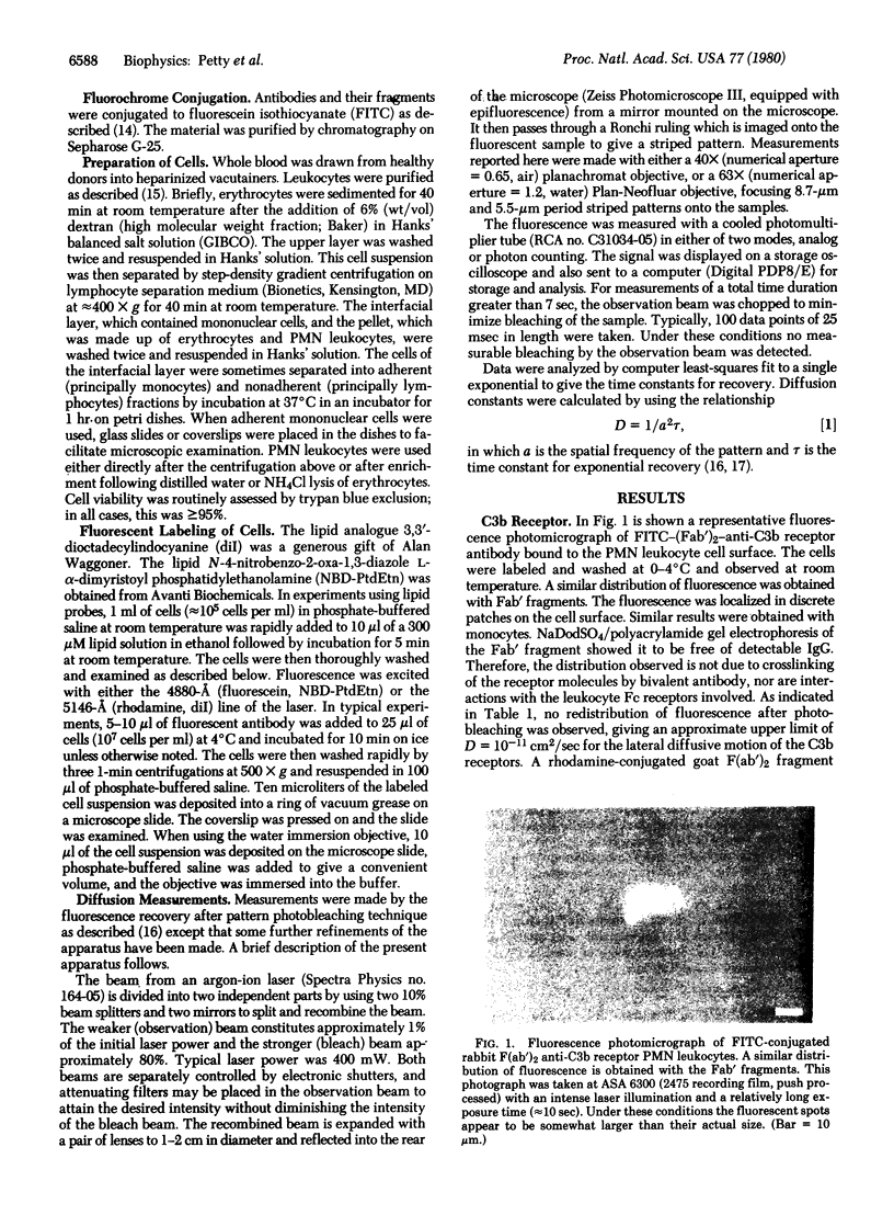
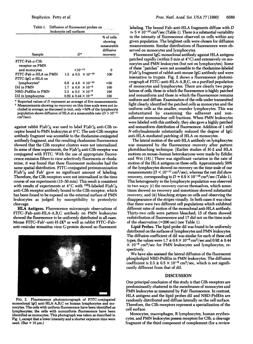
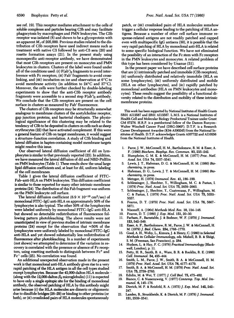
Fluorescence microscopy and fluorescence redistribution after pattern photobleaching have been used to measure the distribution and motion of a number of fluorescent molecules bound to the plasma membranes of human leukocytes. The fluorescent molecules include fluorescein-labeled F(ab')2 and Fab' fragments of an anti-C3b receptor antibody, fluorescein-labeled IgG and Fab fragments of a monoclonal anti-HLA antibody, and the two lipid probes 3,3'-dioctadecylindocyanine and N-4-nitrobenzo-2-oxa-1,3-diazole L-alpha-dimyristoyl phosphatidylethanolamine. From these studies we have concluded that the C3b receptors on human polymorphonuclear leukocytes and monocytes are predominantly present in discrete clusters. No diffusive motion of these clusters could be detected. In contrast, HLA antigens hve a dispersed distribution and have diffusion coefficients between 10(-9) and 10(-10) cm2/sec in these peripheral blood leukocytes. Cell-membrane-bound monoclonal IgG anti-HLA undergoes a rapid patching in all these leukocytes except lymphocytes. In about 50% of the lymphocytes, fluoresceinated IgG diffuses slowly, if at all, whereas in the other 50%, fluoresceinated IgG diffuses with a diffusion coefficient of 6.9 X 10(-10) cm2/sec. The two above-mentioned fluorescent lipid probes have the usual rapid lipid diffusion coefficient (approximately 10(-8) cm2/sec) in leukocytes.
Full text
PDF
Images in this article
Selected References
These references are in PubMed. This may not be the complete list of references from this article.
- Bianco C., Nussenzweig V. Complement receptors. Contemp Top Mol Immunol. 1977;6:145–176. doi: 10.1007/978-1-4684-2841-4_5. [DOI] [PubMed] [Google Scholar]
- Cherry R. J. Rotational and lateral diffusion of membrane proteins. Biochim Biophys Acta. 1979 Dec 20;559(4):289–327. doi: 10.1016/0304-4157(79)90009-1. [DOI] [PubMed] [Google Scholar]
- Cresswell P., Dawson J. R. Dimeric and monomeric forms of HL-A antigens solubilized by detergent. J Immunol. 1975 Jan;114(1 Pt 2):523–525. [PubMed] [Google Scholar]
- Dierich M. P., Reisfeld R. A. C3-mediated cytoadherence. Formation of C3 receptor aggregates as prerequisite for cell attachment. J Exp Med. 1975 Jul 1;142(1):242–247. doi: 10.1084/jem.142.1.242. [DOI] [PMC free article] [PubMed] [Google Scholar]
- Dragsten P., Henkart P., Blumenthal R., Weinstein J., Schlessinger J. Lateral diffusion of surface immunoglobulin, Thy-1 antigen, and a lipid probe in lymphocyte plasma membranes. Proc Natl Acad Sci U S A. 1979 Oct;76(10):5163–5167. doi: 10.1073/pnas.76.10.5163. [DOI] [PMC free article] [PubMed] [Google Scholar]
- Edidin M., Wei T. Y. Diffusion rates of cell surface antigens of mouse-human heterokaryons. I. Analysis of the population. J Cell Biol. 1977 Nov;75(2 Pt 1):475–482. doi: 10.1083/jcb.75.2.475. [DOI] [PMC free article] [PubMed] [Google Scholar]
- Esser A. F., Bartholomew R. M., Parce J. W., McConnell H. M. The physical state of membrane lipids modulates the activation of the first component of complement. J Biol Chem. 1979 Mar 25;254(6):1768–1770. [PubMed] [Google Scholar]
- Fearon D. T. Regulation of the amplification C3 convertase of human complement by an inhibitory protein isolated from human erythrocyte membrane. Proc Natl Acad Sci U S A. 1979 Nov;76(11):5867–5871. doi: 10.1073/pnas.76.11.5867. [DOI] [PMC free article] [PubMed] [Google Scholar]
- Humphries G. M., McConnell H. M. Membrane-controlled depletion of complement activity by spin-label-specific IgM. Proc Natl Acad Sci U S A. 1977 Aug;74(8):3537–3541. doi: 10.1073/pnas.74.8.3537. [DOI] [PMC free article] [PubMed] [Google Scholar]
- Landen B., Strunkheide K., Dierich M. P. C3-mediated cytoadherence. II. Dependence of cell attachment on similar topographic distribution of the receptors (for C3) and the ligands (C3/C3b). J Immunol. 1978 Dec;121(6):2539–2541. [PubMed] [Google Scholar]
- Mardiney M. R., Jr, Müller-Eberhard H. J., Feldman J. D. Ultrastructural localization of the third and fourth components of complement on complement-cell complexes. Am J Pathol. 1968 Aug;53(2):253–261. [PMC free article] [PubMed] [Google Scholar]
- Metzger H. The IgE-mast cell system as a paradigm for the study of antibody mechanisms. Immunol Rev. 1978;41:186–199. doi: 10.1111/j.1600-065x.1978.tb01465.x. [DOI] [PubMed] [Google Scholar]
- NISONOFF A. ENZYMATIC DIGESTION OF RABBIT GAMMA GLOBULIN AND ANTIBODY AND CHROMATOGRAPHY OF DIGESTION PRODUCTS. Methods Med Res. 1964;10:134–141. [PubMed] [Google Scholar]
- Parce J. W., McConnell H. M., Bartholomew R. M., Esser A. F. Kinetics of antibody-dependent activation of the first component of complement on lipid bilayer membranes. Biochem Biophys Res Commun. 1980 Mar 13;93(1):235–242. doi: 10.1016/s0006-291x(80)80271-3. [DOI] [PubMed] [Google Scholar]
- Parham P., Barnstable C. J., Bodmer W. F. Use of a monoclonal antibody (W6/32) in structural studies of HLA-A,B,C, antigens. J Immunol. 1979 Jul;123(1):342–349. [PubMed] [Google Scholar]
- Parish C. R. Separation and functional analysis of subpopulations of lymphocytes bearing complement and Fc receptors. Transplant Rev. 1975;25:98–120. doi: 10.1111/j.1600-065x.1975.tb00727.x. [DOI] [PubMed] [Google Scholar]
- Peterson P. A., Rask L., Sege K., Klareskog L., Anundi H., Ostberg L. Evolutionary relationship between immunoglobulins and transplantation antigens. Proc Natl Acad Sci U S A. 1975 Apr;72(4):1612–1616. doi: 10.1073/pnas.72.4.1612. [DOI] [PMC free article] [PubMed] [Google Scholar]
- Petty H. R., Smith B. A., Ware B. R., Rocklin R. E. Alterations of polymorphonuclear leukocyte surface charge by stimulated lymphocyte supernatants. Cell Immunol. 1980 Sep 1;54(2):435–444. doi: 10.1016/0008-8749(80)90223-3. [DOI] [PubMed] [Google Scholar]
- Schlessinger J., Shechter Y., Cuatrecasas P., Willingham M. C., Pastan I. Quantitative determination of the lateral diffusion coefficients of the hormone-receptor complexes of insulin and epidermal growth factor on the plasma membrane of cultured fibroblasts. Proc Natl Acad Sci U S A. 1978 Nov;75(11):5353–5357. doi: 10.1073/pnas.75.11.5353. [DOI] [PMC free article] [PubMed] [Google Scholar]
- Schlessinger J., Shechter Y., Willingham M. C., Pastan I. Direct visualization of binding, aggregation, and internalization of insulin and epidermal growth factor on living fibroblastic cells. Proc Natl Acad Sci U S A. 1978 Jun;75(6):2659–2663. doi: 10.1073/pnas.75.6.2659. [DOI] [PMC free article] [PubMed] [Google Scholar]
- Schreiner G. F., Unanue E. R. Membrane and cytoplasmic changes in B lymphocytes induced by ligand-surface immunoglobulin interaction. Adv Immunol. 1976;24:37–165. doi: 10.1016/s0065-2776(08)60329-6. [DOI] [PubMed] [Google Scholar]
- Shearer G. M., Polisson R. P. Mutual recognition of parental and F1 lymphocytes. Selective abrogation of cytotoxic potential of F1 lymphocytes by parental lymphocytes. J Exp Med. 1980 Jan 1;151(1):20–31. doi: 10.1084/jem.151.1.20. [DOI] [PMC free article] [PubMed] [Google Scholar]
- Smith B. A., McConnell H. M. Determination of molecular motion in membranes using periodic pattern photobleaching. Proc Natl Acad Sci U S A. 1978 Jun;75(6):2759–2763. doi: 10.1073/pnas.75.6.2759. [DOI] [PMC free article] [PubMed] [Google Scholar]
- Smith L. M., Parce J. W., Smith B. A., McConnell H. M. Antibodies bound to lipid haptens in model membranes diffuse as rapidly as the lipids themselves. Proc Natl Acad Sci U S A. 1979 Sep;76(9):4177–4179. doi: 10.1073/pnas.76.9.4177. [DOI] [PMC free article] [PubMed] [Google Scholar]


