Abstract
Reprogramming of cellular metabolism is a key event during tumorigenesis. Despite being known for decades (Warburg effect), the molecular mechanisms regulating this switch remained unexplored. Here, we identify SIRT6 as a novel tumor suppressor that regulates aerobic glycolysis in cancer cells. Importantly, loss of SIRT6 leads to tumor formation without activation of known oncogenes, while transformed SIRT6-deficient cells display increased glycolysis and tumor growth, suggesting that SIRT6 plays a role in both establishment and maintenance of cancer. Using a conditional SIRT6 allele, we show that SIRT6 deletion in vivo increases the number, size and aggressiveness of tumors. SIRT6 also functions as a novel regulator of ribosome metabolism by co-repressing MYC transcriptional activity. Lastly, SIRT6 is selectively downregulated in several human cancers, and expression levels of SIRT6 predict prognosis and tumor-free survival rates, highlighting SIRT6 as a critical modulator of cancer metabolism. Our studies reveal SIRT6 to be a potent tumor suppressor acting to suppress cancer metabolism.
INTRODUCTION
Cancer cells are characterized by the acquisition of several characteristics that enable them to become tumorigenic (Hanahan and Weinberg, 2000). Among them, the ability to sustain uncontrolled proliferation represents the most fundamental trait of cancer cells. This hyperproliferative state involves the deregulation of proliferative signaling pathways as well as loss of cell cycle regulation. In addition, tumor cells need to readjust their energy metabolism to fuel cell growth and division. This metabolic adaptation is directly regulated by many oncogenes and tumor suppressors, and is required to support the energetic and anabolic demands associated with cell growth and proliferation (Lunt and Vander Heiden, 2011).
Alteration in glucose metabolism is the best-known example of metabolic reprogramming in cancer cells. Under aerobic conditions, normal cells convert glucose to pyruvate through glycolysis, which enters the mitochondria to be further catabolized in the tricarboxylic acid cycle (TCA) to generate adenosine-5’-triphosphate (ATP). Under anaerobic conditions, mitochondrial respiration is abated; glucose metabolism is shifted towards glycolytic conversion of pyruvate into lactate. This metabolic reprogramming is also observed in cancer cells even in the presence of oxygen and was first described by Otto Warburg several decades ago (Warburg, 1956; Warburg et al., 1927). By switching their glucose metabolism towards “aerobic glycolysis”, cancer cells accumulate glycolytic intermediates that will be used as building blocks for macromolecular synthesis (Vander Heiden et al., 2009). Most cancer cells exhibit increased glucose uptake, which is due, in part, to the upregulation of glucose transporters, mainly GLUT1 (Yamamoto et al., 1990; Younes et al., 1996). Moreover, cancer cells display a high expression and activity of several glycolytic enzymes, including phospho-fructose kinase (PFK)-1, pyruvate kinase M2, lactate dehydrogenase (LDH)-A and pyruvate dehydrogenase kinase (PDK)-1 (Lunt and Vander Heiden, 2011), leading to the high rate of glucose catabolism and lactate production characteristic of these cells. Importantly, downregulation of either LDH-A or PDK1 decreases tumor growth (Bonnet et al., 2007; Fantin et al., 2006; Le et al., 2010) suggesting an important role for these proteins in the metabolic reprogramming of cancer cells.
Traditionally, cancer-associated alterations in metabolism have been considered a secondary response to cell proliferation signals. However, growing evidence has demonstrated that metabolic reprogramming of cancer cells is a primary function of activated oncogenes and inactivated tumor suppressors (Dang et al., 2012; DeBerardinis et al., 2008; Ward and Thompson, 2012). Despite this evidence, whether the metabolic reprogramming observed in cancer cells is a driving force for tumorigenesis remains as yet poorly understood.
Sirtuins are a family of NAD+-dependent protein deacetylases involved in stress resistance and metabolic homeostasis (Finkel et al., 2009). In mammals, there are seven members of this family (SIRT1-7). SIRT6 is a chromatin-bound factor that was first described as a suppressor of genomic instability (Mostoslavsky et al., 2006). SIRT6 also localizes to telomeres in human cells and controls cellular senescence and telomere structure by deacetylating histone H3 lysine 9 (H3K9) (Michishita et al., 2008). However, the main phenotype SIRT6 deficient mice display is an acute and severe metabolic abnormality. At 20 days of age, they develop a degenerative phenotype that includes complete loss of subcutaneous fat, lymphopenia, osteopenia, and acute onset of hypoglycemia, leading to death in less than ten days (Mostoslavsky et al., 2006). Recently, we have demonstrated that the lethal hypoglycemia exhibited by SIRT6 deficient mice is caused by an increased glucose uptake in muscle and brown adipose tissue (Zhong et al., 2010). Specifically, SIRT6 co-represses HIF-1α by deacetylating H3K9 at the promoters of several glycolytic genes and, consequently, SIRT6 deficient cells exhibit increased glucose uptake and upregulated glycolysis even under normoxic conditions (Zhong et al., 2010). Such a phenotype, reminiscent of the “Warburg Effect” in tumor cells, prompted us to investigate whether SIRT6 may protect against tumorigenesis by inhibiting glycolytic metabolism.
Here, we demonstrate that SIRT6 is a novel tumor suppressor that regulates aerobic glycolysis in cancer cells. Strikingly, SIRT6 acts as a first hit tumor suppressor and lack of this chromatin factor leads to tumor formation even in non-transformed cells. Notably, inhibition of glycolysis in SIRT6 deficient cells completely rescues their tumorigenic potential, suggesting that enhanced glycolysis is the driving force for tumorigenesis in these cells. Furthermore, we provide new data demonstrating that SIRT6 regulates cell proliferation by acting as a co-repressor of c-Myc, inhibiting the expression of ribosomal genes. Finally, SIRT6 expression is downregulated in human cancers, strongly reinforcing the idea that SIRT6 is a novel tumor suppressor.
RESULTS
SIRT6 deficient cells are tumorigenic
We have previously shown that SIRT6 protects from genomic instability (Mostoslavsky et al., 2006) and regulates aerobic glycolysis (Zhong et al., 2010), key features of cancer cells. Therefore, we hypothesized that SIRT6 deficiency could lead to tumorigenesis. To study this possibility, we obtained mouse embryonic fibroblasts (MEFs) from Sirt6 WT and KO embryos and immortalized them using a standard 3T3 protocol. We found that Sirt6 KO MEFs showed increased proliferation (Figure 1A) and formed larger colonies when plated at very low density, compared to Sirt6 WT cells (Figure 1B), indicating that SIRT6 deficiency confers a growth advantage. Next, we injected Sirt6 WT and KO MEFs into the flanks of SCID mice to assess the ability of these cells to form tumors in vivo. Immortalized MEFs never develop tumors in this setting unless they are transformed with an activated oncogene, such as Ras or Myc (“second hit”). Strikingly, Sirt6 KO MEFs readily formed tumors (Figure 1C), indicating that SIRT6 deficiency is sufficient to induce transformation of immortalized MEFs. To confirm that this was not due to non-specific effects of the immortalization process, we immortalized Sirt6 WT and KO MEFs by knocking-down p53 in passage 3 primary MEFs (Figure S1A). Again, Sirt6 KO MEFs showed increased proliferation (Figure S1B) and were able to form tumors when injected into SCID mice (Figure S1C). Together, these results suggest that SIRT6 acts as a tumor suppressor and, upon loss of cell-cycle checkpoint control, SIRT6 deficiency can lead to tumorigenesis in mice.
Figure 1. SIRT6 deficient cells are tumorigenic.
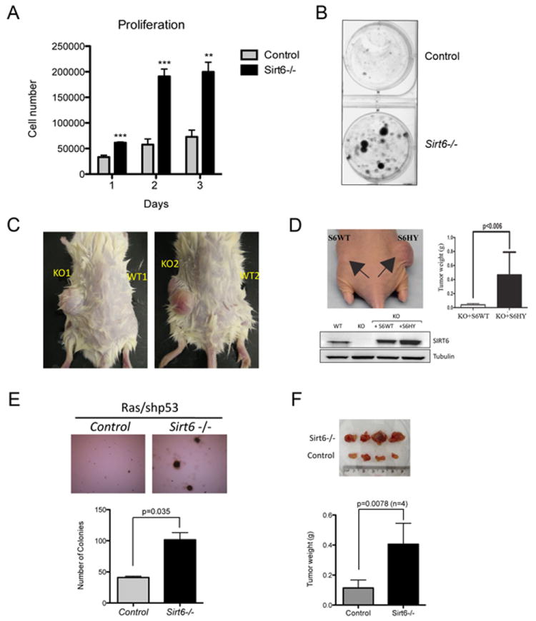
A) Sirt6 WT and Sirt6 KO immortalized MEFs (two independent cell lines for each) were plated and cells counted at the indicated times. Errors bars indicate SEM. B) Sirt6 WT and KO cells were plated at very low confluency and assayed for colony formation. C) Two independent immortalized cell lines of the indicated genotypes were injected into the flanks of SCID mice to assess their tumorigenic potential. D) Sirt6 KO immortalized MEFs were transduced with lentiviruses encoding Flag-SIRT6 (WT and HY) and assayed for in vivo tumor formation as in C. E) Anchorage-independent cell growth of Sirt6 WT and KO H-RasV12/shp53 transformed MEFs (error bars indicate SD). F) The same cells as in D were injected into the flanks of SCID mice (n=4) and the tumors harvested and weighted (error bars indicate SD). See also Figure S1.
Genomic instability can induce transformation by activating oncogenes or inactivating tumor suppressors. Therefore, we first analyzed whether the genomic instability exhibited by SIRT6 deficient cells was responsible for their transformed phenotype. For this purpose, we re-expressed SIRT6 in KO MEFs and injected these cells into nude mice. If genomic instability causes SIRT6-dependent transformation, we would expect tumor formation even in the presence of SIRT6; the effect of mutations occurring on any oncogene or tumor suppressor pathway would not be reverted by simply re-expressing SIRT6 (“mutator phenotype”). However, re-expression of SIRT6 in KO MEFs completely abolished the ability of these cells to form tumors (Figure 1D), suggesting an alternative mechanism for tumor suppression mediated by SIRT6. Furthermore, re-expression of the catalytically inactive SIRT6-H133Y mutant was not able to rescue the tumorigenic phenotype (Figure 1D). Altogether, these results confirm that the tumorigenic capacity of Sirt6 KO MEFs is specific to the lack of this chromatin regulator and that SIRT6 activity is required for its tumor suppressor function.
Next, we analyzed whether SIRT6 influences tumor growth in the presence of activating oncogenes. For this purpose, we transformed Sirt6 WT and KO MEFs by expressing an activated form of H-Ras (H-RasV12) and knocking-down p53 expression (shp53). We found that even in the presence of a strong oncogenic signal such as H-RasV12, Sirt6 KO cells exhibited increased anchorage-independent growth in soft agar (Figure 1E) and formed bigger tumors when injected into SCID mice compared to Sirt6 WT cells (Figure 1F), indicating that SIRT6 deficiency also confers a growth advantage to transformed cells. Taken together, these results strongly suggest that SIRT6 is a tumor suppressor involved in both cancer initiation and tumor growth.
SIRT6 deficient cells and tumors exhibit enhanced aerobic glycolysis
To identify the mechanism underlying the tumorigenic phenotype associated with SIRT6 deficiency, we focused on SIRT6-dependent regulation of glucose metabolism. Immortalized Sirt6 KO MEFs showed increased glucose uptake and lactate production (Figure 2A). In addition, re-expression of SIRT6 in these cells reduced glucose consumption (Figure 2B) as well as tumor formation (Figure 1D), suggesting that a switch towards aerobic glycolysis may be involved in the tumorigenic process. Furthermore, acute deletion of SIRT6 by adeno-Cre infection in MEFs derived from SIRT6 KO conditional mice (Figure 7) increased glucose uptake in these cells (Figure S2A), confirming that this phenotype is specific to the absence of this chromatin factor. To further characterize the glycolytic phenotype of these cells, we measured expression levels of key glycolytic enzymes that are direct targets for SIRT6 (Zhong et al., 2010). When compared to WT MEFs, Sirt6 KO MEFs expressed higher levels of Glut1, Pfk1, Pdk1 and Ldha (Figure 2C). Importantly, the expression of these glycolytic genes was further increased in cells derived from Sirt6 deficient tumors (Figure 2C, tumor bar). This may indicate a selective advantage within Sirt6 KO tumors for cells with increased glycolytic activity. To confirm the reliance of Sirt6 KO cells on glycolysis, we analyzed their survival post glucose starvation. While nearly all Sirt6 WT MEFs survived glucose withdrawal, a significant percentage of Sirt6 KO cells died under these conditions (Figure 2D), indicating that SIRT6 deficiency promotes a state of glucose-addiction, a hallmark of cancers undergoing aerobic glycolysis.
Figure 2. SIRT6 deficient cells and tumors exhibit enhanced aerobic glycolysis.
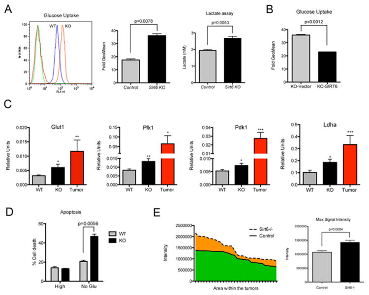
A) Glucose uptake (left and middle panels) and lactate production (right panel) of Sirt6 WT and Sirt6 KO immortalized MEFs (two independent cell lines, error bars indicate SEM). B) Glucose uptake of Sirt6 KO immortalized MEFs transduced with either an empty vector or a plasmid encoding for Flag-SIRT6 (error bars indicate SD). C) Real-Time PCR showing the expression of the indicated genes in Sirt6 WT and Sirt6 KO (n=20 experiments from two independent lines) immortalized MEFs and in the cells derived from Sirt6 KO tumors (n=8 experiments from three independent lines) (error bars indicate SEM). D) The same cells as in A were cultured with or without glucose for 6 days and cell death assayed by AnnexinV staining (error bars indicate SEM). E) 18FDG-Glucose uptake in Sirt6 WT and KO H-RasV12/shp53 tumors. Left panel shows FDG-PET intensity of the 5 sections of each tumor (total of 6 tumors for each genotype) showing the highest intensity. Right panel shows the average of 30 FDG-PET signals (6 tumors, 5 sections per tumor) for the indicated genotypes (error bars indicate SD). See also Figure S2.
Figure 7. SIRT6 functions as a tumor suppressor in vivo.
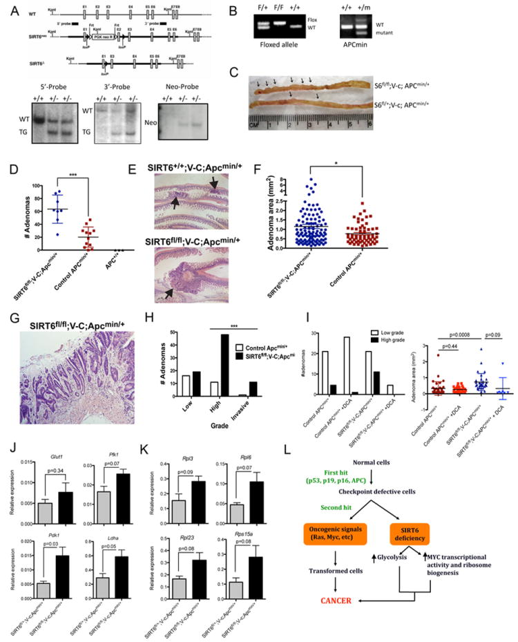
A) Strategy to target the SIRT6 locus (upper panel). Southern blot (5’, 3’ and Neo probes) of KpnI-digested genomic DNA showing the targeted allele in the heterozygous cells (+/-) (lower panels). B) PCR showing the presence of the SIRT6 floxed allele (left) and the mutant APC allele (right). C) Representative image of a intestine section from SIRT6fl/fl;V-c;APCmin/+ and SIRT6fl/+;V-c;APCmin/+ mice. Arrows indicate the presence of polyps. D) Adenoma number in the intestines of mice of the indicated genotype. E) H&E staining showing the adenoma size in the indicated mice. F) Adenoma area in SIRT6fl/fl;V-c;APCmin/+ and Control;APCmin/+ mice. G) Representative image and (H) quantification of the grade of the tumors in the indicated mice. I) Grade (righ panel) and area (left panel) of the adenomas in SIRT6fl/fl;V-c;APCmin/+ and Control;APCmin/+ mice untreated or treated with DCA (5g/L). J) and K) Expression of several glycolytic and ribosomal genes in adenomas (n=3) of SIRT6fl/fl;V-c;APCmin/+ and SIRT6fl/+;V-c;APCmin/+ mice (error bars indicate SEM). See also Figure S7.
Our results indicate that SIRT6 deficiency plays a crucial role in tumor initiation and growth (Figure 1F). To assess if increased glycolysis is also responsible for the tumor growth phenotype, we analyzed the glycolytic activity of these cells. We found that Sirt6KO/H-RasV12/shp53 transformed MEFs uptake more glucose (Figure S2B) and produce more lactate in culture (Figure S2C) when compared to Sirt6WT/H-RasV12/shp53 controls. Next, we injected these cells into SCID mice and analyzed glucose uptake in vivo by 18F-fluorodeoxyglucose-positron emission tomography (FDG-PET). Importantly, tumors derived from transformed Sirt6 KO cells exhibited increase FDG intensity compared to Sirt6 WT cells (Figure 2E) indicating that SIRT6 deficiency in tumors increases glucose uptake and glycolysis both in vitro and in vivo. Together, these results strongly suggest that SIRT6 may act as a tumor suppressor by repressing aerobic glycolysis.
Oncogene-independent transformation in Sirt6 KO cells
The data presented above suggests a role for a SIRT6-dependent glycolytic switch in cancer initiation and progression. Nonetheless, SIRT6 deficiency might promote tumor formation via activation of an oncogenic pathway. To test this possibility, we analyzed the activation of oncogenic signaling pathways in SIRT6 deficient cells. Since deregulation of most oncogenes leads to the activation of the downstream ERK and AKT pathways, we focused on these signaling pathways. Phospho-ERK and phospho-AKT levels were similar in Sirt6 WT and KO MEFs (Figure 3A, left panels).
Figure 3. Oncogene-independent, glycolysis-dependent transformation of SIRT6 deficient cells.
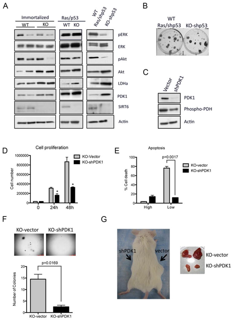
A) Western blots showing the activation of ERK and AKT pathways as well as PDK1 and LDHa expression in Sirt6 WT and KO immortalized and transformed MEFs. B) Colony formation assay with the indicated cell lines. C) Western blot of PDK expression and PDH-E1a-Ser293 phosphorylation in Sirt6 KO-shPDK1 cells D) Cell proliferation of Sirt6 KO-shVector and Sirt6 KO-shPDK1 (error bars indicate SD). E) Glucose starvation-induced cell death of Sirt6 KO-shVector and Sirt6 KO-shPDK1 cells (error bars indicate SD). F) Anchorage-independent cell growth of the same cells as in E (error bars indicate SD). F) The same cells as in F were injected into the flanks of SCID mice (n=2) and the tumors harvested and photographed. See also Figure S3.
In addition, primary MEFs immortalized by knocking down p53 exhibited the same phenotype (Figure S3A), ruling out non-specific effect of the immortalization process. Moreover, activation of these pathways in H-RasV12/shp53 transformed MEFs was similar in the presence or absence of SIRT6 (Figure 3A, middle panels). These results suggest that tumorigenesis in Sirt6 KO cells is oncogene-independent. Importantly, PDK1 and LDHa protein levels were specifically upregulated in both immortalized and transformed Sirt6 KO cells (Figure 3A and S3A) confirming that these cells are highly glycolytic.
In order to better understand the transformation process in SIRT6 deficient cells we directly compared Ras-transformed Sirt6 WT cells with immortalized Sirt6 KO cells. To this end, we obtained primary MEFs from WT and KO embryos and infected them in parallel with viruses expressing H-RasV12 plus shp53 or shp53 alone, respectively. As expected, analysis of the ERK and AKT pathways showed strong activation of these proteins in H-RasV12/shp53-transformed Sirt6 WT cells (Figure 3A, right panels). However, these oncogenic pathways were not activated in shp53-immortalized KO cells, despite their transformation capability. Importantly, LDHa and PDK1 expression was higher in Sirt6 KO cells (Figure 3A) suggesting that enhanced glycolysis, rather than oncogene activation, may be the driving force for tumorigenesis in SIRT6-deficient cells. Further supporting this notion, a colony formation assay indicates similar growth in shp53-immortalized Sirt6 KO cells and H-RasV12/shp53-transformed WT cells (Figure 3B). Similar to the 3T3 immortalized SIRT6-deficient cells, shp53-immortalized Sirt6 KO cells gave rise to tumors when injected into SCID mice (Figure S1C).
Inhibition of glycolysis suppresses tumorigenesis in Sirt6 KO cells
The above results indicate that SIRT6 acts as a tumor suppressor potentially by inhibiting a switch towards aerobic glycolysis (Warburg Effect). We reasoned that, if this was the case, inhibition of glycolysis would abolish the tumorigenic potential of Sirt6 KO cells. Since conversion of pyruvate to lactate is rate-limiting and represents a downstream step in the glycolytic pathway, we aimed to modulate glycolytic activity in SIRT6 deficient cells by controlling this step. For this purpose, we knocked down the expression of Pdk1 using short hairpin RNAs (shPDK1) (Figure 3C). As expected, PDK1 downregulation reduced PDH phosphorylation (Figure 3C). In addition, Sirt6 KO-shPDK1 cells exhibited reduced proliferative capacity (Figure 3D). Notably, these cells were no longer “glucose-addicted”, as reflected by the complete rescue of glucose starvation-induced cell death (Figure 3E and S3B). Moreover, PDK1 downregulation in Sirt6 KO MEFs inhibited the anchorage-independent cell growth in soft agar (Figure 3F) and severely diminished tumor formation in vivo (Figure 3G). Together, these results demonstrate that SIRT6 may act as a tumor suppressor by blocking a switch towards aerobic glycolysis. In addition, inhibition of glycolysis in SIRT6 deficient cells is sufficient to revert this phenotype, further confirming that these cells have not acquired cancer-driving mutations, but rather rely fully on glycolysis for growth.
SIRT6 controls cancer cell proliferation by co-repressing Myc transcriptional activity
In most cancer cells, increased glycolysis per se is not sufficient to provide a growth advantage, suggesting that SIRT6 might be controlling proliferating genes as well. In order to determine whether this is the case, we used datasets from SIRT6 chromatin immunoprecipitation followed by massively parallel DNA sequencing (ChIP-seq). These data include chromatin maps from two independent cell lines: K562 erythroleukemia cells and human embryonic stem cells (hESCs) (Ram et al., 2011). Gene ontology analysis of SIRT6-bound genomic regions revealed significant enrichment of ribosomal and ribonucleoprotein genes (Figure 4A). Interestingly, the transcription factor MYC has recently been described as a global regulator of ribosome biogenesis (Arabi et al., 2005; Dai and Lu, 2008; Grandori et al., 2005; Grewal et al., 2005), leading us to speculate that SIRT6 and MYC could cooperate in the regulation of ribosomal gene expression. To study this possibility, we first compared the SIRT6 genome-wide binding maps with a published MYC ChIP-seq dataset (Ram et al., 2011, Raha et al., 2010) to identify commonly bound genes. Notably, we found that a significantly high percentage of MYC-target genes were also enriched for SIRT6 binding (75%; 752/top 1000 bound genes) (Figure 4B and Table S1). The correlation between SIRT6 and MYC bound promoters (0.63) was very similar to the one exhibited by the MYC interactors FOS (0.76) and JUN (0.86) (Figure S4A). In contrast, no correlation was observed between Sirt6 and other chromatin modulators, such as EZH2, similar to what was observed for Myc (Figure S4B). Moreover, we analyzed the MSigDB gene set collection for their enrichment of overlapping SIRT6-MYC target genes using the hypergeometric test. We find clear enrichment for ribosome biosynthesis (p=9×10⁻⁸), structural constituent of ribosome (p=0) and translation (p=1.2×10⁻¹⁴)(Figure 4B yellow dots- and Figure 4C), suggesting that SIRT6 might be involved in the regulation of MYC-dependent ribosomal gene expression. Remarkably, ChIP-seq analysis for SIRT6 and MYC in mouse ES cells showed similar co-binding patterns (data not shown), strongly indicating that such mechanism is evolutionary conserved. We analyzed the co-binding of SIRT6 and MYC on the ribosomal protein genes Rpl3, Rpl6, Rpl23 and Rps15a (four of the top hits in overlapping SIRT6/MYC target genes) (Table S1). As expected, MYC binding exhibited a sharp peak on the promoters, co-localizing with the signal of H3K4me3 (a mark of activated as well as poised promoters) (Schneider et al., 2004; Zhou et al., 2011)(Figure 4D). Strikingly, SIRT6 also showed significant enrichment on the promoters of these genes (Figure 4D), suggesting that MYC and SIRT6 are sitting together on the promoter region of ribosomal protein genes. Interestingly, SIRT6 binding extended into the intragenic regions, arguing that SIRT6 may play a role beyond transcriptional initiation, as suggested by our previous work (Zhong et al., 2010).
Figure 4. SIRT6 inhibits ribosomal gene expression by co-repressing MYC transcriptional activity.
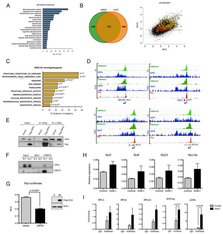
A) Gene Ontology clustering of SIRT6-bound promoters B) Overlapping of the top 1000 SIRT6- and MYC-bound promoters. C) Gene Set Enrichment Analysis for the overlapping genes described in B. D) H3K4me3, SIRT6 and MYC ChIP signal in the indicated genomic regions in K562 cells and human ES cells (H1). E) Flag-SIRT6 and cMYC IPs showing physical interaction between SIRT6 and MYC. F) Endogenous SIRT6 was immunoprecipitated and the interaction with MYC analyzed by western blot. F) A luciferase reporter gene under the regulation of a MYC-responsive element was contrasfected with empty vector or Flag-SIRT6 plasmids in 293T cells and luciferase expression analyzed 24h later (error bars indicate SEM). G) Expression of the indicated genes in Sirt6 WT and KO H-RasV12/shp53 tumors (n=4) (error bars indicate SEM). I) ChIP analysis of H3K56 acetylation levels in Sirt6 WT and KO H-RasV12/shp53 MEFs (n=4, error bars indicate SEM). See also Figure S4 and Table S1.
The co-occupancy of ribosomal gene promoters by MYC and SIRT6 suggests that these two proteins may interact to coordinate expression of target genes. Indeed, Flag-SIRT6 IP in U2OS pulled down MYC, indicating that SIRT6 and MYC can interact (Figure S4C). Although both SIRT2 and SIRT5 exhibited weak interaction with MYC as well, SIRT6 showed the strongest interaction (Figure S4C). Similar results were obtained in 293T cells overexpressing Flag-SIRT6, where MYC was detected in the Flag-IP and viceversa (Figure 4E). To confirm that these proteins interact under physiological conditions, we performed endogenous SIRT6 IP in ES cells.
Importantly, MYC was specifically pulled-down in the SIRT6 IP (Figure 4F). All together, the above results indicate that SIRT6 and MYC interact on the promoter region of ribosomal protein genes. MYC has been described as a transcriptional activator of genes involved in ribosome biogenesis (van Riggelen et al., 2010), whereas we and others have described a role for SIRT6 as a transcriptional repressor (Kawahara et al., 2009; Zhong et al., 2010). Thus, we hypothesized that SIRT6 might act as a co-repressor of MYC activity in the context of ribosomal gene expression. To study this possibility, we first tested whether SIRT6 could influence MYC-dependent expression of a luciferase reporter. Indeed, expression of SIRT6 in 293T cells carrying a MYC-luciferase reporter dramatically reduced luciferase expression (Figure 4G) indicating that SIRT6 co-represses MYC activity in this setting. In line with this, we found increased expression of Rpl3, Rpl6, Rpl23 and Rps15a in SIRT6 deficient tumors (Figure 4H). Interestingly, the expression of all these genes is not upregulated in immortalized Sirt6 KO MEFs (Figure S4B), suggesting that the increase in ribosome biogenesis may be a late event during the tumorigenic process in SIRT6 deficient cells. Consistent with this idea, ribosomal gene expression was found to be upregulated in cells derived from Sirt6 KO tumors (Figure S4D, tumor bar). Similarly, glutamine uptake and glutaminase (Gls) expression, which are also regulated by MYC in cancer cells (Dang et al., 2012), are not upregulated in Sirt6 KO immortalized MEFs (Figure S4E and S4F), while Sirt6 KO H-RasV12/shp53-transformed MEFs exhibited increased glutamine uptake (Figure S4E).
We next studied in detail the molecular mechanism by which SIRT6 regulates MYC transcriptional activity. MYC expression and protein stability are not affected by SIRT6 deficiency (Figure S5A and S5B). Similarly, MYC acetylation levels are not changed in SIRT6 KO cells (Figure S5C). Although we cannot completely rule out by western blot that SIRT6 may deacetylate MYC in a specific residue, this result strongly suggests that SIRT6 is not a main deacetylase for MYC. Moreover, SIRT6 is not regulating the recruitment of MYC to its target promoters, since MYC binding to ribosomal genes promoters was not affected in Sirt6 KO cells (Figure S5D). As mentioned above, SIRT6 has been described as a H3K9 deacetylase. Thus, we analyzed by ChIP the acetylation levels of this histone mark on the promoter region of ribosomal protein genes. Surprisingly, the levels of H3K9 acetylation were not changed on these promoters, in contrast with what we observed on glycolytic genes promoters (Figure S5E) (Zhong et al., 2010). However, we found an increase in H3K56 acetylation on the promoter region of ribosomal protein genes in SIRT6-deficient cells (Figure 4I). H3K56Ac is a direct substrate of SIRT6 (Yang et al., 2009; Michishita et al., 2009) and this histone mark has been involved in transcriptional regulation (Xie et al., 2009), indicating that this residue might be a specific substrate of SIRT6 in the context of ribosomal gene expression.
Finally, to fully test whether MYC-dependent gene expression was important for the tumorigenic phenotype in the absence of SIRT6, we knocked-down the expression of cMyc in Sirt6 KO immortalized MEFs (Figure 5A) and found that, indeed, MYC downregulation in these cells reduced their proliferation (Figure 5B) and, more importantly, dampened tumor growth (Figure 5C). In addition, MYC knock-down decreased ribosomal protein gene expression as well as Gls expression (Figure 5D). However, glucose uptake and glycolytic gene expression was unaffected in Sirt6 KO-shMYC cells. These results indicate that MYC is controlling tumor growth in SIRT6-deficient cells specifically by regulating ribosome and glutamine metabolism, whereas SIRT6 effect on glycolysis likely depend on its function as a HIF-corepressor (Zhong et al., 2010; see Discussion).
Figure 5. MYC regulates tumor growth of SIRT6-deficient cells.
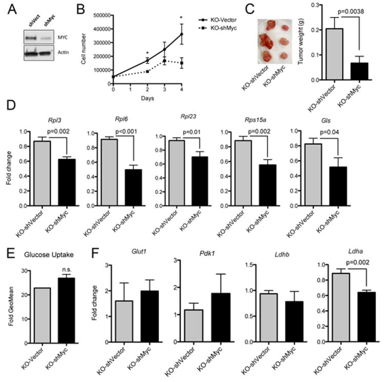
A) Western blot showing MYC levels in Sirt6 KO-shVector and shMYC cells. B) 5×10⁵ MEFs were plated in triplicate and cells counted at the indicated time points (error bars indicate SD). C) 5×10⁶ cells of the indicated genotypes were injected into SCID mice and the tumors harvested and weighted (error bars indicate SD). D) Expression of the indicated genes in Sirt6 KO-shVector and KO-shMYC cells (n=9) (error bars indicate SEM). E) Glucose uptake was analyzed in the same cells as in A (error bars indicate SD). F) The same samples as in D were used to analyze the expression of the indicated genes (error bars indicate SEM). See also Figure S5.
Sirt6 expression is downregulated in human cancers
The above results indicate a putative role for SIRT6 as a tumor suppressor regulating glycolytic metabolism, suggesting that its expression or activity might be decreased in human cancers. To study this possibility, we analyzed Sirt6 gene copy number across multiple cancer types using the Tumorscape database (Beroukhim et al., 2010). Strikingly, Sirt6 lies within a region in chromosome 19 significantly deleted across the entire dataset (Figure 6A)(q-value=0.00011). Additionally, The Cancer Genome Atlas (TCGA) database revealed that SIRT6 is deleted in 20% of all cancers analyzed (q-value= 3.87E-110) and, importantly, that it is located within a peak of deletion in almost 8% of colorectal cancers (Figure S6A)(q-value=0.0119). Next, we used the Cancer Cell Line Encyclopedia (CCLE) (Barretina et al., 2012) to further study gene-copy alterations of Sirt6 in human cancers. We found that the Sirt6 locus is deleted in 35% of approximately 1000 cancer cell lines collected in this dataset and, importantly, in 62.5% and 29% of pancreatic and colorectal cancer cell lines, respectively (Figure 6B and Figure S6B). In accordance with our model, SIRT6 is not amplified in any of the pancreatic cancer cell lines and only in 4% of colorectal cancer cell lines analyzed (Figure 6B and S6B).
Figure 6. SIRT6 expression is downregulated in human cancers.
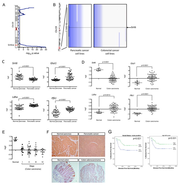
A) Analysis of gene copy number loss in chromosome 19. Blue line indicates deletion significance (-log10(q value), 0.6 –dotted line- is the significance threshold for deletion). SIRT6 location within the chromosome is indicated. B) SIRT6 copy number data for pancreatic (left graph, n=40) and colorectal (right graph, n=51) cancer cell lines. Color bars indicate degree of copy number loss (blue) or gain (red). C) Gene expression of the indicated genes in human pancreatic cancer (GEO dataset GSE15471). D) Gene expression of the indicated genes in human colon carcinoma (GEO dataset GSE31905). E) SIRT6 expression in the same colon carcinoma dataset as D but classified by stage. F) IHC showing SIRT6 expression in pancreatic cancer and colon adenocarcinoma compared to normal tissue. G) Kaplan-Meier curves showing disease free survival rates in patients with node positive tumors (left) or high CRP serum levels (right) with high and low levels of nuclear SIRT6. See also Figure S6.
The significant loss of the Sirt6 locus in pancreatic and colorectal cancer suggests that SIRT6 expression might be downregulated in these types of cancer. Indeed, we found that SIRT6 expression is downregulated in a pancreatic ductal adenocarcinoma dataset of 36 individual cases (Badea et al., 2008) compared to their matched normal tissue (Figure 6C, p<0.0001). Moreover, the analysis of a dataset containing 55 colorectal carcinomas (Anders et al., 2011) also showed decreased SIRT6 expression when compared to normal colon samples (Figure 6D, p<0.0001). Remarkably, the expression of the SIRT6-target genes Glut1, Ldha and Pfk1is significantly upregulated in these samples.
Although we cannot rule out that activation or inhibition of other pathways could also be responsible for the increased glycolytic gene expression, our results indicate that the pancreatic and colorectal tumors analyzed are highly glycolytic, and such increase in glycolysis strongly correlate with selective downregulation of Sirt6 expression in these tumors. Furthermore, the analysis of additional datasets (Oncomine and GEO) also reveals decreased expression of SIRT6 in pancreatic and colon cancer as well as in rectal adenocarcinoma (Figure S6C)(p<0.0001). These observations suggest a general role for SIRT6 as a tumor suppressor in these carcinoma types. Interestingly, SIRT6 expression is also downregulated in pancreatic intraepithelial neoplasia and colon adenomas (p<0.0001), a phenotype that correlates with high expression of glycolytic genes in these samples (Figure S6C and S6D). This further suggests that SIRT6 downmodulation is an early event during tumorigenesis, thereby indicating that this glycolytic switch may play a role in initiation of tumor development. In line with this evidence, classification of the 55 colorectal carcinomas described above showed that SIRT6 expression is downregulated in early stages and, importantly, its low expression is maintained during cancer progression, indicating that SIRT6 downregulation might be required for both tumor initiation and maintenance (Figure 6E).
To further validate these observations, we used immunohistochemistry to analyze SIRT6 expression in a set of human pancreatic and colorectal cancers. While normal pancreatic ducts and colon crypts exhibited strong SIRT6 staining, pancreatic ductal adenocarcinoma and colorectal carcinoma tissues had a clear decrease in SIRT6 protein levels (Figure 6F). Taken together, the results derived from human datasets strongly indicate that SIRT6 may act as a tumor suppressor in human pancreatic and colorectal cancer. Furthermore, selective downregulation of SIRT6 in tumors may provide an important selective advantage through modulation of glycolytic metabolism.
In order to determine whether SIRT6 expression levels could be correlated with cancer progression and/or survival, we performed IHC for SIRT6 expression in samples from 253 colorectal carcinomas (CRCs), collected over a period of eleven years at the Department of Surgery, Western Infirmary, Glasgow. Two independent observers scored tumors using the histoscore method (see Experimental Procedures). When patients were categorized according to nodal status, there was no significant difference in patient outcome due to SIRT6 expression levels in node negative patients. However, in node-positive patients, low levels of SIRT6 were associated with shorter time to relapse (p=0.021, 96 months vs 128 months, Figure 6G, left panel). Furthermore, these patients were 2.3 times more likely to relapse than those patients whose tumors expressed high levels of SIRT6 (p=0.024). Patients were also categorized by C-Reactive Protein (CRP) serum levels, a known marker of colon cancer progression. In the subgroup with high levels of CRP, patients with low levels of nuclear SIRT6 had shorter time to relapse than those patients with high levels of nuclear SIRT6 (p=0.031, 101 vs 131 months, Figure 6G, right panel). These patients were 2.2 times more likely to relapse than patients whose tumors expressed high levels of SIRT6 (p=0.036). These results suggest that decreased disease-free survival time is associated with low tumor levels of nuclear SIRT6 in patients with more aggressive tumors (node-positive tumors and high CRP serum levels).
SIRT6 acts as a tumor suppressor in vivo
The above results strongly indicate that SIRT6 functions as a tumor suppressor, suggesting that its absence would lead to tumorigenesis in vivo. However, Sirt6 germline KO mice die early in life (Mostoslavsky et al., 2006), thus preventing the use of this mouse model to experimentally confirm this hypothesis. To overcome this issue, we took advantage of conditional gene-targeting technology to inactivate SIRT6 in a tissue-specific manner and generated mice with one or both floxed alleles for Sirt6 (Figure 7B, left panel; Figure 7A-C, I-J). In parallel, we analyzed a previously described mouse strain with a floxed Sirt6 allele with similar results (Figure 7D-H)(Kim et al, 2010).
To determine the role of SIRT6 in tumorigenesis in vivo, we focused on a model of colorectal adenomatosis, utilizing the well-established APCmin mouse (see methods) (Moser et al., 1990; Su et al., 1992). We have generated mouse lines carrying the APC mutation in the presence or specific-intestinal deletion of SIRT6, hereafter referred to as control (SIRT6+/+ or SIRT6fl/+);V-C;APCmin/+ and SIRT6fl/fl;V-C;APCmin/+, respectively (Figure 7B, right panel). Strikingly, we found that SIRT6fl/fl;V-C;APCmin/+ mice developed a three-fold increase in the number of adenomas when compared to APCmin/+ control animals (Figure 7C and 7D)(p<0.0001) and that these adenomas were on average two-fold larger than those observed in control mice (p=0.017) (Figure 7E and 7F). Furthermore, pathologic analysis of the polyps showed that the lesions were of higher grade in the absence of SIRT6, resulting in many invasive tumors, a phenotype rarely observed in APCmin/+ animals (Figure 7G and 7H). Importantly, glucose uptake (measured by FDG-PET scanning) and expression of glycolytic genes, were upregulated in the adenomas from SIRT6fl/fl;V-C;APCmin/+ mice (Figure S7A and Figure 7J), suggesting that SIRT6 suppresses intestinal tumorigenesis by inhibiting glycolysis. Remarkably, treatment with the PDK1 small-molecule inhibitor dichloroacetate (DCA) (Bonnet et al., 2007) specifically inhibited tumor formation in SIRT6fl/fl;V-C;APCmin/+ mice, as we observed less, smaller and lower grade tumors compared to untreated animals (Figure 7I and S7B). In contrast, DCA-treatment had little to no effect on control V-C;APCmin/+ mice, strongly indicating that glycolysis plays a dominant and driving role in SIRT6-deficient tumors. Finally, ribosomal gene expression and Gls expression were also upregulated in the adenomas from SIRT6fl/fl;V-C;APCmin/+ mice (Figure 7K and Figure S4G). Together, our results demonstrate that SIRT6 inhibits the initiation and progression of colorectal cancer in vivo by repressing aerobic glycolysis and ribosomal gene expression (Figure 7L).
DISCUSSION
The data presented here reveals a novel role for SIRT6 as a tumor suppressor. Using a combination of in vitro and in vivo studies, as well as data from several human cancer databases, we have demonstrated that loss of SIRT6 leads to tumorigenesis and its expression is selectively downregulated in several human cancers. Mechanistically, SIRT6 represses aerobic glycolysis (Warburg effect) dampening cancer initiation and growth. Moreover, we describe a key role of this sirtuin in controlling cancer cell proliferation by co-repressing MYC transcriptional activity and the expression of ribosomal genes.
Given their absolute dependency on NAD+, sirtuins have evolved as critical modulators of stress responses, DNA repair and metabolism, sensing changes in metabolic cues in order to exert adaptive responses (Finkel et al., 2009). In this context, these proteins represent good candidates to control tumorigenesis and cancer growth. Indeed, SIRT1, SIRT2 and SIRT3 have been described to have tumor suppressive activity by controlling genomic stability and cellular metabolism (Kim et al., 2011; Martinez-Pastor and Mostoslavsky, 2012). Here, we show that SIRT6 functions as a first-hit tumor suppressor, and lack of this chromatin factor leads to tumor initiation and growth. Several lines of evidence support this model. First, SIRT6 deficiency, even in non-transformed cells, causes tumorigenesis (Figure 1C). Importantly, this appears to be specific to SIRT6, since the tumor suppressive effect of other sirtuins has been observed only in transformed cells (Bell et al., 2011; Fang and Nicholl, 2011; Finley et al., 2011; Kim et al., 2011). Although immortalized SIRT2-deficient cells also exhibit tumorigenic potential (Kim et al., 2011), this phenotype might be related to the accumulation of genomic instability in these cells, leading to the activation of oncogenic signals, a phenotype not observed in Sirt6 KO cells (as discussed below). Second, SIRT6 deficiency promotes tumor growth in transformed cells (H-RasV12/shp53) (Figure 1F), indicating that SIRT6 is also controlling the proliferation of cancer cells. Third, SIRT6 gene copy number, as well as mRNA expression, are downregulated in several human cancer databases (Figure 6), arguing for a positive selection within the tumor for cells that exhibit low levels of SIRT6. Strikingly, analysis of colon carcinomas from patients followed over a span of eleven years showed that low levels of SIRT6 correlated with shorter relapse even in more aggressive tumors. Finally, deletion of SIRT6 in an in vivo model of colon carcinoma increases adenoma number and size and, strikingly, promotes aggressiveness (Figure 7), fully confirming the role of SIRT6 as a tumor suppressor. Interestingly, it has been shown that male mice overexpressing SIRT6 have increased lifespan compared to control animals (Kanfi et al., 2012). Our results, in combination with those of Kanfi et al, suggest that SIRT6 overexpression may extend lifespan at least in part by acting as a tumor suppressor. In contrast, a decline in NAD+ levels during aging could potentially decrease SIRT6 activity, thus leading to increased susceptibility to tumor formation.
SIRT6 is a chromatin-bound factor that was first described as a suppressor of genomic instability by promoting base excision DNA repair (BER) (Mostoslavsky et al., 2006). Recent studies have demonstrated that SIRT6 is involved in DNA double-strand break (DSB) repair by regulating the activity of C-terminal binding protein (CtBP) interacting protein (CtIP) (Kaidi et al., 2010) and poly[adenosine diphosphate (ADP)-ribose] polymerase 1 (PARP1) (Mao et al., 2011), further supporting a role for SIRT6 as a key DNA repair factor. Genomic instability is a known characteristic of cancer cells. Surprisingly, our data indicate that chromosome instability likely does not account for the increased tumorigenic potential in SIRT6-deficient cells, since reintroduction of SIRT6 in Sirt6 KO cells completely abolishes tumor formation (Figure 1D). Furthermore, we have not observed activation of known oncogenic pathways in SIRT6-deficient cells (Figure 3A). Although we cannot rule out activation of other signaling pathways, our data strongly suggest that the genomic instability observed in SIRT6-deficient cells is not the major driving force for tumorigenesis in this setting.
We have recently shown that SIRT6 is a master regulator of glucose homeostasis (Zhong et al., 2010). Here, we further extend these observations and demonstrate that SIRT6 represses tumorigenesis by inhibiting a glycolytic switch (Warburg effect), recently proposed as a “new hallmark” of cancer cells (Hanahan and Weinberg, 2012; Ward and Thompson, 2012). In support of this model, we have found that, similar to what we observe in normal cells (Zhong et al., 2010), SIRT6 deficiency in transformed cells also increases aerobic glycolytic metabolism, and such effect is specifically selected by cancer cells in order to proliferate (Figure 2E and Figure S2). This phenotype is likely HIF-1α-dependent, as previously described (Zhong et al., 2010). However, pinpointing its precise contribution to the glycolytic phenotype observed in SIRT6 KO transformed cells may be difficult. HIF-1α is involved in multiple processes -besides controlling glycolysis- that may impact in tumorigenesis. Moreover, HIF-1α and HIF-2α have overlapping functions, and how these two factors influence tumorigenesis remains still highly controversial (Keith et al., 2012). Interestingly, SIRT3 has been also described as a tumor suppressor regulating mitochondrial ROS production and, indirectly, HIF-1α stability and aerobic glycolysis (Finley et al., 2011; Bell et al., 2011; Kim et al., 2010b). However, SIRT6 expression is not downregulated in human breast cancers with low levels of SIRT3 (Finley et al., 2011) (Figure S6E), suggesting that loss of expression of these two sirtuins might be mutually exclusive in the context of human cancer. Importantly, the role of Sirt3 as a tumor suppressor regulating aerobic glycolysis is only observed in already transformed cells (Finley et al., 2011), whereas activation of this glycolytic switch in non-transformed SIRT6-deficient cells also leads to tumor formation in vivo. Furthermore, we demonstrated that inhibition of glycolysis -by means of knocking-down Pdk1 and inhibiting PDK1 activity- completely inhibited tumor formation in the context of SIRT6 deficiency (Figure 3G and 7I), confirming that increased aerobic glycolysis is the driving force for tumorigenesis in SIRT6-deficient cells (and further arguing against a mutator phenotype behind this phenotype). Thus, it is reasonable to hypothesize that SIRT6 deficiency promotes both tumor establishment and progression by modulating glucose metabolism. Further supporting this idea, SIRT6 levels are downregulated in pancreatic and colon premalignant lesions (Figure S6C and S6D) and low SIRT6 expression is selectively maintained in late stages of colon cancer (Figure 6E). Interestingly, SIRT1 has also been involved in colorectal cancer by modulating the activity of β-catenin (Firestein et al., 2008), suggesting that sirtuins might have evolved to regulate different aspects of the tumorigenic process.
In addition to controlling glucose metabolism in cancer cells, our current work unravels a novel function of SIRT6 as a regulator of ribosomal gene expression. One of the main features of cancer cells is their high proliferative potential. In order to proliferate, cancer cells readjust their metabolism to generate biosynthetic precursors for macromolecular synthesis (Deberardinis et al., 2008). However, protein synthesis also requires the activation of a transcriptional program leading to ribosome biogenesis and mRNA translation (van Riggelen et al., 2010). As a master regulator of cell proliferation, MYC regulates ribosome biogenesis and protein synthesis by controlling the transcription and assembly of ribosome components as well as translation initiation (Dang et al., 2012; van Riggelen et al., 2010). Our results show that SIRT6 specifically regulates the expression of ribosomal genes. In keeping with this, SIRT6-deficient tumor cells exhibit high levels of ribosomal protein gene expression. Beyond ribosome biosynthesis, MYC regulates glucose and glutamine metabolism (Dang et al., 2012). Our results show that glutamine - but not glucose - metabolism is rescued in SIRT6-deficient/MYC knockdown cells, suggesting that SIRT6 and MYC might have redundant roles in regulating glucose metabolism.
Overall, our results indicate that SIRT6 represses tumorigenesis by inhibiting a glycolytic switch required for cancer cell proliferation. Inhibition of glycolysis in SIRT6-deficient cells abrogates tumor formation, providing proof of concept that inhibition of glycolytic metabolism in tumors with low SIRT6 levels could provide putative alternative approaches to modulate cancer growth. Furthermore, we uncover a new role for SIRT6 as a regulator of ribosome biosynthesis by co-repressing MYC transcriptional activity. Our results indicate that SIRT6 sits at a critical metabolic node, modulating both glycolytic metabolism and ribosome biosynthesis (Figure 7L). SIRT6 deficiency deregulates both pathways, leading to robust metabolic reprogramming that is sufficient to promote tumorigenesis bypassing major oncogenic signaling pathway activation.
EXPERIMENTAL PROCEDURES
All experimental procedures are described in detail in the Extended Experimental Procedures (supplementary information)
Immortalized and transformed MEFs
Primary MEFs were generated from 13.5-days-old embryos as described (Mostoslavsky et al., 2006). These cells were immortalized by using the standard 3T3 protocol or, alternatively, by knocking down p53 expression. Primary MEFs were transformed by expressing H-RasV12 and knocking down p53 expression.
Xenograft studies
5×10⁶ cells in 200μl 50% matrigel were injected subcutaneously into the flanks of SCID mice (Taconic Farms, Inc., Hudson, NY, USA) or athymic nude (Foxn1nu/Foxn1nu) mice (Jackson Laboratories). Mice were checked for the appearance of tumors twice a week and the tumors harvested when they reached 10mm in size.
Genome-wide overlap of SIRT6 and MYC binding
Briefly, ChIP-sequencing datasets (aligned to hg19) for SIRT6 and MYC were obtained from Ram et al. and Raha et al., respectively. The 1000 top bound genes in the SIRT6 and MYC datasets were selected and the overlapping genes subjected to hypergeometric test analysis, using the MSigDB gene set collection C5 (GO gene sets- Broad Institute). H3K4me3, SIRT6 and MYC ChIP graphs were done by using the CRome Software (Broad Institute).
Human datasets
SIRT6 gene copy number data was obtained from the Tumorscape (Beroukhim et al., 2010) and Cancer Cell Line Encyclopedia (Barretina et al., 2012) (Broad Institute) by using the Integrated Genomics Viewer (IGV). Expression levels of SIRT6 and glycolytic genes in human cancer were obtained from datasets collected in GEO-NCBI and Oncomine portals.
Supplementary Material
Highlights.
SIRT6 is a tumor suppressor that regulates cancer metabolism
SIRT6 can lead to tumor formation even in the absence of oncogene activation.
SIRT6 levels predict tumor-free survival time in human cancers.
Inhibition of glycolysis in SIRT6-deficient cells abrogates tumor formation.
Acknowledgments
This work was supported by NIH awards GM093072-01 (R.M.) and GM101171 (D.B.L.), the Sidney Kimmel Cancer Research Foundation (R.M.), the Richard D. and Katherine M. O’Connor Research Fund of the University of Michigan Comprehensive Cancer Center (D.B.L.), the Nathan Shock Center (AG013283; D.B.L.) and the Pardee Foundation (D.B.L.). R.M is a Howard Goodman Scholar and an MGH Research Scholar. D.B.L. is a New Scholar in Aging of the Ellison Medical Foundation. C.S. is the recipient of a Beatriu de Pinos Postdoctoral Fellowship (Generalitat de Catalunya). M.A.G. is supported by a National Defense Science & Engineering Graduate Fellowship. D.T. is the recipient of the Brain Power for Israel Foundation. C.C. is supported by a Fellowship from the Fondazione Umberto Veronesi. A.R. is supported by a P50HG006193 from the NHGRI Center of Excellence in Genome Science. D.B.L. would like to thank Dr. Chuxia Deng (NIDDK/NIH) for the floxed SIRT6 mouse strain. We thank Nabeel Bardeesy, Eric Fearon, Alexandros Tzatsos, Polina Paskaleva, Kevin Haigis, Agustina D’Urso, Marco Bezzi and the Mostoslavsky and Lombard labs for technical advice, reagents and helpful discussions. We thank the ENCODE Chromatin Project at the Broad Institute for data sharing.
Footnotes
Publisher's Disclaimer: This is a PDF file of an unedited manuscript that has been accepted for publication. As a service to our customers we are providing this early version of the manuscript. The manuscript will undergo copyediting, typesetting, and review of the resulting proof before it is published in its final citable form. Please note that during the production process errors may be discovered which could affect the content, and all legal disclaimers that apply to the journal pertain.
References
- Anders M, Fehlker M, Wang Q, Wissmann C, Pilarsky C, Kemmner W, Hocker M. Microarray meta-analysis defines global angiogenesis-related gene expression signatures in human carcinomas. Mol Carcinog. 2011 doi: 10.1002/mc.20874. [DOI] [PubMed] [Google Scholar]
- Arabi A, Wu S, Ridderstrale K, Bierhoff H, Shiue C, Fatyol K, Fahlen S, Hydbring P, Soderberg O, Grummt I, et al. c-Myc associates with ribosomal DNA and activates RNA polymerase I transcription. Nat Cell Biol. 2005;7:303–310. doi: 10.1038/ncb1225. [DOI] [PubMed] [Google Scholar]
- Badea L, Herlea V, Dima SO, Dumitrascu T, Popescu I. Combined gene expression analysis of whole-tissue and microdissected pancreatic ductal adenocarcinoma identifies genes specifically overexpressed in tumor epithelia. Hepatogastroenterology. 2008;55:2016–2027. [PubMed] [Google Scholar]
- Barna M, Pusic A, Zollo O, Costa M, Kondrashov N, Rego E, Rao PH, Ruggero D. Suppression of Myc oncogenic activity by ribosomal protein haploinsufficiency. Nature. 2008;456:971–975. doi: 10.1038/nature07449. [DOI] [PMC free article] [PubMed] [Google Scholar]
- Barretina J, Caponigro G, Stransky N, Venkatesan K, Margolin AA, Kim S, Wilson CJ, Lehar J, Kryukov GV, Sonkin D, et al. The Cancer Cell Line Encyclopedia enables predictive modelling of anticancer drug sensitivity. Nature. 2012;483:603–607. doi: 10.1038/nature11003. [DOI] [PMC free article] [PubMed] [Google Scholar]
- Bell EL, Emerling BM, Ricoult SJ, Guarente L. SirT3 suppresses hypoxia inducible factor 1alpha and tumor growth by inhibiting mitochondrial ROS production. Oncogene. 2011;30:2986–2996. doi: 10.1038/onc.2011.37. [DOI] [PMC free article] [PubMed] [Google Scholar]
- Beroukhim R, Mermel CH, Porter D, Wei G, Raychaudhuri S, Donovan J, Barretina J, Boehm JS, Dobson J, Urashima M, et al. The landscape of somatic copy-number alteration across human cancers. Nature. 2010;463:899–905. doi: 10.1038/nature08822. [DOI] [PMC free article] [PubMed] [Google Scholar]
- Bonnet S, Archer SL, Allalunis-Turner J, Haromy A, Beaulieu C, Thompson R, Lee CT, Lopaschuk GD, Puttagunta L, Harry G, et al. A mitochondria-K+ channel axis is suppressed in cancer and its normalization promotes apoptosis and inhibits cancer growth. Cancer Cell. 2007;11:37–51. doi: 10.1016/j.ccr.2006.10.020. [DOI] [PubMed] [Google Scholar]
- Dai MS, Lu H. Crosstalk between c-Myc and ribosome in ribosomal biogenesis and cancer. J Cell Biochem. 2008;105:670–677. doi: 10.1002/jcb.21895. [DOI] [PMC free article] [PubMed] [Google Scholar]
- Dang CV. MYC on the path to cancer. Cell. 2012;149:22–35. doi: 10.1016/j.cell.2012.03.003. [DOI] [PMC free article] [PubMed] [Google Scholar]
- DeBerardinis RJ, Lum JJ, Hatzivassiliou G, Thompson CB. The biology of cancer: metabolic reprogramming fuels cell growth and proliferation. Cell Metab. 2008;7:11–20. doi: 10.1016/j.cmet.2007.10.002. [DOI] [PubMed] [Google Scholar]
- Fantin VR, St-Pierre J, Leder P. Attenuation of LDH-A expression uncovers a link between glycolysis, mitochondrial physiology, and tumor maintenance. Cancer Cell. 2006;9:425–434. doi: 10.1016/j.ccr.2006.04.023. [DOI] [PubMed] [Google Scholar]
- Finkel T, Deng CX, Mostoslavsky R. Recent progress in the biology and physiology of sirtuins. Nature. 2009;460:587–591. doi: 10.1038/nature08197. [DOI] [PMC free article] [PubMed] [Google Scholar]
- Firestein R, Blander G, Michan S, Oberdoerffer P, Ogino S, Campbell J, Bhimavarapu A, Luikenhuis S, de Cabo R, Fuchs C, et al. The SIRT1 deacetylase suppresses intestinal tumorigenesis and colon cancer growth. PLoS One. 2008;3:e2020. doi: 10.1371/journal.pone.0002020. [DOI] [PMC free article] [PubMed] [Google Scholar]
- Grandori C, Gomez-Roman N, Felton-Edkins ZA, Ngouenet C, Galloway DA, Eisenman RN, White RJ. c-Myc binds to human ribosomal DNA and stimulates transcription of rRNA genes by RNA polymerase I. Nat Cell Biol. 2005;7:311–318. doi: 10.1038/ncb1224. [DOI] [PubMed] [Google Scholar]
- Grewal SS, Li L, Orian A, Eisenman RN, Edgar BA. Myc-dependent regulation of ribosomal RNA synthesis during Drosophila development. Nat Cell Biol. 2005;7:295–302. doi: 10.1038/ncb1223. [DOI] [PubMed] [Google Scholar]
- Hanahan D, Weinberg RA. Hallmarks of cancer: the next generation. Cell. 2012;144:646–674. doi: 10.1016/j.cell.2011.02.013. [DOI] [PubMed] [Google Scholar]
- Hanahan D, Weinberg RA. The hallmarks of cancer. Cell. 2000;100:57–70. doi: 10.1016/s0092-8674(00)81683-9. [DOI] [PubMed] [Google Scholar]
- Kaidi A, Weinert BT, Choudhary C, Jackson SP. Human SIRT6 promotes DNA end resection through CtIP deacetylation. Science. 329:1348–1353. doi: 10.1126/science.1192049. [DOI] [PMC free article] [PubMed] [Google Scholar] [Retracted]
- Kanfi Y, Naiman S, Amir G, Peshti V, Zinman G, Nahum L, Bar-Joseph Z, Cohen HY. The sirtuin SIRT6 regulates lifespan in male mice. Nature. 2012;483:218–221. doi: 10.1038/nature10815. [DOI] [PubMed] [Google Scholar]
- Kawahara TL, Michishita E, Adler AS, Damian M, Berber E, Lin M, McCord RA, Ongaigui KC, Boxer LD, Chang HY, et al. SIRT6 links histone H3 lysine 9 deacetylation to NF-kappaB-dependent gene expression and organismal life span. Cell. 2009;136:62–74. doi: 10.1016/j.cell.2008.10.052. [DOI] [PMC free article] [PubMed] [Google Scholar]
- Keith B, Johnson RS, Simon MC. HIF1alpha and HIF2alpha: sibling rivalry in hypoxic tumour growth and progression. Nat Rev Cancer. 2012;12:9–22. doi: 10.1038/nrc3183. [DOI] [PMC free article] [PubMed] [Google Scholar]
- Kim HS, Patel K, Muldoon-Jacobs K, Bisht KS, Aykin-Burns N, Pennington JD, van der Meer R, Nguyen P, Savage J, Owens KM, et al. SIRT3 is a mitochondria-localized tumor suppressor required for maintenance of mitochondrial integrity and metabolism during stress. Cancer Cell. 2010b;17:41–52. doi: 10.1016/j.ccr.2009.11.023. [DOI] [PMC free article] [PubMed] [Google Scholar]
- Kim HS, Vassilopoulos A, Wang RH, Lahusen T, Xiao Z, Xu X, Li C, Veenstra TD, Li B, Yu H, et al. SIRT2 maintains genome integrity and suppresses tumorigenesis through regulating APC/C activity. Cancer Cell. 2011;20:487–499. doi: 10.1016/j.ccr.2011.09.004. [DOI] [PMC free article] [PubMed] [Google Scholar]
- Kim HS, Xiao C, Wang RH, Lahusen T, Xu X, Vassilopoulos A, Vazquez-Ortiz G, Jeong WI, Park O, Ki SH, et al. Hepatic-specific disruption of SIRT6 in mice results in fatty liver formation due to enhanced glycolysis and triglyceride synthesis. Cell Metab. 2010;12:224–236. doi: 10.1016/j.cmet.2010.06.009. [DOI] [PMC free article] [PubMed] [Google Scholar]
- Le A, Cooper CR, Gouw AM, Dinavahi R, Maitra A, Deck LM, Royer RE, Vander Jagt DL, Semenza GL, Dang CV. Inhibition of lactate dehydrogenase A induces oxidative stress and inhibits tumor progression. Proc Natl Acad Sci U S A. 2010;107:2037–2042. doi: 10.1073/pnas.0914433107. [DOI] [PMC free article] [PubMed] [Google Scholar]
- Lunt SY, Vander Heiden MG. Aerobic glycolysis: meeting the metabolic requirements of cell proliferation. Annu Rev Cell Dev Biol. 2011;27:441–464. doi: 10.1146/annurev-cellbio-092910-154237. [DOI] [PubMed] [Google Scholar]
- Madison BB, Dunbar L, Qiao XT, Braunstein K, Braunstein E, Gumucio DL. Cis elements of the villin gene control expression in restricted domains of the vertical (crypt) and horizontal (duodenum, cecum) axes of the intestine. J Biol Chem. 2002;277:33275–33283. doi: 10.1074/jbc.M204935200. [DOI] [PubMed] [Google Scholar]
- Mao Z, Hine C, Tian X, Van Meter M, Au M, Vaidya A, Seluanov A, Gorbunova V. SIRT6 promotes DNA repair under stress by activating PARP1. Science. 2011;332:1443–1446. doi: 10.1126/science.1202723. [DOI] [PMC free article] [PubMed] [Google Scholar]
- Michishita E, McCord RA, Berber E, Kioi M, Padilla-Nash H, Damian M, Cheung P, Kusumoto R, Kawahara TL, Barrett JC, et al. SIRT6 is a histone H3 lysine 9 deacetylase that modulates telomeric chromatin. Nature. 2008;452:492–496. doi: 10.1038/nature06736. [DOI] [PMC free article] [PubMed] [Google Scholar]
- Moser AR, Pitot HC, Dove WF. A dominant mutation that predisposes to multiple intestinal neoplasia in the mouse. Science. 1990;247:322–324. doi: 10.1126/science.2296722. [DOI] [PubMed] [Google Scholar]
- Mostoslavsky R, Chua KF, Lombard DB, Pang WW, Fischer MR, Gellon L, Liu P, Mostoslavsky G, Franco S, Murphy MM, et al. Genomic instability and aging-like phenotype in the absence of mammalian SIRT6. Cell. 2006;124:315–329. doi: 10.1016/j.cell.2005.11.044. [DOI] [PubMed] [Google Scholar]
- Raha D, Wang Z, Moqtaderi Z, Wu L, Zhong G, Gerstein M, Struhl K, Snyder M. Close association of RNA polymerase II and many transcription factors with Pol III genes. Proc Natl Acad Sci U S A. 2010;107:3639–3644. doi: 10.1073/pnas.0911315106. [DOI] [PMC free article] [PubMed] [Google Scholar]
- Ram O, Goren A, Amit I, Shoresh N, Yosef N, Ernst J, Kellis M, Gymrek M, Issner R, Coyne M, et al. Combinatorial patterning of chromatin regulators uncovered by genome-wide location analysis in human cells. Cell. 2011;147:1628–1639. doi: 10.1016/j.cell.2011.09.057. [DOI] [PMC free article] [PubMed] [Google Scholar]
- Schneider R, Bannister AJ, Myers FA, Thorne AW, Crane-Robinson C, Kouzarides T. Histone H3 lysine 4 methylation patterns in higher eukaryotic genes. Nat Cell Biol. 2004;6:73–77. doi: 10.1038/ncb1076. [DOI] [PubMed] [Google Scholar]
- Shachaf CM, Felsher DW. Tumor dormancy and MYC inactivation: pushing cancer to the brink of normalcy. Cancer Res. 2005;65:4471–4474. doi: 10.1158/0008-5472.CAN-05-1172. [DOI] [PubMed] [Google Scholar]
- Su LK, Kinzler KW, Vogelstein B, Preisinger AC, Moser AR, Luongo C, Gould KA, Dove WF. Multiple intestinal neoplasia caused by a mutation in the murine homolog of the APC gene. Science. 1992;256:668–670. doi: 10.1126/science.1350108. [DOI] [PubMed] [Google Scholar]
- van Riggelen J, Yetil A, Felsher DW. MYC as a regulator of ribosome biogenesis and protein synthesis. Nat Rev Cancer. 2010;10:301–309. doi: 10.1038/nrc2819. [DOI] [PubMed] [Google Scholar]
- Vander Heiden MG, Cantley LC, Thompson CB. Understanding the Warburg effect: the metabolic requirements of cell proliferation. Science. 2009;324:1029–1033. doi: 10.1126/science.1160809. [DOI] [PMC free article] [PubMed] [Google Scholar]
- Warburg O. On the origin of cancer cells. Science. 1956;123:309–314. doi: 10.1126/science.123.3191.309. [DOI] [PubMed] [Google Scholar]
- Warburg O, Wind F, Negelein E. The Metabolism of Tumors in the Body. J Gen Physiol. 1927;8:519–530. doi: 10.1085/jgp.8.6.519. [DOI] [PMC free article] [PubMed] [Google Scholar]
- Ward PS, Thompson CB. Metabolic Reprogramming: A Cancer Hallmark Even Warburg Did Not Anticipate. Cancer Cell. 2012;21:297–308. doi: 10.1016/j.ccr.2012.02.014. [DOI] [PMC free article] [PubMed] [Google Scholar]
- Wu CH, Sahoo D, Arvanitis C, Bradon N, Dill DL, Felsher DW. Combined analysis of murine and human microarrays and ChIP analysis reveals genes associated with the ability of MYC to maintain tumorigenesis. PLoS Genet. 2008;4:e1000090. doi: 10.1371/journal.pgen.1000090. [DOI] [PMC free article] [PubMed] [Google Scholar]
- Yamamoto T, Seino Y, Fukumoto H, Koh G, Yano H, Inagaki N, Yamada Y, Inoue K, Manabe T, Imura H. Over-expression of facilitative glucose transporter genes in human cancer. Biochem Biophys Res Commun. 1990;170:223–230. doi: 10.1016/0006-291x(90)91263-r. [DOI] [PubMed] [Google Scholar]
- Younes M, Lechago LV, Somoano JR, Mosharaf M, Lechago J. Wide expression of the human erythrocyte glucose transporter Glut1 in human cancers. Cancer Res. 1996;56:1164–1167. [PubMed] [Google Scholar]
- Zhong L, D’Urso A, Toiber D, Sebastian C, Henry RE, Vadysirisack DD, Guimaraes A, Marinelli B, Wikstrom JD, Nir T, et al. The histone deacetylase Sirt6 regulates glucose homeostasis via Hif1alpha. Cell. 2010;140:280–293. doi: 10.1016/j.cell.2009.12.041. [DOI] [PMC free article] [PubMed] [Google Scholar]
- Zhou VW, Goren A, Bernstein BE. Charting histone modifications and the functional organization of mammalian genomes. Nat Rev Genet. 2011;12:7–18. doi: 10.1038/nrg2905. [DOI] [PubMed] [Google Scholar]
Associated Data
This section collects any data citations, data availability statements, or supplementary materials included in this article.


