Summary
Brain-derived neurotrophic factor (BDNF) is a key positive regulator of neural plasticity, promoting for example, the actions of stimulant drugs of abuse such as cocaine. We discovered a surprising opposite role for BDNF in countering responses to chronic morphine. The suppression of BDNF in the ventral tegmental area (VTA) enhanced the ability of morphine to increase dopamine (DA) neuron excitability and promote reward. In contrast, optical stimulation of VTA DA terminals in nucleus accumbens (NAc) completely reversed the suppressive effect of BDNF on morphine reward. Furthermore, we identified numerous genes in NAc, a major target region of VTA DA neurons, whose regulation by BDNF in the context of chronic morphine exposure mediated this counteractive function. These findings provide insight into the molecular basis of morphine-induced neuroadaptations in the brain’s reward circuitry.
BDNF is a positive modulator of many forms of neural plasticity throughout the adult nervous system (1, 2). In the context of drug addiction, BDNF is best characterized for its role in promoting the neural and behavioral plasticity induced by cocaine or other stimulants via actions on the mesolimbic DA system, a key reward circuit in brain, where the BDNF pathway is engaged in a feed-forward loop that promotes further actions of stimulant drugs (3–8).
Opiates also act on the VTA-NAc to produce reward acutely and addiction chronically. However, there are differences in the cellular actions of opiates vs. stimulants on this reward circuit. Stimulants promote DAergic signaling in NAc primarily by acting on DAergic terminals in this region to increase extracellular DA levels. In contrast, opiates promote DAergic signaling in NAc by inhibiting local GABAergic interneurons in VTA, which then disinhibits (activates) VTA DA neurons (9); opiates also act via DA-independent mechanisms (10). Chronic opiates induce some unique biochemical and morphological alterations in VTA. While the effect of opiates on BDNF expression in VTA is inconsistent (10–12), opiates downregulate intracellular BDNF signaling cascades and reduce the soma size of VTA DA neurons (13–16), effects not seen with stimulants. Some of these biochemical and morphological changes in VTA are reversed by direct administration of BDNF into this brain region (13, 16). This suggests that, opposite to the situation for cocaine and other stimulants, opiate and BDNF actions might converge by producing counteractive effects on VTA DA neurons. This led us to hypothesize an antagonistic role for endogenous BDNF-TrkB signaling in modulating adaptive responses of the VTA-NAc pathway to chronic opiate exposure.
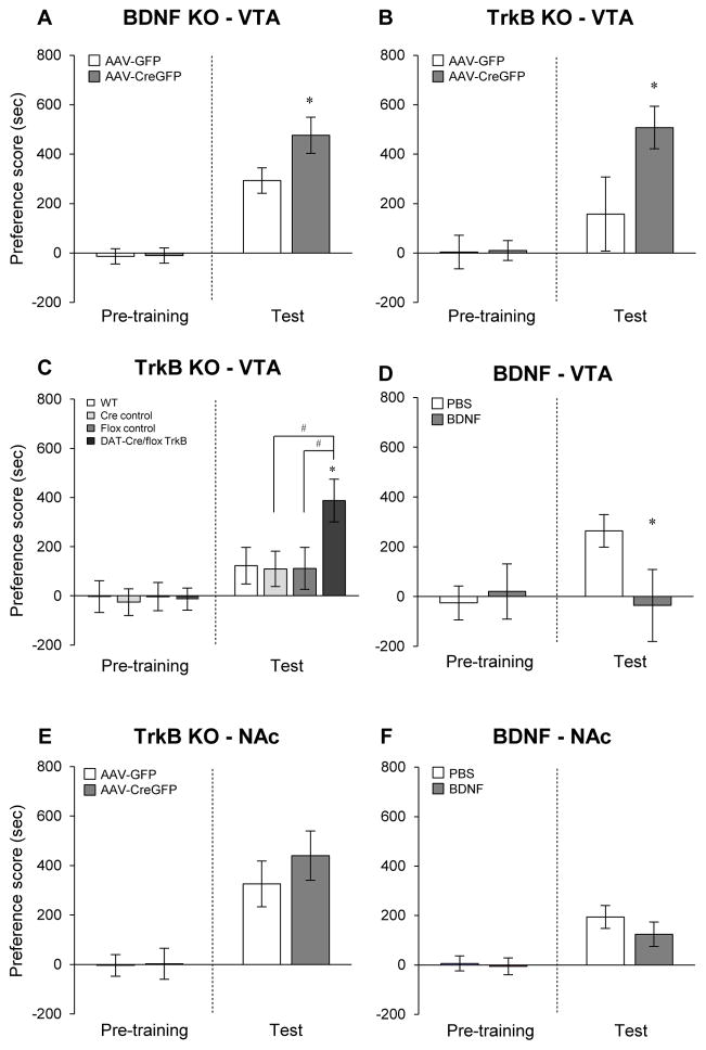
We first demonstrated that chronic morphine, whether given by subcutaneous pellets or intermittent IP injections, decreased BDNF expression in VTA (fig. S1). Next, we examined the role of BDNF-TrkB signaling in the VTA-NAc in morphine action by performing morphine conditioned place preference (CPP), which provides an indirect measure of drug reward. Morphine doses were selected based on an initial dose-response analysis (fig. S2). Infusion of an AAV vector encoding Cre recombinase fused to GFP (AAV-CreGFP) into VTA of floxed BDNF (flBDNF) or floxed TrkB (flTrkB) mice produced highly localized CreGFP expression in this brain region (fig. S3A). This induced a 40–60% reduction of BDNF or TrkB mRNA levels in VTA, compared with control animals injected with AAV-GFP (fig. S3, B and C). Localized knockdown of BDNF enhanced the rewarding effect of morphine at both sub-threshold (5 mg/kg; fig. S3D) and higher (15 mg/kg; Fig. 1A) doses compared with AAV-GFP controls. Equivalent effects were seen for local VTA knockdown of TrkB (Fig. 1B, 15 mg/kg). There were no differences in baseline preference scores prior to conditioning among the flBDNF and flTrkB groups.
Fig. 1. Effects of BDNF-TrkB signaling within the VTA-NAc on morphine reward.
Localized knockout (KO) of BDNF (A) or TrkB (B) from VTA neurons enhances morphine conditioned place conditioning (CPP, 15 mg/kg, sc). Student’s t-test, *p < 0.05, n = 8–12. (C) DAT-cre/flTrkB (TrkBlx/lx;DATcre/wt) mice also displayed enhanced morphine CPP (10 mg/kg, ip). One-way ANOVA, Fisher’s PLSD post-hoc test, *p < 0.05 compared to controls; #p < 0.05 compared with TrkBlx/lx;DATcre/wt mice, n = 6–11. (D) A single infusion of BDNF into VTA (0.25 μg/side) suppressed morphine CPP (15 mg/kg, sc). t-test, *p < 0.05, n = 7–8. (E) Localized TrkB KO in NAc and (F) intra-NAc BDNF infusion (1.0 μg/side) had no effect on morphine CPP (15 mg/kg, sc), n = 8–9.
Because the viral-mediated knockdown procedure affects both DAergic and non-DAergic VTA neurons, we used a complementary approach to knockout TrkB selectively from DA neurons by crossing flTrkB mice with DA transporter (DAT)-Cre mice (TrkBlx/lx; DATcre/wt) (17). Selective ablation of TrkB from DA neurons in TrkBlx/lx; DATcre/wt mice increases morphine reward (Fig. 1C). There were no differences in baseline or morphine CPP among several control groups examined, which included wild-type mice (TrkBw/w; DATwt/wt), floxed control mice (TrkBlx/lx; DATwt/wt), and Cre control mice (TrkBw/w; DATcre/wt), indicating that the increase in morphine reward seen upon selective TrkB ablation in DA neurons does not result from allele-specific effects. In contrast, a single intra-VTA infusion of BDNF (0.25 μg/side) decreases morphine CPP compared with vehicle-infused control animals (Fig. 1D).
BDNF synthesized in VTA DA neurons can undergo anterograde transport and release in NAc to activate TrkB receptors on NAc neurons (18). We therefore tested the effect of localized deletion of TrkB receptors in NAc of flTrkB mice and of intra-NAc infusions of BDNF (1.0 μg/side) on morphine reward. In contrast to VTA, TrkB knockdown (Fig. 1E) and BDNF infusion (Fig. 1F) in NAc had no effect on morphine CPP. It is thus local BDNF signaling in VTA DA neurons that is responsible for regulation of morphine reward.
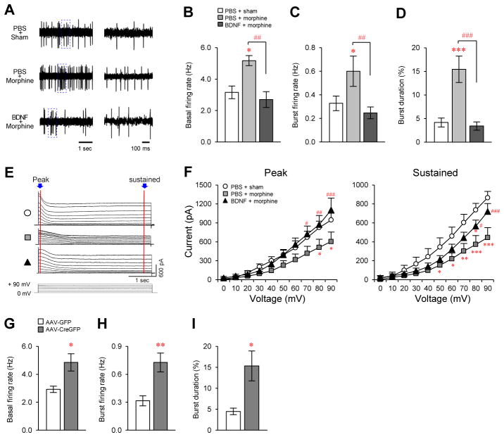
We have recently shown that chronic morphine decreases the expression of certain K+ channels in VTA, such as kcnab2, kcnj2, and girk3, and that such adaptations are associated with increased excitability of DA neurons (14). Viral-mediated BDNF KO in VTA similarly suppressed mRNA levels of these and some additional K+ channels (fig. S4A). These findings raise the possibility that morphine increases VTA DA neuron excitability via downregulation of BDNF and the subsequent reduction in K+ channel expression. To test this hypothesis, we obtained extracellular single unit recordings from VTA DA neurons in three groups of anesthetized mice: controls, chronic morphine, and chronic morphine+intra-VTA BDNF-infused (Fig. 2A). Consistent with previous ex vivo findings from brain slices (14), morphine increased the in vivo spontaneous firing rates of VTA DA neurons by 44%. Intra-VTA infusion of BDNF normalized this morphine-induced firing rate increase (Fig. 2B). Analysis of burst phasic firing, which substantially increases DA release compared to single-spike tonic firings (19, 20), revealed that overall bursting events were increased by chronic morphine and restored by intra-VTA BDNF (Fig. 2, C and D; fig. S4, B and C). Conversely, localized VTA BDNF KO alone (in morphine-naïve mice) increased the spontaneous burst firing of DA neurons (Fig. 2, G to I).
Fig. 2. Regulation of VTA DA neuron excitability by morphine and BDNF.
(A) Sample traces of in vivo recordings from VTA DA neurons from control (top), morphine-treated (middle), and BDNF+morphine-treated mice (bottom). (B to E) Morphine (25 mg pellet, sc; animals analyzed 48 hr later) increases (B) basal firing rate, (C) burst firing rate, and (D) burst duration in VTA DA neurons, which were normalized by intra-VTA infusion of BDNF (0.25 μg/side). One-way ANOVA, Fisher’s PLSD test, *p < 0.05, ***p < 0.001, compared with control; ##p < 0.01, ###p < 0.001, compared with morphine group. n = 4–6. (E) Sample traces of K+ conductance recorded from VTA DA neurons in brain slices from control, morphine-treated, and BDNF+morphine-treated mice. (F) Morphine treatment as in (A) significantly decreased both peak and sustained phases of K+ currents in VTA DA neurons, an effect that was reversed by BDNF. Two-way ANOVA, Fisher’s PLSD test, *p < 0.05, **p < 0.01, ***p < 0.001, compared with control; #p < 0.05, ##p < 0.01, ###p < 0.001, compared with morphine group. n = 5–9. Localized BDNF KO from VTA increases (G) basal firing rate, (H) burst firing rate, and (I) burst duration in VTA DA neurons. t-test, *p < 0.05, **p < 0.01 compared with AAV-GFP controls, n = 7.
Next, we studied possible ionic mechanisms underlying these changes using standard whole-cell voltage-clamp recordings. Different components (peak and sustained) of K+ currents in VTA DA neurons were reduced by morphine treatment, which were blocked by intra-VTA BDNF (Fig. 2, E and F). These data demonstrate that morphine and BDNF differentially regulate K+ currents in these neurons, which is consistent with our molecular findings above. The ability of chronic morphine to excite VTA DA neurons may be mediated via decreased AKT signaling (14), which would be an expected downstream consequence of withdrawal of BDNF support. Prior work suggested as well that downregulation of BDNF signaling in VTA would excite VTA DA neurons further by reducing GABAA receptor responses, also downstream of reduced AKT signaling (21). Our findings demonstrate that BDNF additionally controls the intrinsic excitability of VTA DA neurons via altered K+ channel expression, and thereby opposes morphine-induced DA neuron excitability through a homeostatic scaling mechanism (22, 23).
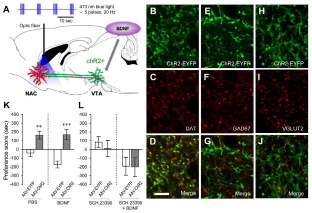
Given these direct links between VTA BDNF expression and VTA DA neuron excitability in morphine action, we next determined whether BDNF-regulated activity of VTA DA neurons is important for BDNF’s influence on behavioral responses to morphine. We stereotaxically delivered AAV-ChR2 (channel rhodopsin)-EYFP or AAV-EYFP into mouse VTA as described (24). Two-three weeks later, when AAV expression is maximal, we infused BDNF into VTA and implanted cannulae in NAc for optic fiber placement (Fig. 3A and fig. S5A). Animals were studied one week later. 86% of ChR2-EYFP positive cells in VTA co-localized with TH, a marker of DA neurons (fig. S5B). ChR2-EYFP immunoreactivity in NAc co-localized with DAT, a marker of DA nerve terminals (Fig. 3, B to D), but not with GAD67 (Fig. 3, E to G) or VGLUT2 (Fig. 3, H to J), markers of GABAergic or glutamatergic terminals, respectively, showing selective expression of ChR2-EYFP in DAergic nerve terminals in NAc. Mice that express AAV-ChR2-EYFP or AAV-EYFP in VTA were studied in the morphine CPP paradigm by conditioning them with sub-threshold morphine doses (10 mg/kg) plus 20 Hz phasic pulses delivered to NAc in one chamber, and with saline and no light in the opposite chamber. In non-BDNF-infused animals, this optogenetic protocol induced significant morphine CPP in mice expressing ChR2-EYFP but not EYFP alone (Fig. 3K). Such stimulation also completely prevented the inhibitory effect of intra-VTA BDNF infusion on morphine CPP (Fig. 3K, compare to Fig. 1D and fig. S6A). In this paradigm, light stimulation of DA terminals in NAc—at 20 Hz or several other frequencies—in the absence of morphine did not induce a CPP regardless of intra-VTA BDNF infusion (fig. S6, C–E). The effect of light stimulation was mediated by D1 DA receptors in NAc, as intra-NAc injection of a D1 receptor antagonist (SCH 23390, 1 μg), at a dose known to be behaviorally active (25, 26), completely blocked the ability of light stimulation to enhance morphine CPP regardless of intra-VTA BDNF infusion (Fig. 3L). In contrast, intra-NAc injection of behaviorally active doses (27–29) of a D2 (eticlopride, 1 or 4 μg) or glutamate (DNQX, 1 or 4 μg; or NBQX, 400 ng) receptor antagonist had no effect (fig. S7). These data demonstrate that BDNF impairment of morphine reward can be rescued by phasic stimulation of the VTA-NAc DA pathway, specifically through D1 receptors in NAc. This is consistent with evidence showing that DA released by burst-like phasic firing selectively binds to D1 receptors (19, 30). In contrast to the NAc, optical stimulation of DA terminals in medial prefrontal cortex had no effect on morphine CPP (fig. S8).
Fig. 3. Modulation of morphine reward by optogenetic activation of DA terminals in NAc.
(A) Experimental paradigm for optogenetic stimulation during morphine CPP. Mice were conditioned to a morphine/light chamber and a saline/no-light chamber for 30 min. 470 nm phasic light pulses (20 Hz, 5 pulses, 40 ms duration) were delivered during the 30-min conditioning session. (B to J) Immunostaining for ChR2-EYFP (B, E, and H, green), DAT (C, red), GAD67 (F, red), and VGLUT2 (I, red) in NAc. (D, G, and J) Confocal microscopy shows that ChR2-EYFP puncta in NAc co-label for DAT, but not GAD67 or VGLUT2. Scale bar, 10 μm. (K) In vivo optogenetic stimulation of VTA DA nerve terminals in NAc enhances morphine reward (10 mg/kg, ip) and prevents VTA BDNF-induced impairment of morphine reward, (L) while D1 receptor antagonism (SCH 23390, 1 μg) blocks light potentiation, and VTA BDNF-induced impairment, of morphine reward. t-test, **p < 0.01, **p < 0.001, n = 7–11.
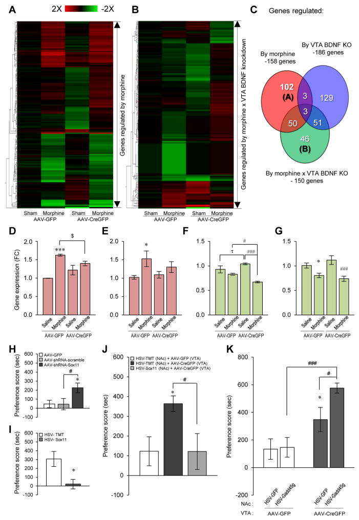
We investigated the downstream consequences of VTA BDNF and chronic morphine on gene expression in the NAc. We performed microarray analysis on NAc from mice in which VTA BDNF was virally knocked down, with half of the animals then treated chronically with morphine. We identified clusters of NAc genes that are regulated by morphine or by knockdown of VTA BDNF and analyzed interactive effects between the two to investigate the molecular mechanisms underlying BDNF regulation of morphine responses. Three main categories of genes were identified (Table S1): Category A, genes regulated by morphine where that regulation is lost upon knockdown of VTA BDNF (Fig. 4A); Category B, genes whose regulation by morphine was uncovered upon knockdown of VTA BDNF (Fig. 4A); and Category C, genes regulated by morphine regardless of VTA BDNF knockdown (Fig. 4B) (see Table S2 for complete gene lists). Among the genes significantly regulated in NAc under these conditions are several that have previously been implicated in morphine action: e.g., zfp40, xdh, nt5e, sult1a1, gadd45g, rbm3, and zbtb16 (31–34). We also found a significantly greater transcriptional effect of chronic morphine in NAc of mice lacking BDNF in VTA: ~3-fold more genes (151 vs. 46) were regulated by morphine in VTA BDNF knockdown mice vs. control mice (fig. S9, A to C). Similarly, ~5-fold more genes (185 vs. 39) in NAc were regulated upon knockdown of BDNF in VTA in morphine-treated mice compared with sham mice (fig. S9, D to F) (see Table S3 for complete gene lists). These findings extend our behavioral and electrophysiological evidence that BDNF in VTA antagonizes chronic morphine actions on the VTA-NAc circuit.
Fig. 4. Morphine-regulated NAc gene expression after VTA BDNF deletion: Identification of novel NAc mediators of BDNF-morphine interactions.
Microarray analysis was performed on NAc of sham- and morphine-pelleted mice under control or VTA BDNF knockdown conditions. (A) Heatmap of up (red)- or down (green)-regulated NAc genes upon knockdown of VTA BDNF. (B) Heatmap of up- or down-regulated NAc genes by morphine regardless of knockdown of VTA BDNF. (C) Venn diagrams of genes that were uniquely regulated by morphine (red) or by knockdown of VTA BDNF (blue), and of genes that were regulated by morphine and knockdown of VTA BDNF in an interactive manner (green). (D to G) Alterations of sox11 (D and E) and gadd45g (F and G) expression in NAc from heatmap of microarray analysis (D and F) and qRT-PCR validation (E and G). One-way ANOVA for qRT-PCR validation, Fisher’s PLSD test, τp < 0.1, *p < 0.05, ***p < 0.001 compared to sham+AAV-GFP controls; $p < 0.1, #p < 0.05, ###p < 0.001 compared to sham+AAV-CreGFP mice, n = 9–12. (H) Reduction of sox11 expression using AAV-shRNA-Sox11 increases morphine CPP (10 mg/kg, sc). Fisher’s PLSD test, *p < 0.05 compared with AAV-GFP controls; #p < 0.05 compared with AAV-scrambled controls, n = 11–12. (I) Sox11 overexpression in NAc using HSV-Sox11 decreases morphine CPP (15 mg/kg, sc). t-test, *p < 0.05, n = 8. (J) Enhancement of morphine reward (15 mg/kg, sc) induced by knockdown of VTA TrkB is counteracted by sox11 overexpression in NAc. Fisher’s PLSD test, *p < 0.05 compared with HSV-tomato (TMT) (NAc)+AAV-GFP (VTA) controls; #p < 0.05 compared with HSV-TMT (NAc)+AAV-CreGFP (VTA) mice. (K) Enhancement of morphine reward (12.5 mg/kg, sc) induced by knockdown of VTA TrkB is further enhanced by gadd45g overexpression in NAc. Fisher’s PLSD test, *p < 0.05 compared to HSV-GFP+AAV-GFP controls; #p < 0.05, ###p < 0.001 compared to HSV-Gadd45g+AAV-CreGFP.
We selected two genes, sox11 and gadd45g, from Categories A and B, respectively, for further study and validated their expression patterns using RT-PCR analysis (Fig. 4, D to G). SOX11 is a transcription factor involved in embryonic neurogenesis and tissue remodeling (35). The function of the gene in the adult nervous system remains unknown, although its regulation was observed in a previous microarray study on NAc (36). We found that sox11 gene expression levels in NAc were induced by chronic morphine and that this increase was prevented by deletion of BDNF in VTA (Fig. 4, D and E).
To test directly whether alterations in sox11 expression in NAc influence morphine reward, we generated an AAV vector that expresses a short hairpin RNA (shRNA) against sox11 to knock it down in NAc selectively and a Herpes Simplex Virus (HSV) vector to overexpress sox11 in this region (fig. S10A). After first validating the AAV-shRNA-Sox11 vector in cultured Neuro2A cells (fig. S10, B and C), we demonstrated that intra-NAc infusion of this vector reduced sox11 expression in NAc (fig. S10D). In contrast, intra-NAc infusion of HSV-Sox11 robustly augmented sox11 mRNA levels in this region (fig. S10E). We observed that AAV-shRNA-Sox11 in NAc increased morphine reward: a sub-threshold dose of morphine (10 mg/kg) induced significant CPP, an effect not seen in animals treated with control AAV-GFP or AAV-scrambled shRNA vectors (Fig. 4H). Conversely, HSV-Sox11 in NAc decreased CPP to a higher dose of morphine (15 mg/kg) as compared to HSV-Tomato infused control animals (Fig. 4I). Furthermore, the ability of locally knocking down BDNF-TrkB signaling in VTA to enhance morphine’s rewarding effects was completely normalized upon HSV-mediated sox11 overexpression in NAc (Fig. 4J). No changes were observed in baseline levels of place preference in these experiments.
Another gene implicated in these interactions by our microarray data is gadd45g, a stress-responsive immediate early gene, which is involved in DNA repair, cell growth arrest, and apoptosis (37). Here, gadd45g gene expression levels were more robustly and consistently suppressed in NAc by chronic morphine in mice with deletion of VTA BDNF (Fig. 4, F and G). We then tested whether such regulation of gadd45g expression in NAc influences the rewarding effects of morphine using HSV-Gadd45g that we developed and validated for gadd45g overexpression (fig. S11). HSV-Gadd45g infusion into NAc of normal mice did not affect the rewarding actions of a moderate dose of morphine (12.5 mg/kg). However, gadd45g overexpression in NAc of mice with local VTA knockdown of TrkB enhanced morphine CPP compared to HSV-GFP treated mice (Fig. 4K). No changes were observed in baseline levels of place preference.
We observed the opposite effect of BDNF-TrkB signaling in the VTA-NAc on morphine reward compared to earlier observations with cocaine and other stimulants. Prior work has shown that knockdown of BDNF from VTA, or knockdown of BDNF or TrkB from NAc, antagonizes behavioral responses to cocaine, while knockdown of TrkB in VTA has no effect (3). These findings identified the NAc as the primary site of action of BDNF-TrkB signaling in regulating cocaine reward. In contrast, we show here that the primary site of action of BDNF-TrkB signaling in regulating morphine reward is the VTA, because knockdown of either BDNF or TrkB in VTA promotes behavioral responses to morphine, while knockdown of TrkB in NAc and BDNF administration into NAc was without effect. On the other hand, our findings that optical stimulation of VTA DA nerve terminals in NAc completely reverses the ability of BDNF, acting in VTA, to impair morphine reward demonstrate that BDNF’s influence on VTA DA neuron excitability is responsible for its behavioral effects reported here (fig. S12). These optogenetic experiments also identify the NAc as the key neural site where VTA BDNF’s influence on morphine reward is ultimately mediated, and we show that this occurs via DA activation of D1 receptors on NAc neurons. For this reason, we analyzed morphine-induced changes in gene expression in NAc as a function of VTA BDNF, and demonstrate the importance of two genes, sox11 and gadd45g, among numerous other putative genes regulated in a similar fashion, in behavioral responses to morphine (fig. S12). The results of the present study differ from an earlier report (10), which found that exogenous BDNF increases opiate reward in rats by altering GABAA receptor function in VTA GABAergic neurons (fig. S12). The different findings could be due to the different species, drug treatment regimens, or behavioral paradigms used.
Consistent with the differences in BDNF’s influence on cocaine vs. morphine action are the very different ways in which the drugs initially affect the VTA-NAc pathway. In addition, while opiate and stimulant drugs of abuse induce many common molecular and cellular adaptations in both the VTA and NAc (38), some notable differences are seen as well with respect to synaptic plasticity (39, 40) and to VTA BDNF expression (fig. S1B). Further studies are now needed to directly investigate whether the opposite interactions between BDNF and stimulants vs. opiates are related to these adaptations. Given substance-specific features of drug addiction syndromes, it will be important to further explore the downstream functional consequences of the BDNF-stimulant feed-forward loop vs. the BDNF-opiate negative feedback loop in addiction, and study how they influence polydrug use.
Supplementary Material
Acknowledgments
We thank Rebecca E Burger-Caplan and Nii Mensah for excellent technical assistance. This work was supported by grants from the National Institute on Drug Abuse (EJN, MMR) and a Rubicon Grant from the Dutch Scientific Organization (CSL).
Footnotes
Materials and Methods
References (41–53)
References and Notes
- 1.Thoenen H. Science. 1995;270:593. doi: 10.1126/science.270.5236.593. [DOI] [PubMed] [Google Scholar]
- 2.Pierce RC, Bari AA. Rev Neurosci. 2001;12:95. doi: 10.1515/revneuro.2001.12.2.95. [DOI] [PubMed] [Google Scholar]
- 3.Graham DL, et al. Biol Psychiatry. 2009;65:696. doi: 10.1016/j.biopsych.2008.09.032. [DOI] [PMC free article] [PubMed] [Google Scholar]
- 4.Hall FS, Drgonova J, Goeb M, Uhl GR. Neuropsychopharmacology. 2003;28:1485. doi: 10.1038/sj.npp.1300192. [DOI] [PubMed] [Google Scholar]
- 5.Horger BA, et al. J Neurosci. 1999;19:4110. doi: 10.1523/JNEUROSCI.19-10-04110.1999. [DOI] [PMC free article] [PubMed] [Google Scholar]
- 6.Lu L, Dempsey J, Liu SY, Bossert JM, Shaham Y. J Neurosci. 2004;24:1604. doi: 10.1523/JNEUROSCI.5124-03.2004. [DOI] [PMC free article] [PubMed] [Google Scholar]
- 7.Lobo MK, et al. Science. 2010;330:385. doi: 10.1126/science.1188472. [DOI] [PMC free article] [PubMed] [Google Scholar]
- 8.Graham DL, et al. Nat Neurosci. 2007;10:1029. doi: 10.1038/nn1929. [DOI] [PubMed] [Google Scholar]
- 9.Hyman SE, Malenka RC, Nestler EJ. Annu Rev Neurosci. 2006;29:565. doi: 10.1146/annurev.neuro.29.051605.113009. [DOI] [PubMed] [Google Scholar]
- 10.Vargas-Perez H, et al. Science. 2009;324:1732. doi: 10.1126/science.1168501. [DOI] [PMC free article] [PubMed] [Google Scholar]
- 11.Chu NN, et al. Addict Biol. 2008;13:47. doi: 10.1111/j.1369-1600.2007.00092.x. [DOI] [PubMed] [Google Scholar]
- 12.Numan S, et al. J Neurosci. 1998;18:10700. doi: 10.1523/JNEUROSCI.18-24-10700.1998. [DOI] [PMC free article] [PubMed] [Google Scholar]
- 13.Berhow MT, et al. Neuroscience. 1995;68:969. doi: 10.1016/0306-4522(95)00207-y. [DOI] [PubMed] [Google Scholar]
- 14.Mazei-Robison MS, et al. Neuron. 2011;72:977. doi: 10.1016/j.neuron.2011.10.012. [DOI] [PMC free article] [PubMed] [Google Scholar]
- 15.Russo SJ, et al. Nat Neurosci. 2007;10:93. doi: 10.1038/nn1812. [DOI] [PubMed] [Google Scholar]
- 16.Sklair-Tavron L, et al. Proc Natl Acad Sci U S A. 1996;93:11202. doi: 10.1073/pnas.93.20.11202. [DOI] [PMC free article] [PubMed] [Google Scholar]
- 17.Kramer ER, et al. PLoS Biol. 2007;5:e39. doi: 10.1371/journal.pbio.0050039. [DOI] [PMC free article] [PubMed] [Google Scholar]
- 18.Altar CA, et al. Nature. 1997;389:856. doi: 10.1038/39885. [DOI] [PubMed] [Google Scholar]
- 19.Grace AA, Floresco SB, Goto Y, Lodge DJ. Trends Neurosci. 2007;30:220. doi: 10.1016/j.tins.2007.03.003. [DOI] [PubMed] [Google Scholar]
- 20.Floresco SB, West AR, Ash B, Moore H, Grace AA. Nat Neurosci. 2003;6:968. doi: 10.1038/nn1103. [DOI] [PubMed] [Google Scholar]
- 21.Krishnan V, et al. Biol Psychiatry. 2008;64:691. doi: 10.1016/j.biopsych.2008.06.003. [DOI] [PMC free article] [PubMed] [Google Scholar]
- 22.Rutherford LC, Nelson SB, Turrigiano GG. Neuron. 1998;21:521. doi: 10.1016/s0896-6273(00)80563-2. [DOI] [PubMed] [Google Scholar]
- 23.Desai NS, Rutherford LC, Turrigiano GG. Learn Mem. 1999;6:284. [PMC free article] [PubMed] [Google Scholar]
- 24.Tsai HC, et al. Science. 2009;324:1080. doi: 10.1126/science.1168878. [DOI] [PMC free article] [PubMed] [Google Scholar]
- 25.Abrahao KP, Quadros IM, Souza-Formigoni ML. Int J Neuropsychopharmacol. 2011;14:175. doi: 10.1017/S1461145710000441. [DOI] [PubMed] [Google Scholar]
- 26.Stuber GD, et al. Nature. 2011;475:377. doi: 10.1038/nature10194. [DOI] [PMC free article] [PubMed] [Google Scholar]
- 27.Kaddis FG, Uretsky NJ, Wallace LJ. Brain Res. 1995;697:76. doi: 10.1016/0006-8993(95)00786-p. [DOI] [PubMed] [Google Scholar]
- 28.Vialou V, et al. Nat Neurosci. 2010;13:745. doi: 10.1038/nn.2551. [DOI] [PMC free article] [PubMed] [Google Scholar]
- 29.Boye SM, Grant RJ, Clarke PB. Neuropharmacology. 2001;40:792. doi: 10.1016/s0028-3908(01)00003-x. [DOI] [PubMed] [Google Scholar]
- 30.Goto Y, Grace AA. Nat Neurosci. 2005;8:805. doi: 10.1038/nn1471. [DOI] [PubMed] [Google Scholar]
- 31.Korostynski M, Piechota M, Kaminska D, Solecki W, Przewlocki R. Genome Biol. 2007;8:R128. doi: 10.1186/gb-2007-8-6-r128. [DOI] [PMC free article] [PubMed] [Google Scholar]
- 32.Piechota M, et al. Genome Biol. 2010;11:R48. doi: 10.1186/gb-2010-11-5-r48. [DOI] [PMC free article] [PubMed] [Google Scholar]
- 33.Sanchis-Segura C, Lopez-Atalaya JP, Barco A. Neuropsychopharmacology. 2009;34:2642. doi: 10.1038/npp.2009.125. [DOI] [PubMed] [Google Scholar]
- 34.Spijker S, et al. FASEB J. 2004;18:848. doi: 10.1096/fj.03-0612fje. [DOI] [PubMed] [Google Scholar]
- 35.Jankowski MP, Cornuet PK, McIlwrath S, Koerber HR, Albers KM. Neuroscience. 2006;143:501. doi: 10.1016/j.neuroscience.2006.09.010. [DOI] [PMC free article] [PubMed] [Google Scholar]
- 36.McClung CA, Nestler EJ. Nat Neurosci. 2003;6:1208. doi: 10.1038/nn1143. [DOI] [PubMed] [Google Scholar]
- 37.Ying J, et al. Clin Cancer Res. 2005;11:6442. doi: 10.1158/1078-0432.CCR-05-0267. [DOI] [PubMed] [Google Scholar]
- 38.Nestler EJ. Nat Neurosci. 2005;8:1445. doi: 10.1038/nn1578. [DOI] [PubMed] [Google Scholar]
- 39.Badiani A, Belin D, Epstein D, Calu D, Shaham Y. Nat Rev Neurosci. 2011;12:685. doi: 10.1038/nrn3104. [DOI] [PMC free article] [PubMed] [Google Scholar]
- 40.Russo SJ, et al. Trends Neurosci. 2010;33:267. doi: 10.1016/j.tins.2010.02.002. [DOI] [PMC free article] [PubMed] [Google Scholar]
Associated Data
This section collects any data citations, data availability statements, or supplementary materials included in this article.


