Abstract
Many systemic diseases affect oral health. The aim of this research was to conduct a systematic review on the association between dental erosion (DE) and gastroesophageal reflux disease (GERD) and the effect of saliva's flow rate, buffering capacity and oral microbial changes caused by GERD. All descriptive, analytical studies up to December 2011 that have relevant objectives, proper sampling method and sufficient results were included by searching PubMed and Scopus electronic data bases. Fifteen studies were selected according to our inclusion criteria (10 in adult and 5 in children population). There was a strong association between DE and GERD in the adult population, and the relationship in the children population was found to be of less importance. Early diagnosis and treatment of refluxed acid in both age groups through lifestyle changes and medications can prevent further damage and tooth loss.
Keywords: Dental erosion, gastroesophageal reflux disease, saliva, systematic review
INTRODUCTION
Many systemic diseases such as AIDS, graft-versus-host disease, leukemia, and diabetes mellitus may primarily manifest themselves in the oral cavity. Yet, many systemic diseases affect oral health in indistinct ways and, thus, the oral cavity remains an often underestimated window on manifestations of these diseases.[1] One example is gastroesophageal reflux disease (GERD). Classic reflux symptoms are not always present in patients with GERD. A significant number of patients with GERD present atypical or extraesophagael symptoms[2] such as dental erosion (DE).[1] The relationship between the GERD and DE has been first reported first by Howden et al.[3] However, only in the last decade this subject has been more intensively studied. Myllarniemi and Saario[4] stated that, tooth erosions may even be a diagnostic sign for previous acid reflux. DE is an important cause of tooth damage both in children and in adults.[5] It can be defined as a chemical dissolution of tooth substances by non-cariogenic acids with multifactorial etiology.[6,7] The outcome of premature loss of tooth tissues may result in dentin hypersensitivity, pulpal irritation, and the need for long and expensive restorative treatment.[8] The extent of teeth damage may range from a barely noticeable enamel lesion to the partial or complete exposure of dentine with its characteristic yellow color through the thinned overlying enamel.[9,10] DE is caused by the presence of intrinsic and extrinsic acid of non-bacterial origin in the mouth, combined or separately.[11] Intrinsic causes of acid are known to be regurgitation, vomiting, GERD or rumination. Extrinsic sources of acid are mostly dietary acids, and a number of Medications.[12,13] As stated in the recently published Montreal Criteria, dealing with a global classification of GERD: ‘The prevalence of DEs, especially on the lingual and palatal tooth surfaces, is increased in patients with GERD.[14] Examination of the oral cavity, in search for ‘atypical’ DEs, should be an integral part of the physical examination of the patient with suspected GERD.[15] On the other hand, other authors have denied, at least in children, that DEs may represent a relevant problem in GERD patients.[16]
The poor buffering capacity of saliva in patients with GERD in conjunction with the higher prevalence of tooth wear in this group of people might suggest that saliva may play an important role in tooth wear.[17]
The aim of this systemic review was to analyze the association between DE and GERD and the effect of flow rate, buffering capacity of saliva, and oral microbial changes caused by GERD on the severity of the lesions. In addition, this review provides background knowledge for oral health track of the “Study on the Epidemiology of Psychological, Alimentary Health and Nutrition” (SEPAHAN).[18] The data of this study will determine the impact of masticatory dysfunction and oral health on GERD and functional gastrointestinal disorders and will be published later by the same study group.
MATERIALS AND METHODS
Search strategy
For electronic searching we used “GERD”, “DE”, “oral health” as keywords for title and abstracts in MeSH word search. References of each article were also reviewed.
Electronic databases
This review study has been searched up to 31 December 2011 in PubMed, Scopus, and Cochrane library for systematic reviews by searching mentioned keywords in various combinations. Three hundred and three studies were found by searching in PubMed. PubMed query translation was “tooth erosion” (MeSH Terms) and (“gastroesophageal reflux” [MeSH Terms] or GERD [Text Word]). The search through Scopus listed out 39 articles, which all overlapped with the results found in PubMed.
Inclusion criteria
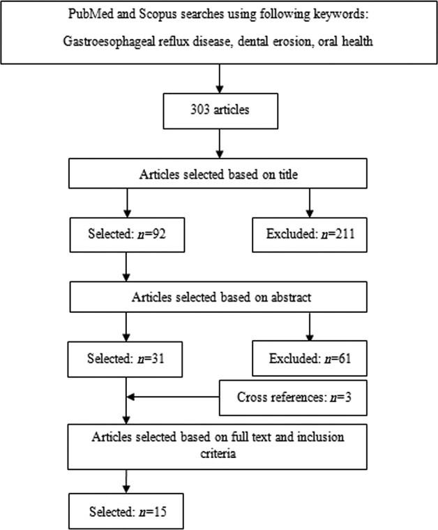
Inclusion criteria were limited to the journal articles in human, clinical trials, cohort, review, and case-control studies in English language. Ninety two studies were selected first based on title, of these, 31 articles were chosen by abstract outlined in Figure 1. Finally, 12 articles were selected based on full text; and the additional search through cross-references resulted in three more citations. This left a total of 15 studies [Figure 1].[5,15–17,19–29]
Figure 1.

Diagram of the systematic review and searches
Data extraction
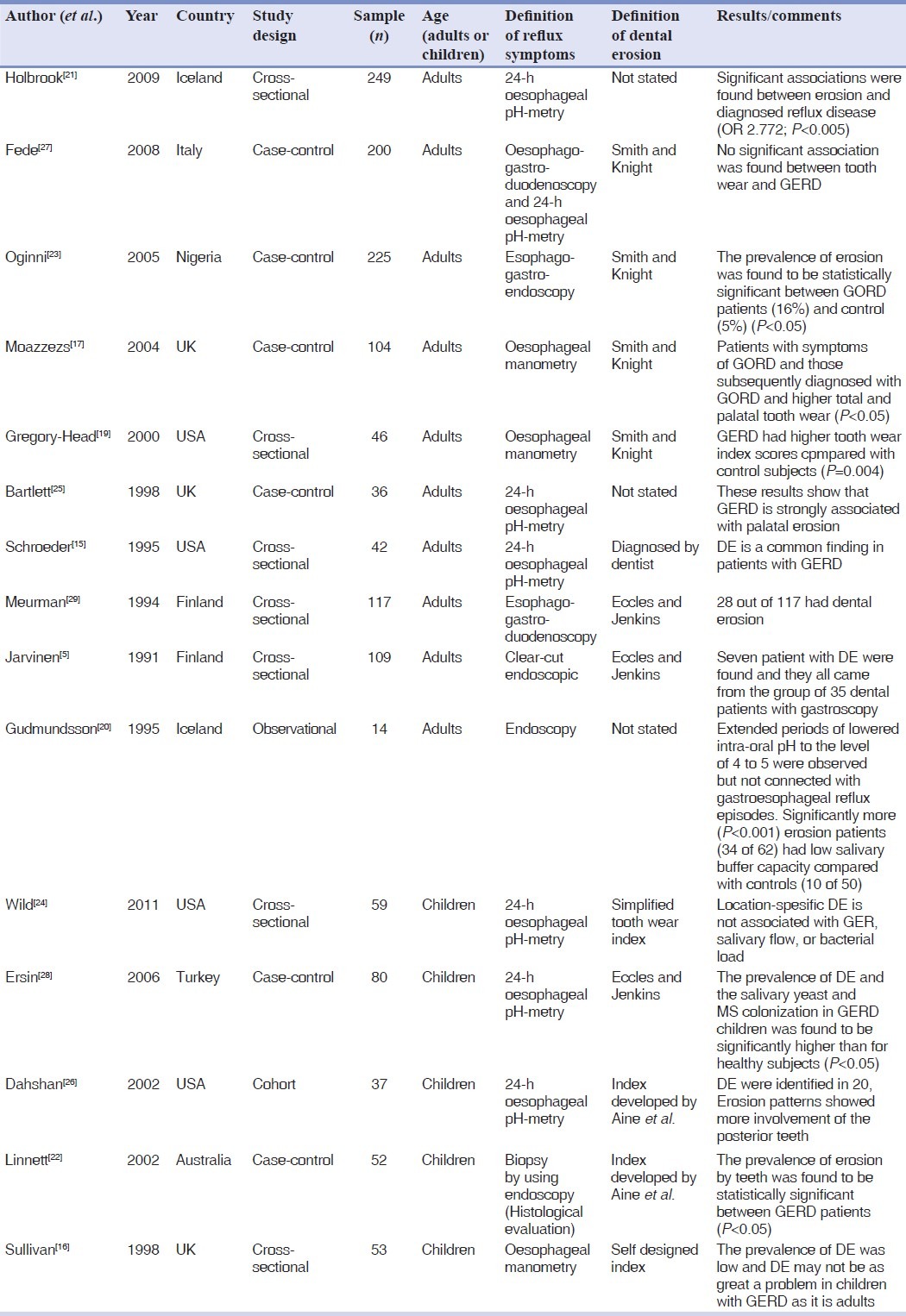
For data extraction, we designed a check lists, regarding DE and GERD including: Author, year, country, sample size, study design, method of data collection, and definition of extra-oesophageal symptom and main results of each study [Table 1].
Table 1.
Summery of studied original research articles

RESULTS
The PubMed, Scopus searches identified 303 articles, of these 211 articles were excluded based on title. A further 61 more were excluded based on abstract and finally 12 articles were selected based on full texts and inclusion criteria, an additional 3 more articles were added by search through cross-references. This left a total of 15 eligible studies.[5,15–17,19–29]
Studies carried out on adults
Overall 10 original studies were found on adults,[5,15,17,19–21,23,25,27,29] of these 3 studies reported significant association between DE and GERD.[19,21,23] The study conducted by Schroeder et al.[15] had both a dental group, which was screened for GERD, and a gastroenterological group, referred for dental evaluation with a prevalence of 83% DE. Meurman et al.[29] investigated the occurrence of DE in patients with long-term reflux disease. Fede et al.[27] could not find any relationship between DE and GERD.
Gregory-Head et al.[19] studied 20 patients, 10 subjects were diagnosed with GERD and 10 subjects had manometry scores below the level indicating GERD. Overall, subjects diagnosed with GERD had significantly higher tooth wear index (TWI) scores compared with control subjects (mean difference=0.6554; P=0.004).
Jarvinen et al.[5] studied the oro-dental status, particularly DE in 109 patients with upper gastrointestinal symptoms. Seven patients with DE were found and they all came from the group of 35 patients with reflux esophagitis or duodenal ulcer. There was no direct association between the frequency of regurgitation symptoms and the severity of erosive lesions.
Moazzez et al.[17] investigated tooth wear, stimulated salivary flow rate (SFR) and buffering capacity and symptoms of GERD in 100 patients. Patients with symptoms of GERD and those subsequently diagnosed with GERD had higher total and palatal tooth wear (P<0.05).
Oginni et al.[23] studied a total of 225 subjects (100 volunteers and 125 patients diagnosed with GERD), of these 20 patients with GERD presented with DE in the maxillary anterior teeth with TWI scores ranging from 1–3. The prevalence of erosion was found to be statistically significant between GERD patients (16%) and controls (5%) (P<0.05).
Bartlett et al.[25] concluded that, GERD is strongly associated with palatal erosion and the patients with palatal DE should be assessed for GERD as a possible cause, even in the absence of clinical symptoms of reflux.
Holbrook et al. performed a study involving 249 Icelandic individuals and significant associations were found between tooth erosion and acid reflux.[21]
Gudmundsson et al.[20] found no changes in oral pH in a total of 339 acid reflux episodes, not even in long supine reflux episodes. Extended periods of lowered intra-oral pH to the level of 4 to 5 were observed, but not connected with gastroesophageal reflux episodes.
Studies carried out on children
A total of 5 studies were found regarding children[16,22,24,26,28] with a prevalence of DE ranging from 14% to 83% in GERD patients. While 2 studies indicated that DE may not be as great problem in children with GERD as it is in adults.[16] Three studies showed statistically significant higher DE in GERD patients.[22,26,28]
Ersin et al.[28] investigated stimulated SFR and buffer capacity, and salivary Streptococcus mutans, lactobacilli, and yeast colonization. The prevalence of DE and the salivary yeast and Streptococcus mutans colonization in GERD children was found to be significantly higher than healthy individuals (P<0.05).
Influence of saliva
Jarvinen et al.[5] reported that the mean salivary secretion rate and pH were normal in patient and control groups, while salivary buffer capacity was lower in patients. There were no particular differences in the salivary microbial counts among the both groups.
Meurman et al.[29] reported that salivary analyses showed no statistically significant difference observed in the mean values between the 2 patient groups. The number of patients with low buffering capacity, however, was relatively high in GERD patients. Moazzez et al.[17] showed greater buffering capacity of the stimulated saliva in control subjects than patients with symptoms of GERD (P<0.001). There were no statistical significant differences for the stimulated SFR.
DISCUSSION
The lowest prevalence of DE in GERD in adults was reported in studies conducted by Jarvinen et al.[5] and Fede et al.[27] with a percentage of 5% and 9%, respectively. Other studies showed a prevalence of 63%.[15,19,20,23,25]
The severity of DE may depend on the frequency of regurgitation and duration of the gastro-esophageal reflux.[23] This is in contrast with the findings of Jarvinen et al.[5]
Of the 5 studies regarding children population, Wild et al.[16] and O’sullivan et al.[24] showed no difference in the prevalence of DE in children with and without GERD. In contrary, a high prevalence of 76% and 87% of DE was reported by others.[28,30] The difference in results among the studies may be due to differences in age and sample sizes. The differences in subject ages, and the relative lengths of time of exposure of the gastric acid may also account for the differences.[22] It is also possible that, some of the very early lesions of erosion are difficult to diagnose.[22]
Saliva is considered one of the major protective mechanisms against gastric reflux and its qualitative and quantitative abnormalities has been linked to GERD pathogenesis.[31,32] In 5 of the studies reviewed low buffering capacity with normal flow rate in GERD patients was reported.[5,17,20,21,29] None of the studies reviewed, supported the study conducted by Wöltgens et al.[33] stating that DE may be associated with a reduced saliva flow.[5,17,20,21,29]
Out of the 15 articles reviewed, only 2 had examined microbial changes in their studies.[22,28] In the study conducted by Ersin et al.[28] the prevalence of salivary yeast and Streptococci mutans colonization in GERD children was found to be significantly higher than healthy subjects (P<0.05). According to the reports of the study carried out by Linnet et al.[22] there were more subjects with Streptococci mutans in GERD patients, although the difference was not statistically significant.
Various methods have been used in the investigations of GERD, 9 studies,[15–17,19–21,24,25,28] used 24 h esophageal pH monitoring which is considered to be the gold standard investigation of GERD,[23] 4 used endoscopy,[5,23,26,29] and one study[27] used both methods for the determining the reflux symptoms.
Our study limitations were as followed:
We looked for relevant articles only in PubMed and Scopus which may cause some restriction in the articles found.
We narrowed our keywords in order to focus on the prevalence of DE in GERD, and overlooked the rest of the variables in association with GERD.
According to the present review, GERD and DE in adults are strongly associated. However, this association is not very strong in young population. Early diagnosis and treatment of refluxed acid, through lifestyle changes and medications can prevent further damage and tooth loss. The primary care physician and the gastroenterologist should pay more attention to the oral examinations and diagnosis of DE.
Footnotes
Source of Support: Nil.
Conflict of Interest: None declared.
REFERENCES
- 1.Scheutzel P. Etiology of dental erosion-intrinsic factors. Eur J Oral Sci. 1996;104:178–90. doi: 10.1111/j.1600-0722.1996.tb00066.x. [DOI] [PubMed] [Google Scholar]
- 2.Pace F, Pallotta S, Tonini M, Vakil N, Bianchi Porro G. Systematic review: Gastro-oesophageal reflux disease and dental lesions. Aliment Pharmacol Ther. 2008;27:1179–86. doi: 10.1111/j.1365-2036.2008.03694.x. [DOI] [PubMed] [Google Scholar]
- 3.Howden GF. Erosion as the presenting symptom in hiatus hernia. A case report. Br Dent J. 1971;131:455–6. doi: 10.1038/sj.bdj.4802772. [DOI] [PubMed] [Google Scholar]
- 4.Myllarniemi H, Saario I. A new type of sliding hiatus hernia. Ann Surg. 1985;202:159–61. doi: 10.1097/00000658-198508000-00004. [DOI] [PMC free article] [PubMed] [Google Scholar]
- 5.Jarvinen VK, Rytomaa II, Heinonen OP. Risk factors in dental erosion. J Dent Res. 1991;70:942–7. doi: 10.1177/00220345910700060601. [DOI] [PubMed] [Google Scholar]
- 6.Lennard-Jones J. Provision of gastrointestinal endoscopy and related services for a district general hospital. Working Party of the Clinical Services Committee of the British Society of Gastroenterology. Gut. 1991;32:95–105. doi: 10.1136/gut.32.1.95. [DOI] [PMC free article] [PubMed] [Google Scholar]
- 7.Blum AL. Therapeutic approach to ulcer healing. Am J Med. 1985;79:8–14. doi: 10.1016/0002-9343(85)90565-0. [DOI] [PubMed] [Google Scholar]
- 8.Filipi K, Halackova Z, Filipi V. Oral health status, salivary factors and microbial analysis in patients with active gastro-oesophageal reflux disease. Int Dent J. 2011;61:231–7. doi: 10.1111/j.1875-595X.2011.00063.x. [DOI] [PMC free article] [PubMed] [Google Scholar]
- 9.Pindborg J. Chemical and physical injuries. Philadelphia: WB Saunders; 1970. [Google Scholar]
- 10.Shafer W, Hine M, Levy B. A text book of oral pathology. 3rd ed. Philadelphia: WB Saunders; 1974. [Google Scholar]
- 11.Lazarchik DA, Filler SJ. Dental erosion: Predominant oral lesion in gastroesophageal reflux disease. Am J Gastroenterol. 2000;95(8 Suppl):S33–8. doi: 10.1016/s0002-9270(00)01076-5. [DOI] [PubMed] [Google Scholar]
- 12.Lussi A, Jaeggi T. Dental erosion in children. Monogr Oral Sci. 2006;20:140–51. doi: 10.1159/000093360. [DOI] [PubMed] [Google Scholar]
- 13.Mahoney EK, Kilpatrick NM. Dental erosion: Part 1. Aetiology and prevalence of dental erosion. N Z Dent J. 2003;99:33–41. [PubMed] [Google Scholar]
- 14.Vakil N, van Zanten SV, Kahrilas P, Dent J, Jones R. The Montreal definition and classification of gastroesophageal reflux disease: A global evidence-based consensus. Am J Gastroenterol. 2006;101:1900–20. doi: 10.1111/j.1572-0241.2006.00630.x. quiz 43. [DOI] [PubMed] [Google Scholar]
- 15.Schroeder PL, Filler SJ, Ramirez B, Lazarchik DA, Vaezi MF, Richter JE. Dental erosion and acid reflux disease. Ann Intern Med. 1995;122:809–15. doi: 10.7326/0003-4819-122-11-199506010-00001. [DOI] [PubMed] [Google Scholar]
- 16.O’Sullivan EA, Curzon ME, Roberts GJ, Milla PJ, Stringer MD. Gastroesophageal reflux in children and its relationship to erosion of primary and permanent teeth. Eur J Oral Sci. 1998;106:765–9. doi: 10.1046/j.0909-8836.1998.eos106302.x. [DOI] [PubMed] [Google Scholar]
- 17.Moazzez R, Bartlett D, Anggiansah A. Dental erosion, gastro-oesophageal reflux disease and saliva: How are they related? J Dent. 2004;32:489–94. doi: 10.1016/j.jdent.2004.03.004. [DOI] [PubMed] [Google Scholar]
- 18.Adibi P, Keshteli AH, Esmaillzadeh A, Afshar H, Roohafza H, Bagherian-Sararoudi H, et al. The Study on the Epidemiology of Psychological, Alimentary Health and Nutrition (SEPAHAN): Overview of methodology. J Res Med Sci. 2012;17(5) [In Press] [Google Scholar]
- 19.Gregory-Head BL, Curtis DA, Kim L, Cello J. Evaluation of dental erosion in patients with gastroesophageal reflux disease. J Prosthet Dent. 2000;83:675–80. [PubMed] [Google Scholar]
- 20.Gudmundsson K, Kristleifsson G, Theodors A, Holbrook WP. Tooth erosion, gastroesophageal reflux, and salivary buffer capacity. Oral Surg Oral Med Oral Pathol Oral Radiol Endod. 1995;79:185–9. doi: 10.1016/s1079-2104(05)80280-x. [DOI] [PubMed] [Google Scholar]
- 21.Holbrook WP, Furuholm J, Gudmundsson K, Theodors A, Meurman JH. Gastric reflux is a significant causative factor of tooth erosion. J Dent Res. 2009;88:422–6. doi: 10.1177/0022034509336530. [DOI] [PubMed] [Google Scholar]
- 22.Linnett V, Seow WK, Connor F, Shepherd R. Oral health of children with gastro-esophageal reflux disease: A controlled study. Aust Dent J. 2002;47:156–62. doi: 10.1111/j.1834-7819.2002.tb00321.x. [DOI] [PubMed] [Google Scholar]
- 23.Oginni AO, Agbakwuru EA, Ndububa DA. The prevalence of dental erosion in Nigerian patients with gastro-oesophageal reflux disease. BMC Oral Health. 2005;5:1. doi: 10.1186/1472-6831-5-1. [DOI] [PMC free article] [PubMed] [Google Scholar]
- 24.Wild YK, Heyman MB, Vittinghoff E, Dalal DH, Wojcicki JM, Clark AL, et al. Gastroesophageal reflux is not associated with dental erosion in children. Gastroenterology. 2011;141:1605–11. doi: 10.1053/j.gastro.2011.07.041. [DOI] [PMC free article] [PubMed] [Google Scholar]
- 25.Bartlett D. Regurgitated acid as an explanation for tooth wear. Br Dent J. 1998;185:210. doi: 10.1038/sj.bdj.4809775. [DOI] [PubMed] [Google Scholar]
- 26.Dahshan A, Patel H, Delaney J, Wuerth A, Thomas R, Tolia V. Gastroesophageal reflux disease and dental erosion in children. J Pediatr. 2002;140:474–8. doi: 10.1067/mpd.2002.123285. [DOI] [PubMed] [Google Scholar]
- 27.Di Fede O, Di Liberto C, Occhipinti G, Vigneri S, Lo Russo L, Fedele S, et al. Oral manifestations in patients with gastro-oesophageal reflux disease: A single-center case-control study. J Oral Pathol Med. 2008;37:336–40. doi: 10.1111/j.1600-0714.2008.00646.x. [DOI] [PubMed] [Google Scholar]
- 28.Ersin NK, Oncag O, Tumgor G, Aydogdu S, Hilmioglu S. Oral and dental manifestations of gastroesophageal reflux disease in children: A preliminary study. Pediatr Dent. 2006;28:279–84. [PubMed] [Google Scholar]
- 29.Meurman JH, Toskala J, Nuutinen P, Klemetti E. Oral and dental manifestations in gastroesophageal reflux disease. Oral Surg Oral Med Oral Pathol. 1994;78:583–9. doi: 10.1016/0030-4220(94)90168-6. [DOI] [PubMed] [Google Scholar]
- 30.Aine L, Baer M, Maki M. Dental erosions caused by gastroesophageal reflux disease in children. ASDC J Dent Child. 1993;60:210–4. [PubMed] [Google Scholar]
- 31.Kongara K, Varilek G, Soffer EE. Salivary growth factors and cytokines are not deficient in patients with gastroesophageal reflux disease or Barrett's esophagus. Dig Dis Sci. 2001;46:606–9. doi: 10.1023/a:1005615703009. [DOI] [PubMed] [Google Scholar]
- 32.Korsten MA, Rosman AS, Fishbein S, Shlein RD, Goldberg HE, Biener A. Chronic xerostomia increases esophageal acid exposure and is associated with esophageal injury. Am J Med. 1991;90:701–6. [PubMed] [Google Scholar]
- 33.Woltgens JH, Vingerling P, de Blieck-Hogervorst JM, Bervoets DJ. Enamel erosion and saliva. Clin Prev Dent. 1985;7:8–10. [PubMed] [Google Scholar]


