Abstract
Aim: Describe the use of 320 row detector CT scanner for 4 Dimensional CT acquisition on a specialized platform designed for wrist kinematic evaluation and to demonstrate the utility of 4 Dimensional CT in the assessment of wrist biomechanics before and after surgical repair. Materials and Methods: Six wrists (1 volunteer and 4 patients) were uniformly imaged with conventional X-rays and 4 Dimensional CT on a 320 row detector CT scanner (Aquilion one, Toshiba, Tokyo, Japan). A dedicated custom designed wrist platform was used for kinematic imaging. Three subjects (3 wrists) had prior fixation of the complex wrist injury. Clinical correlations were obtained. Results: All subjects were successfully scanned in various wrist motions. 4 Dimensional CT image quality was adequate and carpal kinematic behavior was easily assessed in various wrist motions both before and after surgical repair. The normal and altered carpal kinematic behaviors correlated well with the clinical findings. In the operated wrists, while X-rays demonstrated slight gapping after scapholunate ligament repair, kinematic imaging demonstrated no abnormal widening on dynamic motion and showed normal dorsally hinged “scissoring” type scapholunate motion with active wrist motion. However, normal mid-carpal initiated wrist motion on flexion/extension was replaced by radiocarpal initiated motion, likely because of midcarpal stiffness/scarring. Conclusion: 4 Dimensional CT provides adequate and novel assessment of wrist biomechanics both before and after surgical repair.
The human wrist, which is central to the majority of activities of daily living, is a complex arrangement of multiple, small bones, which allows for a significant degree of physiologic motion. Twenty-eight percent of all injuries to the musculoskeletal are hand/wrist injuries1 and the prevalence of wrist pain in all athletes is approximately 8%.2 The inherently unstable carpal structure is balanced by a hierarchy of primary and secondary ligaments.3-9 In the healthy state, there are no substantial differences in the dynamic motion and stepwise static alignment of the carpal bones.10 However, pathology and injury (eg, acute/chronic trauma, arthritis, synovitis, and depositional diseases) can imbalance this complex ligament structure, enabling subtle alterations in the direction and degree of the hand position that ultimately changes the kinematic behavior of the individual carpal bones.11-14 Often this balance disruption is irreversible, causing distortions in force transmission and setting the stage for progression to permanent wrist pathomechanics, chondral loss, and bony productive changes.15 Preventing this progression by reestablishing the healthy ligament balance has been hindered by inadequate clinical diagnostic tools and a lack of knowledge with regard to the exact epidemiology of this spectrum of instabilities. Even after surgical repair or reconstruction, wrist injuries ranging from isolated scapholunate (SL) ligament tears to Mayfield Grade IV lunate dislocations may progress on to symptomatic or asymptomatic instability and arthritis. Conventional X-rays, fluoroscopy, or static CT are usually performed to guide postoperative rehabilitation and function. However, dynamic imaging has not been described yet as a tool for assessing adequacy of ligamentous stability or normalcy of intercarpal motion due to technical demands and expertise needed in its interpretation.
MATERIALS AND METHODS
“4 Dimensional” CT kinematics were obtained on 6 wrists, 1 volunteer and 4 patients (both wrists in 1 patient) (Table 1). All subjects were uniformly imaged with conventional radiographs (frontal and lateral views) and 4D CT on a 320 row detector CT scanner (Aquilion one, Toshiba, Tokyo, Japan). A dedicated custom-designed wrist platform (Fig 1) was used for kinematic imaging that allowed in-scanner unconstrained wrist motions in different directions during gantry rotation and scanning. All subjects were pretrained for various movements by a research fellow, and each motion was completed in approximately 5 seconds. Following a successful scan on a volunteer, another 4 patients were imaged. Three subjects (3 wrists) had prior fixation of the complex wrist injury. Open reduction and internal fixation of the distal radius fracture was performed along with bone anchor repair of scapholunate interosseus ligament injury and a dorsal wrist capsulodesis utilizing a 4-mm slip of dorsal intercarpal ligament as described by Szabo. Percutaneous 0.045 k-wires were used to fixate the scapholunate and scaphocapitate joints for isolated scapholunate interosseus ligament (SLIL) repairs and additionally lunotriquetral and triquetrohamatocapitate pins were placed for Mayfield Grade III and IV injuries. K-wires were cut short and buried beneath the skin and kept in place for as long as tolerated by patients with a goal of 12 weeks. Complete wrist immobilization within a cast or locking brace was maintained until the removal of k-wire. Once removed, motion was instituted 2 weeks later in supervised hand therapy. Dynamic CT imaging was then performed after initiation of wrist motion. A combination of various motions were performed in the subjects namely, radioulnar deviation, flexion-extension, supination-pronation, clenched fist maneuver, and dart-throwing motion leading to 15 to 20 seconds scanning time for each wrist as described in Table 1. 4D CT reconstructions were uniformly obtained by the research fellow using in scanner software and were loaded onto the conventional PACS (picture archiving and storage system, UV Emageon, Alabama). Both X-rays and 4D CT imaging findings were reported by a musculoskeletal radiologist (A.C.—15 years of radiology experience), including the image quality and normal as well as altered wrist motions. The imaging findings were correlated with the clinical findings.
Table 1.
Patient characteristics
| Subject | Age, y | Sex (M/F) | Prior Surgery | Symptoms at Time of 4DCT | X-ray Findings | Clinical Question | 4DCT Maneuvers | Relevant Findings | Final Disposition |
|---|---|---|---|---|---|---|---|---|---|
| Volunteer | 18 | M | None | None | None | None | F-X S-P R-U | Normal | Normal |
| Patient 1 | 62 | M | ORIF of distal radius fracture and SL ligament repair | Normal postoperative stiffness, no pain | Mild SL diastasis on supinated clenched fist view | ? SL instability | F-X S-P Dart | No kinematic instability | 1 year follow-up from surgery-well healed with no instability |
| Patient 2 | 50 | F | ORIF distal radius fracture and SL ligament repair | Normal postoperative stiffness, no pain | Mild SL diastasis on supinated clenched fist view | ? SL instability | F-X S-P Dart | No kinematic instability | 4 months follow-up from surgery-well healed with no instability |
| Patient 3 | 30 | M | ORIF distal radius fracture and SL ligament repair | Normal postoperative stiffness, no pain | Mild SL diastasis on supinated clenched fist view | ? SL instability | F-X S-P Dart Clenched fist | No kinematic instability | 6 months follow-up from surgery-well healed with no instability |
| Patient 4 Right Wrist | 45 | M | None | Diffuse wrist pain, worse with motion | Ulnar subluxation of carpus on outside X-rays | ? Ulnocarpal impaction | F-X R-U Clenched fist | Dynamic ulnocarpal abutment on radial deviation and relaxed phase of clinched fist motion | Surgical fixation planned |
| Patient 4 Left Wrist | 45 | M | None | Diffuse wrist pain, worse with motion | Ulnar subluxation of carpus on outside X-rays | ? Ulnocarpal impaction | F-X R-U Clenched fist | Dynamic ulnocarpal abutment on radial deviation and relaxed phase of clinched fist motion | Surgical fixation planned |
Dart indicates darth throwing motion; F-X, flexion-extension; ORIF, open reduction internal fixation; R-U, radial-ulnar deviation; S-P, supination-pronation; SL, scapholunate; ?, questionable.
Figure 1a, b.
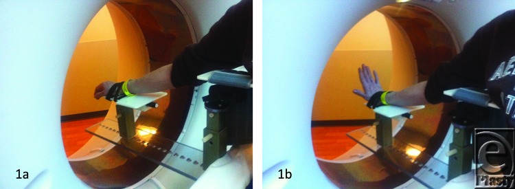
Dedicated “in CT” wrist platform. Pictures taken during unconstrained flexion and extension motions.
RESULTS
The patient demographics, clinical findings, X-ray, and 4D CT imaging findings as well as final patient disposition are depicted in Table 1. All subjects were successfully scanned in various wrist motions (Patient 1-4 videos). 4D CT image quality was adequate in all subjects, and carpal kinematic behavior was easily assessed in various wrist motions both before and after surgical repair. The normal postoperative (Patient 1-3 videos) and altered carpal kinematic behaviors (Patient 4 videos) correlated well with the clinical findings. In the operated wrists (Figs 2-4), while X-rays demonstrated slight gaping after scapholunate ligament repair, kinematic imaging demonstrated no abnormal widening with dynamic motion and showed normal dorsally hinged “scissoring” type scapholunate motion with motion. However, normal mid-carpal initiated wrist motion on flexion/extension was replaced by radiocarpal initiated motion, likely because of midcarpal stiffness/scarring. Clinical suspicion of ulnocarpal abutment was also confirmed in both wrists of Patient 4 (Fig 5).
Figure 2a, b.
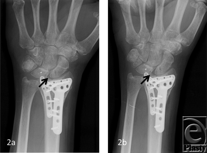
A 62-year-old man with prior ORIF and SLIL ligament repair. Normal SL space (arrow) on posteroanterior X-ray view (a). Notice mild gaping of S-L interval (arrow) on clenched fist posteroanterior X-ray view (b). ORIF indicates open reduction internal fixation; SLIL, scapholunate interosseus ligament.
Figure 4.
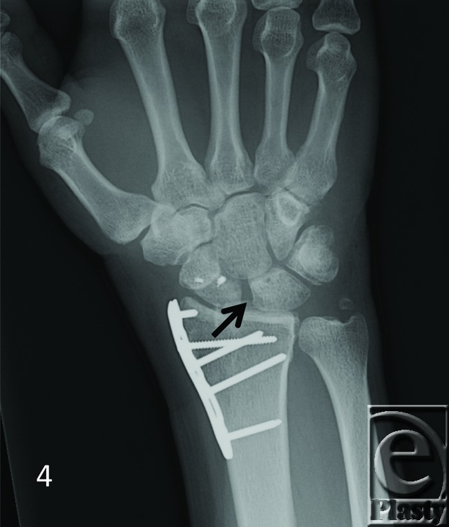
A 30-year-old man with prior ORIF and SLIL ligament repair. Notice mild gaping of S-L interval (arrow) on conventional posteroanterior X-ray view. ORIF indicates open reduction internal fixation; SLIL, scapholunate interosseus ligament.
Figure 5.
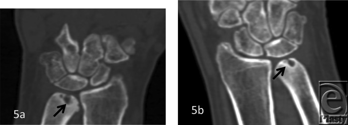
A 45-year-old man with diffuse wrist pain. Outside plain films not available. 2D reconstructions from the kinematic 4D CT volume scan show ulnar sided carpal translocation bilaterally. Also note subchondral cystic changes of ulna bilaterally (arrows) suggesting dynamic ulnocarpal abutment.
DISCUSSION
The human carpus is a complex system of 7 load sharing bones plus a sesamoid (pisiform) linked between the distal radius and ulna and the metacarpal bases with various static (ligament) and dynamic (musculotendinous units) stabilizers. Conventional static X-rays are used to follow patients longitudinally after injury, along with symptom development and physical examination, to determine whether or not a patient may have a ligamentous injury requiring intervention. The early insensitivity of static radiography for scapholunate ligament instability is well known.16 Standard findings of a “DISI” deformity—obtuse scapholunate ligament angle, loss of collinear lunocapitate angle, scapholunate diastasis (“Terry Thomas” sign), or a flexed scaphoid “ring sign”—may not develop for weeks or months after a complete injury to the dorsal limb of the SLIL and has also been associated with the need for not only an injury to the SLIL but also the dorsal intercarpal ligament.
Patients with wrist instability variably present with symptoms of pain, wrist clunk, and decreased grip strength similar to patient 4. Many a times, SLIL or LTIL (lunotriquetral interosseus ligament) injuries may not be fully appreciated until static signs of injury occur weeks to months or longer after an injury. This may diminish the possibility of standard repair of the injury and instead result in the need for more complex and invasive reconstructions or the development of irreversible arthrosis. In the absence of clear radiographic and/or physical examination indicators, magnetic resonance imaging with or without arthrogram, or wrist fluoroscopic evaluation with arthrogram, may be obtained to aid in diagnosis. However, even a partial injury may show positive findings with arthrogram, and the sensitivity of magnetic resonance imaging may vary. In addition, asymptomatic ligament tears are commonly seen on MR imaging or MR arthrograms.17 Although planar stress views and videofluoroscopy has been used to evaluate carpal kinematics,18-20 the measurement of intercarpal angles is difficult and subject to a great degree of variability between examiners.3 The ligament sectioning using 3D animated modeling has been shown to improve the accuracy of detection of intercarpal widening and angular deformations by avoiding overlap among the bony contours.21-27 Yet, these studies have been limited to in vitro models and are not directly comparable to in vivo kinematics because of complex motions which occur during in vivo tendon excursions.19,20,28
Our understanding of the biomechanics involved in allowing stable motion with and without loading continues to develop. Its evolution has progressed substantially from a tightly linked ball-and-socket type joint to the central column theory described by Navarro29 and later Taleisnik,30 to the oval-ring concept proposed by Lichtman,31 which is most currently favored. While our understanding of the bony and ligamentous anatomy seems satisfactory at this point, we continue to learn more about extrinsic stabilizers and how afferent innervation of the known wrist ligaments works with effector muscles, such as, the extrinsic forearm flexors and extensor to stabilize a wrist in motion and under load. However, most biomechanical studies detailing radiocarpal and midcarpal motion are performed on cadaveric specimens devoid of this normal stabilization system of extrinsic muscles, which are just now being appreciated. Motion is studied in this manner without regard to normal protective mechanisms that may limit motion to diminish strain in certain innervated ligaments and thus joints. Whether or not this results in motion with active use that is different than that studied in a cadaveric biomechanical laboratory is unknown. Thus, even using a combination of clinical tests, false-negative results remain common, which result in, continued patient suffering, worsening disability, and mounting treatment costs. Therefore, there is clearly a need for more accurate and precise, in vivo evaluation of carpal bone kinematics in patients presenting with symptoms of instability.
Spiral and multislice imaging built great milestones in the history of CT. 4D kinematic CT offers the next leap forward in CT technology that allows acquisition of isotropic volumes of an entire wrist with a single rotation of the gantry. The ability to acquire the entire wrist from distal forearm to metacarpal heads with one volume scan opens the door to new diagnostic possibilities and can revolutionize patient care. It allows real time visualization of the wrist dynamics during various unconstrained motions, such as supination-pronation, flexion-extension, fist clenching, and radialulnar deviation/dart throwing motions.32,33 A rapid 15- to –20-second study on a 320 slice scanner provides excellent depiction of the wrist motion relative to the forearm and concomitant visualization of the intercarpal translational as well as angular relationships. It was possible in all of our cases with adequate diagnostic image quality. Analyzing in vivo joint motion in such a manner avoids the need for invasive marker techniques.18,19
Patients with obvious findings necessitating surgical intervention such as the radiographic findings already mentioned, or more complex injuries such as a perilunate or lunate dislocation, may have questionable radiographic findings even after surgical repair/reconstruction. A persistent 3- to 4-mm scapholunate gap suggesting persistent ligamentous instability despite repair on multiple unchanging postoperative X-rays following fixation removal can be common finding that delays initiation of wrist motion rehabilitation and return to normal function, as it was observed in patients 1 to 3. For these reasons, we have begun incorporating Dynamic “4D” CT imaging into our postoperative imaging following commencement of wrist motion and rehabilitation for patients with interosseus ligament injuries of the wrist. We could obtain good diagnostic quality imaging and 4D reconstructions in all subjects without significant beam hardening artifacts similar to earlier results on 256-row detector study.34 For this modality to have its highest diagnostic sensitivity and specificity, it requires a thorough knowledge of wrist kinetics to appropriately appreciate what motion is normal and what motion is abnormal. This requires collaboration of hand surgeons and radiologists with study of “normal” dynamic studies and comparison with “abnormal” studies. These cases represent our early experience with this modality and how it has assisted in appreciating the postinjury wrist motion as well as helped guide activity and treatment postoperatively.
The ability to rapidly deliver high-resolution images has made CT a core imaging modality across healthcare today. However, there have been increasing concerns over patient safety due to radiation exposure and ALARA (As Low As Reasonably Achievable) principle should be followed. The extended coverage provided by the 16-cm wide area detector enables high-quality scanning of the entire wrist within one rotation, eliminating the need for helical scanning, which in turns lowers the dose dramatically. Although the extremities are more exposure resistant, there are a number of approaches used in these scans to reduce exposure dose to the patient. These include active collimator, AIDR 3D (Adaptive Iterative Dose Reduction 3D), SUREExposure 3D (automated exposure control) and quantum denoising software. The Active Collimator prevents extra radiation dose by eliminating rays that are not used for image reconstruction. It operates automatically at the start and end of scan range limiting the extent of the X-Ray beam. AIRD 3D is an advanced iterative reconstruction algorithm used in our scanner that reduces noise in both the 3D reconstruction data and raw data domains. SUREExposure 3D is another function that continuously modulates the exposure dose (by altering mA) in all 3 directions based on the patient's wrist shape. With the integration of AIDR 3D into SUREExposure controls, radiation exposure is automatically reduced before the scan, ensuring that the lowest possible dose is employed for the specific diagnostic objective irrespective of the size or shape of the patient. For routine clinical use, with the use of these functions, one is able to remove up to 50% of image noise that corresponds to a dose reduction of up to 75%, ultimately leading to improved spatial resolution and excellent artifact (streak/beam hardening) reduction that are otherwise routinely present at low dose acquisition. There is also minimal penalty in reconstruction times. Quantum Denoising Software is a powerful tool to remove noise in reconstructed images. It works in all dimensions and selectively smoothens out areas of uniform density while preserving the edge information within the image. It also reduces noise and increases the signal-to-noise ratio, while preserving contrast resolution. Finally, Boost3D is a 3D technology that automatically detects streak artifacts, and it eliminates them by reducing the photon starvation effects in the raw data domain. As a result, the image quality is improved in all viewing planes. A technologist training program ensures that operators become proficient in taking advantage of all the dose-reduction technologies available for every scan, providing clinicians with maximum image quality at the lowest possible dose to the patient. We achieved a dose of CT dose index of 10.5mGy and a dose length product of 84.3mGy-cm, close to 2× to 3× that of a static high-resolution scan. The effective dose was only 0.07mSv per scan maneuver (Table 2). With use of low-dose imaging, this novel modality provides an ability to evaluate the effect of muscle loading as well as long-term effects of treatment.28,35 Dynamic evaluation of the carpus promises to be a useful tool when trying to assess for adequacy of repair or reconstruction of complex wrist injuries. Using ALARA principle and the dose-reducing methods described earlier, 4D CT can be used for longitudinal studies without worrying about excessive radiation risks.
Table 2.
4D kinematic CT wrist scanning parameters compared to static high resolution 3D scan
| Scan Mode | Slice thickness | Range | kV | mA | Rotation Time | Acquisition Time | Acquisition Interval | CTDIvol (mGy) | DLP (mGy-cm) | Effective Dose |
|---|---|---|---|---|---|---|---|---|---|---|
| Static high-resolution 3D scan | ||||||||||
| Volume | 0.5 mm × 160 | 80 mm | 120 | 100 | 0.5 s | NA | NA | 4.1 | 32.4 | 0.67 |
| 4D Kinematic Dynamic Volume (Large patient or with metal in situ) | ||||||||||
| Dy-Volume | 0.5 mm × 160 | 80 mm | 100 | 100 | 0.5 s | 5.5 s | Continuous | 26.0 | 207.7 | 0.17 |
| 4D Kinematic Dynamic Volume (Average patient) | ||||||||||
| Dy-Volume | 0.5 mm × 160 | 80 mm | 80 | 80 | 0.5 s | 5.5 s | Continuous | 10.5 | 84.3 | 0.07 |
NA indicates not applicable; CTDIvol, CT dose index volume.
This study had certain limitations. In this pilot study, we did not do reader reliability evaluation or direct comparison of scan quality between 2D, 3D, and 4D reconstruction images, with static high-resolution scan. We did not perform any computational analysis for detection of subtle abnormalities and we only relied on gross visual evaluation of abnormality by the experienced reader. Further longitudinal study is warranted with this technique and should include preoperative and postoperative evaluation of ligamentous injuries in the same subjects for the evaluation of incremental value of this technique over conventional CT examination.
To conclude, kinematic 4 dimensional computed tomography (4DCT) provides adequate and novel assessment of wrist biomechanics both before and after surgical repair.
Figure 3a, b.
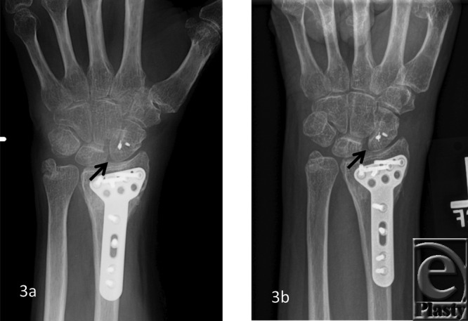
A 50-year-old woman with prior ORIF and SLIL ligament repair. Normal SL space (arrow) on posteroanterior X-ray view (a). Notice mild gaping of S-L interval (arrow) on clenched fist posteroanterior X-ray view (b). ORIF indicates open reduction internal fixation; SLIL, scapholunate interosseus ligament.
Acknowledgments
The authors thank Dr G. K. Thawait and Robert Kim for their support and help during the scans.
REFERENCES
- 1.Aksan AD, Durusoy R, Ada S, Kayalar M, Aksu F, Bal E. Epidemiology of injuries treated at a hand and microsurgery hospital. Acta Orthop Traumatol Turc. 2010;44(5):352–60. doi: 10.3944/AOTT.2010.2372. [DOI] [PubMed] [Google Scholar]
- 2.Jonasson P, Halldin K, Karlsson J, et al. Prevalence of joint-related pain in the extremities and spine in five groups of top athletes. Knee Surg Sports Traumatol Arthrosc. 2011;19(9):1540–6. doi: 10.1007/s00167-011-1539-4. [DOI] [PubMed] [Google Scholar]
- 3.Kuo CE, Wolfe SW. Scapholunate instability: current concepts in diagnosis and management. J Hand Surg Am. 2008;33(6):998–1013. doi: 10.1016/j.jhsa.2008.04.027. [DOI] [PubMed] [Google Scholar]
- 4.Berger RA, Blair WF. The radioscapholunate ligament: a gross and histologic description. Anat Rec. 1984;210(2):393–405. doi: 10.1002/ar.1092100215. [DOI] [PubMed] [Google Scholar]
- 5.Ritt MJ, Berger RA, Kauer JM. The gross and histologic anatomy of the ligaments of the capitohamate joint. J Hand Surg Am. 1996;21(6):1022–8. doi: 10.1016/S0363-5023(96)80310-8. [DOI] [PubMed] [Google Scholar]
- 6.Berger RA. The gross and histologic anatomy of the scapholunate interosseous ligament. J Hand Surg Am. 1996;21(2):170–8. doi: 10.1016/S0363-5023(96)80096-7. [DOI] [PubMed] [Google Scholar]
- 7.Moritomo H, Viegas SF, Nakamura K, Dasilva MF, Patterson RM. The scaphotrapezio-trapezoidal joint. Part 1: an anatomic and radiographic study. J Hand Surg Am. 2000;25(5):899–910. doi: 10.1053/jhsu.2000.4582. [DOI] [PubMed] [Google Scholar]
- 8.Moritomo H, Viegas SF, Elder K, Nakamura K, Dasilva MF, Patterson RM. The scaphotrapezio-trapezoidal joint. Part 2: a kinematic study. J Hand Surg Am. 2000;25(5):911–20. doi: 10.1053/jhsu.2000.8637. [DOI] [PubMed] [Google Scholar]
- 9.Viegas SF, Yamaguchi S, Boyd NL, Patterson RM. The dorsal ligaments of the wrist: anatomy, mechanical properties, and function. J Hand Surg Am. 1999;24(3):456–68. doi: 10.1053/jhsu.1999.0456. [DOI] [PubMed] [Google Scholar]
- 10.Foumani M, Strackee SD, Jonges R, et al. In-vivo three-dimensional carpal bone kinematics during flexion-extension and radio-ulnar deviation of the wrist: dynamic motion versus step-wise static wrist positions. J Biomech. 2009;42(16):2664–71. doi: 10.1016/j.jbiomech.2009.08.016. [DOI] [PubMed] [Google Scholar]
- 11.Moore DC, Crisco JJ, Trafton TG, Leventhal EL. A digital database of wrist bone anatomy and carpal kinematics. J Biomech. 2007;40(11):2537–42. doi: 10.1016/j.jbiomech.2006.10.041. [DOI] [PubMed] [Google Scholar]
- 12.Crisco JJ, Coburn JC, Moore DC, Akelman E, Weiss AP, Wolfe SW. In vivo radiocarpal kinematics and the dart thrower's motion. J Bone Joint Surg Am. 2005;87(12):2729–40. doi: 10.2106/JBJS.D.03058. [DOI] [PubMed] [Google Scholar]
- 13.Crisco JJ, Pike S, Hulsizer-Galvin DL, Akelman E, Weiss AP, Wolfe SW. Carpal bone postures and motions are abnormal in both wrists of patients with unilateral scapholunate interosseous ligament tears. J Hand Surg Am. 2003;28(6):926–37. doi: 10.1016/s0363-5023(03)00422-2. [DOI] [PubMed] [Google Scholar]
- 14.Crisco JJ, McGovern RD, Wolfe SW. Noninvasive technique for measuring in vivo three-dimensional carpal bone kinematics. J Orthop Res. 1999;17(1):96–100. doi: 10.1002/jor.1100170115. [DOI] [PubMed] [Google Scholar]
- 15.Metz VM, Metz-Schimmerl SM, Yin Y. Ligamentous instabilities of the wrist. Eur J Radiol. 1997;25(2):104–11. doi: 10.1016/s0720-048x(97)00041-7. [DOI] [PubMed] [Google Scholar]
- 16.Linscheid RL, Dobyns JH, Beabout JW, Bryan RS. Traumatic instability of the wrist: diagnosis, classification, and pathomechanics. J Bone Joint Surg Am. 2002;84-A(1):142. doi: 10.2106/00004623-200201000-00020. [DOI] [PubMed] [Google Scholar]
- 17.Maizlin ZV, Brown JA, Clement JJ, et al. MR arthrography of the wrist: controversies and concepts. Hand (N Y) 2009;4(1):66–73. doi: 10.1007/s11552-008-9149-4. [DOI] [PMC free article] [PubMed] [Google Scholar]
- 18.Savelberg HH, Otten JD, Kooloos JG, Huiskes R, Kauer JM. Carpal bone kinematics and ligament lengthening studied for the full range of joint movement. J Biomech. 1993;26(12):1389–402. doi: 10.1016/0021-9290(93)90090-2. [DOI] [PubMed] [Google Scholar]
- 19.Wolfe SW, Neu C, Crisco JJ. In vivo scaphoid, lunate, and capitate kinematics in flexion and in extension. J Hand Surg Am. 2000;25(5):860–9. doi: 10.1053/jhsu.2000.9423. [DOI] [PubMed] [Google Scholar]
- 20.Kobayashi M, Berger RA, Nagy L, et al. Normal kinematics of carpal bones: a three-dimensional analysis of carpal bone motion relative to the radius. J Biomech. 1997;30(8):787–93. doi: 10.1016/s0021-9290(97)00026-2. [DOI] [PubMed] [Google Scholar]
- 21.Werner FW, Short WH, Fortino MD, Palmer AK. The relative contribution of selected carpal bones to global wrist motion during simulated planar and out-of-plane wrist motion. J Hand Surg Am. 1997;22(4):708–13. doi: 10.1016/S0363-5023(97)80133-5. [DOI] [PubMed] [Google Scholar]
- 22.Short WH, Werner FW, Green JK, Masaoka S. Biomechanical evaluation of the ligamentous stabilizers of the scaphoid and lunate: part II. J Hand Surg Am. 2005;30(1):24–34. doi: 10.1016/j.jhsa.2004.09.015. [DOI] [PubMed] [Google Scholar]
- 23.Green JK, Werner FW, Wang H, Weiner MM, Sacks JM, Short WH. Three-dimensional modeling and animation of two carpal bones: a technique. J Biomech. 2004;37(5):757–62. doi: 10.1016/j.jbiomech.2003.10.001. [DOI] [PubMed] [Google Scholar]
- 24.Short WH, Werner FW, Green JK, Masaoka S. Biomechanical evaluation of ligamentous stabilizers of the scaphoid and lunate. J Hand Surg Am. 2002;27(6):991–1002. doi: 10.1053/jhsu.2002.35878. [DOI] [PMC free article] [PubMed] [Google Scholar]
- 25.Kaufmann RA, Pfaeffle HJ, Blankenhorn BD, Stabile K, Robertson D, Goitz R. Kinematics of the midcarpal and radiocarpal joint in flexion and extension: an in vitro study. J Hand Surg Am. 2006;31(7):1142–8. doi: 10.1016/j.jhsa.2006.05.002. [DOI] [PubMed] [Google Scholar]
- 26.Tang JB, Gu XK, Xu J, Gu JH. In vivo length changes of carpal ligaments of the wrist during dart-throwing motion. J Hand Surg Am. 2011;36(2):284–90. doi: 10.1016/j.jhsa.2010.11.025. [DOI] [PubMed] [Google Scholar]
- 27.Xu J, Tang JB. In vivo changes in lengths of the ligaments stabilizing the distal radioulnar joint. J Hand Surg Am. 2009;34(1):40–5. doi: 10.1016/j.jhsa.2008.08.006. [DOI] [PubMed] [Google Scholar]
- 28.Moojen TM, Snel JG, Ritt MJ, Kauer JM, Venema HW, Bos KE. Three-dimensional carpal kinematics in vivo. Clin Biomech (Bristol, Avon) 2002;17(7):506–14. doi: 10.1016/s0268-0033(02)00038-4. [DOI] [PubMed] [Google Scholar]
- 29.Navarro A. Anales de Instituto de Clinica Quirurgica y Cirugia Experimental. Montevideo, Uruguay: Imprenta Artistica de Dornaleche Hnos; 1935. [Google Scholar]
- 30.Taleisnik J. AAOS Instr Course Lect. St. Louis: Mosby; 1978. Wrist Anatomy, Function, and Injury; pp. 61–87. [Google Scholar]
- 31.Lichtman DM, Wroten ES. Understanding midcarpal instability. J Hand Surg Am. 2006;31(3):491–8. doi: 10.1016/j.jhsa.2005.12.014. [DOI] [PubMed] [Google Scholar]
- 32.Goh YP, Lau KK. Using the 320-Multidetector computed tomography scanner for four-dimensional functional assessment of the elbow joint. Am J Orthop (Belle Mead NJ) 2012 Feb;41(2):E20–4. [PubMed] [Google Scholar]
- 33.Coolens C, Bracken J, Driscoll B, Hope A, Jaffray D. Dynamic volume vs respiratory correlated 4DCT for motion assessment in radiation therapy simulation. Med Phys. 2012;39(5):2669–81. doi: 10.1118/1.4704498. [DOI] [PubMed] [Google Scholar]
- 34.Kalia V, Obray RW, Filice R, Fayad LM, Murphy K, Carrino JA. Functional joint imaging using 256-MDCT: technical feasibility. AJR Am J Roentgenol. 2009;192(6):W295–9. doi: 10.2214/AJR.08.1793. [DOI] [PubMed] [Google Scholar]
- 35.Neu CP, Crisco JJ, Wolfe SW. In vivo kinematic behavior of the radio-capitate joint during wrist flexion-extension and radio-ulnar deviation. J Biomech. 2001;34(11):1429–38. doi: 10.1016/s0021-9290(01)00117-8. [DOI] [PubMed] [Google Scholar]


