

ABSTRACT
Helicobacter pylori contains four genes that are predicted to encode proteins secreted by the autotransporter (type V) pathway. One of these, the pore-forming toxin VacA, has been studied in great detail, but thus far there has been very little investigation of three VacA-like proteins. We show here that all three VacA-like proteins are >250 kDa in mass and localized on the surface of H. pylori. The expression of the three vacA-like genes is upregulated during H. pylori colonization of the mouse stomach compared to H. pylori growth in vitro, and a wild-type H. pylori strain outcompeted each of the three corresponding isogenic mutant strains in its ability to colonize the mouse stomach. One of the VacA-like proteins localizes to a sheath that overlies the flagellar filament and bulb, and therefore, we designate it FaaA (flagella-associated autotransporter A). In comparison to a wild-type H. pylori strain, an isogenic faaA mutant strain exhibits decreased motility, decreased flagellar stability, and an increased proportion of flagella in a nonpolar site. The flagellar localization of FaaA differs markedly from the localization of other known autotransporters, and the current results reveal an important role of FaaA in flagellar localization and motility.
IMPORTANCE
The pathogenesis of most bacterial infections is dependent on the actions of secreted proteins, and proteins secreted by the autotransporter pathway constitute the largest family of secreted proteins in pathogenic Gram-negative bacteria. In this study, we analyzed three autotransporter proteins (VacA-like proteins) produced by Helicobacter pylori, a Gram-negative bacterium that colonizes the human stomach and contributes to the pathogenesis of gastric cancer and peptic ulcer disease. We demonstrate that these three proteins each enhance the capacity of H. pylori to colonize the stomach. Unexpectedly, one of these proteins (FaaA) is localized to a sheath that overlies H. pylori flagella. The absence of FaaA results in decreased H. pylori motility as well as a reduction in flagellar stability and a change in flagellar localization. The atypical localization of FaaA reflects a specialized function of this autotransporter designed to optimize H. pylori colonization of the gastric niche.
Introduction
Helicobacter pylori is a Gram-negative bacterium that colonizes the stomach in about 50% of humans worldwide (1–4). H. pylori colonization of the stomach results in gastric mucosal inflammation and is a significant risk factor for the development of distal gastric adenocarcinoma and peptic ulcer disease (3–5). One of the major virulence factors of H. pylori is a secreted protein known as vacuolating toxin (VacA) (7–9, 12). VacA is produced as a 140-kDa VacA protoxin that undergoes proteolytic cleavage to yield an 88-kDa protein that exhibits toxin activity (10, 11). The 88-kDa protein is secreted as a soluble protein into the extracellular space, or alternatively, it can remain attached to the bacterial cell surface (12, 13). VacA inserts into membranes to form anion-selective channels and can cause a wide array of alterations in host cells (7–9, 12).
Analysis of H. pylori genomes has revealed the existence of three vacA-like genes (14, 15). In strains 26695 and J99 (the first two H. pylori strains for which complete genome sequences were determined), these are designated HP0289/JHP0274, HP0609-0610/JHP0556, and HP0922/JHP0856 (14, 15). Several large-scale transposon mutagenesis studies provided evidence that two of the vacA-like genes are important for colonization of the rodent stomach by H. pylori. Specifically, a signature-tagged mutagenesis screen identified HP0289/JHP0274 as a gene required for H. pylori colonization of the gerbil stomach (16). HP0289/JHP0274 was required for colonization of the mouse stomach by H. pylori strain LSH100 (a derivative of G27) but not by H. pylori strain SS1 (17). Another transposon mutagenesis screen identified HP0609/JHP0556 as a gene required for H. pylori colonization of the mouse stomach (18). The HP0289/JHP0274 promoter is upregulated upon H. pylori colonization of the mouse stomach compared to H. pylori growth in vitro (6), and a recent study showed that, in comparison to a wild-type H. pylori strain, an HP0289/JHP0274 mutant stimulated greater expression of interleukin 8 (IL-8) and tumor necrosis factor alpha (TNF-α) by gastric epithelial cells; therefore, it was proposed that the protein encoded by HP0289/JHP0274 (also known as ImaA) has immunomodulatory properties (17).
The genes encoding the VacA-like proteins are among the largest in the H. pylori genome and encode proteins with predicted molecular masses of 313 kDa, 348 kDa, and 260 kDa (corresponding to the genes JHP0274, JHP0556, and JHP0856, respectively, in strain J99) (14, 15). Comparison of the sequences of the three VacA-like proteins with that of VacA shows that the highest level of similarity is within the C-terminal domains; other regions of the proteins exhibit very low levels of sequence similarity (15). The C-terminal region of VacA is a β-barrel domain that is required for secretion of VacA through an autotransporter (type V) pathway (19–21). Based on the sequence relatedness of the three VacA-like proteins to VacA within the β-barrel domain, it is presumed that the VacA-like proteins are also secreted by this route. Proteins secreted by the autotransporter pathway constitute the largest family of secreted proteins in Gram-negative bacteria (22–24). These proteins typically consist of three domains: (i) an N-terminal signal peptide, which is required for secretion across the inner membrane, (ii) a passenger domain, and (iii) a C-terminal β-domain, which facilitates translocation of the passenger domain across the outer membrane (15, 22–24). The passenger domains can have a wide variety of functions related to pathogenesis, including adhesion, autoaggregation, invasion, biofilm formation, and cytotoxicity. Structural analyses of several autotransporter passenger domains have revealed a conserved right-handed parallel β-helical fold (25–27). However, the primary amino acid sequences and specific functions of individual passenger domains are quite variable (22–24).
In the present study, we sought to learn more about the expression, subcellular localization, and in vivo roles of the VacA-like proteins. We show that all three vacA-like genes are expressed at increased levels when H. pylori colonizes the mouse stomach compared to H. pylori growth in vitro, and all three VacA-like proteins enhance the capacity of H. pylori to colonize the stomach. Studies of the localization of these proteins indicate that two VacA-like proteins localize in association with the bacterial body, whereas the third protein (which we designate FaaA, for flagella-associated autotransporter A) is detected as a component of the sheath overlying the flagellar filament and bulb. A faaA isogenic mutant exhibits decreased motility, decreased flagellar stability, and mislocalized flagella. Collectively, these results reveal that the localization of FaaA differs markedly from the localization of other known autotransporters and that FaaA has an important role in flagellar functions.
RESULTS
Detection of VacA-like proteins.
H. pylori genomes contain three vacA-like genes, which are designated imaA (17), faaA, and vlpC (defined in Materials and Methods). These three genes, each >7 kb in length, are among the largest in the H. pylori genome. As a first step in analyzing the three VacA-like proteins, we genetically modified H. pylori strain 60190 to yield strains encoding c-Myc-tagged forms of each of these proteins. A schematic diagram of each of these proteins and sites of the c-Myc epitope tag insertion is shown in Fig. 1A. Western blot analysis with an anti-c-Myc antibody revealed that all three VacA-like proteins had a mass of >250 kDa (Fig. 1B). The relative intensity of the VlpC band was consistently lower than that of ImaA or FaaA, and the molecular mass of VlpC was smaller than that of the other VacA-like proteins (Fig. 1B). Genes encoding c-Myc-tagged versions of each VacA-like protein were also introduced into H. pylori strains J99 and X47, and Western blotting yielded results similar to those shown in Fig. 1B. Since the VacA-like proteins are predicted to be secreted by an autotransporter pathway, we hypothesized that these proteins would be localized on the bacterial cell surface. To assess if this is indeed the case, we treated H. pylori strains producing c-Myc-tagged versions of ImaA, FaaA, and VlpC with proteinase K. Western blot analysis showed that all three VacA-like proteins and VacA were susceptible to proteinase K cleavage (Fig. 1C), whereas a negative-control protein (HspB) was not susceptible to proteinase K cleavage. This result provides evidence that all three VacA-like proteins are localized on the surface of H. pylori.
FIG 1 .

Detection of VacA-like proteins. (A) Schematic diagram of the three VacA-like proteins. Numbering corresponds to amino acid positions in the VacA-like proteins encoded by strain J99. Sites of c-Myc epitope tag insertion are shown. (B) H. pylori strain 60190 was engineered to encode c-Myc-tagged forms of the three VacA-like proteins. The WT strain and strains encoding c-Myc-tagged forms of FaaA, VlpC, or ImaA were grown overnight in broth culture, and production of each VacA-like protein was assessed by Western blot analysis using an anti-c-Myc antibody. All three proteins have masses of ≥250 kDa. (C) Strains encoding c-Myc-tagged forms of ImaA, FaaA, or VlpC were treated with 50 µg/ml proteinase K or control buffer at 37°C for 30 min. Susceptibility of the three proteins to proteinase K cleavage was assessed by immunoblotting using an anti-c-Myc antibody. Cleavage of VacA and heat shock protein B (HspB) was monitored as a positive and negative control, respectively, using antisera against these proteins. The experiment was performed three times with similar results.
VacA is secreted as a soluble protein into the broth culture supernatant (10). To determine if the VacA-like proteins are secreted in a similar manner, H. pylori strains expressing c-Myc-tagged forms of these proteins were cultured in broth for 48 h, and intact bacteria were removed by centrifugation at 14,000 × g. ImaA and FaaA were detectable in the culture supernatant by Western blotting, but VlpC was not detected (data not shown), presumably because it was expressed at relatively low levels. To determine whether ImaA and FaaA are released into the culture supernatant as soluble proteins or in another form, preparations of culture supernatant were ultracentrifuged, which resulted in separation of soluble proteins from insoluble components. Western blot analysis indicated that ImaA and FaaA localized mainly to the insoluble fraction (see Fig. S1 in the supplemental material). Thus, in contrast to VacA, we did not detect secretion of VacA-like proteins as soluble proteins into the extracellular space. The presence of these proteins in the insoluble fraction of culture supernatant may be attributable to the release of outer membrane vesicles that contain these proteins (28).
Analysis of imaA, faaA, and vlpC in vivo.
A previous study (using H. pylori strain G27) identified the imaA promoter as one of several promoters that are induced during H. pylori colonization of the mouse stomach compared to growth in vitro (6). Therefore, we hypothesized that the other vacA-like genes might be subject to similar regulation. C57BL/6 mice were orogastrically infected with H. pylori strain G27, and the animals were euthanized 2, 6, and 16 weeks thereafter. Transcription of imaA, faaA, and vlpC by H. pylori in the mouse stomach was analyzed by real-time reverse transcription-PCR (RT-PCR), as described in Materials and Methods. The transcription of all three genes was increased upon H. pylori colonization of the mouse stomach compared to H. pylori growth in vitro (Fig. 2A). The relative increase in transcription (in vivo compared to in vitro) was greatest for imaA at all three time points. These data confirm that imaA expression is increased during bacterial growth in vivo compared to bacterial growth in vitro and indicate that the other vacA-like genes also exhibit increased transcription in vivo.
FIG 2 .
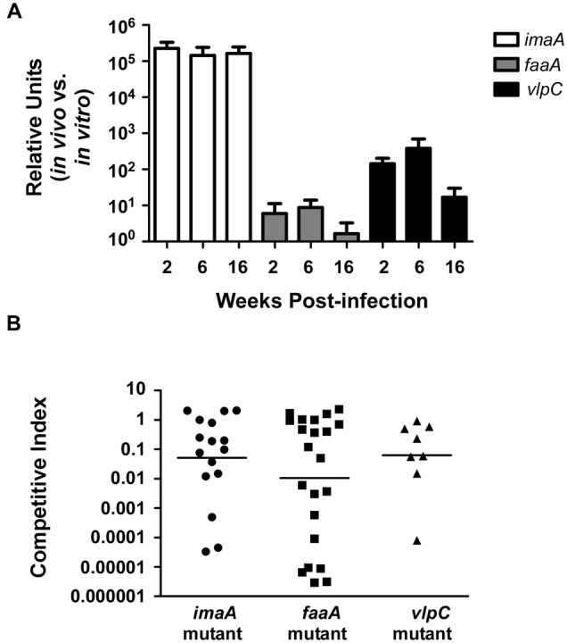
Analysis of imaA, faaA, and vlpC in vivo. (A) C57BL/6 mice were infected with H. pylori strain G27, and gene transcription was analyzed at 2, 6, or 16 weeks postinfection by real-time RT-PCR, as described in Materials and Methods. Expression of the indicated genes is given in relative units, and levels of gene expression in bacteria grown in vitro are assigned a value of 1. All three genes were expressed at higher levels in vivo than during bacterial growth in vitro. (B) Mice were inoculated with 1:1 mixtures of WT H. pylori X47 plus an imaA mutant (n = 16), WT plus a faaA mutant (n = 22), or WT plus a vlpC mutant (n = 8) for 2 weeks. The competitive index was calculated as described in Materials and Methods. Combined data from two independent experiments for FaaA and ImaA and one experiment for VlpC are shown, and each point represents the competitive index for one mouse stomach. WT bacteria outcompeted imaA, faaA, and vlpC mutant bacteria, as indicated by competitive indices below 1 (P ≤ 0.04 for each mutant, Student’s t test). The horizontal lines represent the mean for each group.
A vacA mutant strain was previously reported to have a colonization defect compared to a wild-type strain in mouse infection experiments (29). To assess a potential role of VacA-like proteins in vivo, we performed competition experiments. Since H. pylori strain G27 colonized a relatively low proportion of challenged mice, we used H. pylori strain X47 for these experiments. Mice were coinfected with 1:1 mixtures of the wild-type (WT) strain plus individual isogenic imaA, faaA, or vlpC mutants. After 2 weeks of infection, mice were euthanized, and bacterial colonization was assessed by analyzing bacterial growth on antibiotic-containing media that were selective for either the WT strain (metronidazole resistant) or the mutant strains (chloramphenicol resistant). The total bacterial densities in these animal stomachs ranged from 105 to 106 CFU/g for each animal; the bacterial densities did not differ significantly between animals challenged with different strains. A competitive index was determined as described in Materials and Methods. The WT bacteria outcompeted all three mutant strains (Fig. 2B) (P ≤ 0.04 for each mutant). Collectively, these experiments provide evidence that each of the VacA-like proteins has an important role in vivo.
Localization of FaaA to flagella.
To further investigate the localization of the VacA-like proteins, we first utilized immunofluorescence microscopy to analyze H. pylori strains producing c-Myc-tagged versions of ImaA, FaaA, and VlpC. In these immunofluorescence studies, ImaA and VlpC were localized on the surfaces of the bacteria, as expected based on their sensitivity to proteinase K, and were localized to a bacterial pole (Fig. 3A to D). In contrast, FaaA was localized to a site external to the bacterial body (Fig. 3E and F). Based on the immunofluorescence results, we hypothesized that FaaA might be localized to the flagella. To test this hypothesis, we generated preparations enriched in flagella from an H. pylori strain that produced a c-Myc tagged form of FaaA and from a control strain that produced a c-Myc-tagged form of ImaA. The presence of FaaA and ImaA in cell lysates and in the flagellar preparations was then examined by Western blotting using an anti-c-Myc antibody. As a control, we analyzed the presence of FlaA, the major flagellin subunit. As expected, FlaA was enriched in the flagellar preparations compared to total cell lysates (Fig. 3G), indicating an enrichment of flagellar components in the flagellar preparations. FaaA was also enriched in the flagellar preparations, whereas ImaA was not. These results support the hypothesis that FaaA is associated with flagella.
FIG 3 .
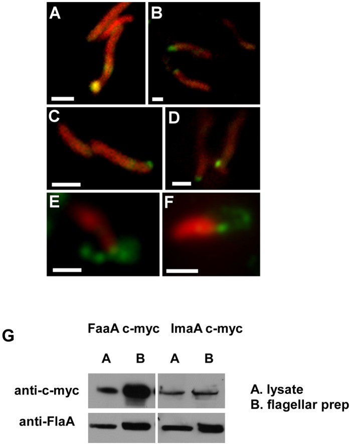
Analysis of the localization of VacA-like proteins. H. pylori strains were immunolabeled with monoclonal mouse anti-c-Myc antibody, followed by labeling with anti-mouse IgG conjugated to Alexa Fluor 488 (green channel). Bacterial cells were then counterstained as described in Materials and Methods (red channel) and visualized by fluorescence microscopy. The experiment was performed three times and a total of ≥200 bacteria were visualized for each experimental group. (A and B) H. pylori strain 60190 ImaA c-Myc; (C and D) H. pylori strain 60190 VlpC c-Myc; (E and F) H. pylori strain 60190 FaaA c-Myc. (G) A preparation enriched in flagella was prepared from H. pylori J99 strains that produced c-Myc-tagged FaaA or ImaA c-Myc. Samples were standardized by protein concentration, and the presence of c-Myc-tagged proteins in total cell lysates and in flagellar preparations was assessed by Western blotting. The presence of FlaA was assessed as a control. FaaA and FlaA were enriched in the flagellar preparations, whereas ImaA was not. The experiment was performed three times with similar results.
We then used immunoelectron microscopy to investigate potential flagellar localization of FaaA. H. pylori is characterized by the presence of multiple unipolar flagella (30, 31), and distinctive features of H. pylori flagella include the presence of a terminal bulb and a flagellar sheath (30–34). Analysis of FaaA localization by EM and immunogold staining revealed that FaaA localized to the flagellar sheath that covers the flagellar filament and the flagellar bulb (Fig. 4A to D). In contrast, ImaA, VlpC, and VacA were not detected in association with flagella (Fig. 4E to G) but were detected at the nonflagellar bacterial pole (see Fig. S2 in the supplemental material). Gold labeling was not detected in any of the negative-control samples; these included H. pylori lacking the c-Myc tag and processed in parallel with the other strains (Fig. 4H) and strains producing c-Myc-tagged versions of ImaA, FaaA, or VlpC that were immunolabeled with secondary antibody conjugated to gold particles alone (primary antibody omitted) (data not shown). Quantitative analyses confirmed that FaaA localizes mainly to the flagella (Table 1) (P < 0.0001). We also used immunofluorescence and electron microscopy to analyze two other H. pylori strains (J99 and X47) that produced c-Myc-tagged versions of ImaA, FaaA, and VlpC. Results in these strains were similar to what was observed in studies of strain 60190 and confirmed that FaaA localized to the flagella (Fig. 4D and data not shown).
FIG 4 .
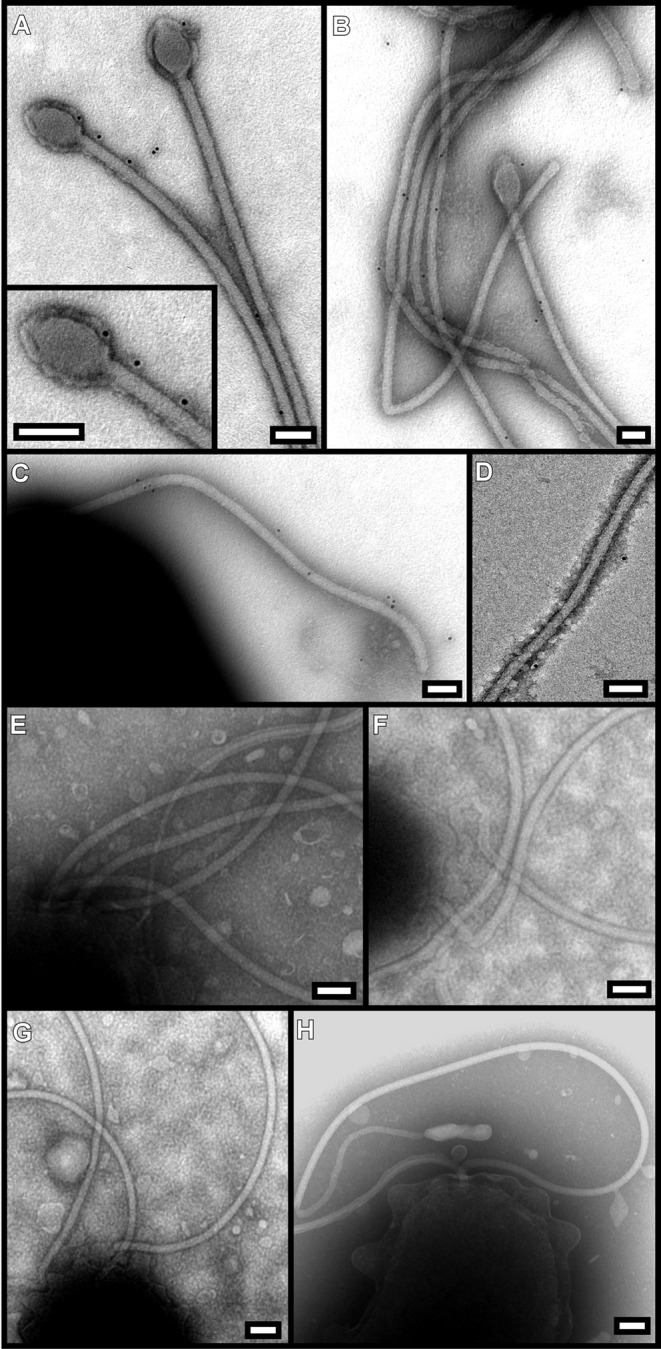
Immunogold EM analysis of FaaA localization. H. pylori strains were immunolabeled with primary antibodies to either VacA or c-Myc, followed by secondary antibodies conjugated to 10-nm immunogold particles. (A to C) 60190 FaaA c-Myc labeled with an anti-c-Myc antibody, demonstrating FaaA localization to the flagellar filament and flagellar bulb. (D) X47 FaaA c-Myc labeled with an anti-c-Myc antibody; (E) 60190 ImaA c-Myc labeled with an anti-c-Myc antibody; (F) 60190 (no c-Myc tag) labeled with an anti-VacA antibody; (G) 60190 VlpC c-Myc labeled with an anti-c-Myc antibody; (H) 60190 (no c-Myc tag) labeled with an anti-c-Myc antibody. FaaA localizes to flagella, whereas VacA, ImaA, and VlpC do not. The experiment was performed three times in multiple strains with similar results. Bars, 100 nm.
TABLE 1 .
| Strain | Mean no. of gold particles per bacterium at site |
|||
|---|---|---|---|---|
| Flagella | Flagellar pole | Nonflagellar pole | Side of bacterium | |
| 60190 FaaA c-Myc | 2.7 ± 0.5 | 0.87 ± 0.18 | 0 | 0.56 ± 0.0.13 |
| 60190 faaA mutant | 0 | 0 | 0 | 0 |
| 60190 | 0 | 0 | 0 | 0.028 ± 0.17 |
FaaA localization in the indicated strains was analyzed by immunogold EM, using an anti-c-Myc antibody followed by a secondary antibody conjugated to immunogold particles. Mean ± SEM values are reported.
The numbers of bacteria that were analyzed to generate the data were 45 for 60190 FaaA c-Myc, 123 for the 60190 faaA mutant, and 37 for 60190.
Flagellar alterations in a faaA mutant strain.
Since FaaA was localized to the flagella, we hypothesized that a faaA mutant strain might have a defect in flagellar morphology. To test this hypothesis, we performed transmission electron microscopy analyses of WT H. pylori strains, isogenic mutants, and mutants with restored alleles. Isogenic vacA, imaA, and vlpC mutants had flagellar morphologies similar to those of the respective WT H. pylori strains (60190, X47, and J99), with multiple flagella localized at the bacterial pole, as expected (Fig. 5A to D and data not shown). In contrast, the majority of the faaA mutant cells had flagellar defects, including absence of flagella, decreased numbers of flagella, or mislocalized flagella (Fig. 5E and F). The mislocalized flagella were found offset from the pole and at the lateral side of the bacterial cell body (Fig. 5E and F). In addition, preparations of the faaA mutants commonly contained broken flagella (Fig. 5E), which were rarely detected in preparations of the WT strain or other mutants; this suggests that the flagella produced by the faaA mutant might be more fragile than WT flagella. Introduction of an intact faaA allele into the faaA mutant reversed these alterations in flagellar localization and morphology (Fig. 5G). Quantitative analyses confirmed that there was a significant reduction in the number of intact flagella in the faaA mutant compared to the WT strain (P < 0.0001) and that there was a significantly increased number of nonpolar flagellar in this mutant (Fig. 5J, P < 0.0001). In summary, these data indicate that FaaA contributes to flagellar stability and proper localization of flagella.
FIG 5 .

Analysis of flagellar morphology and localization. H. pylori strains were analyzed by transmission EM and negative staining, as described in Methods. (A) Strain 60190 (without c-Myc tag); (B) 60190 imaA mutant; (C) 60190 ImaA c-Myc; (D) 60190 vacA-null mutant; (E) 60190 faaA mutant; (F) X47 faaA mutant; (G) 60190 FaaA c-Myc; (H) 60190 vlpC mutant; (I) 60190 VlpC c-Myc. Mislocalized flagella (arrows) were detected in the faaA mutant strains but not in the other mutants. The experiment was performed three times in multiple strains with similar results. Bars, 1 µm. (J) Graph showing quantification of the number of flagella per bacterium (based on analysis of 87 bacteria for 60190 FaaA c-Myc, 64 bacteria for the 60190 faaA mutant, and 19 bacteria for WT 60190). faaA mutant bacteria had significantly fewer flagella per bacterium than either WT bacteria or the mutant with restored faaA (asterisk, P < 0.0001, Kruskal-Wallis test). In addition, the faaA mutant strain had a significantly increased number of nonpolar flagella compared to WT bacteria or the mutant with restored faaA (P < 0.0001, Kruskal-Wallis test).
FaaA plays a role in flagellar stability.
To further examine the role of FaaA in flagellar stability, we examined the production of FlaA (the major flagellin) in a WT strain, in isogenic faaA, imaA and vlpC mutants, and in a mutant with a restored faaA allele. In comparison to WT bacteria, faaA mutant bacteria produced reduced levels of FlaA, based on Western blot analysis (Fig. 6A). In a similar analysis of imaA and vlpC mutants compared to WT bacteria, there was no detectable difference in FlaA production (Fig. 6A). To investigate whether the observed reduction in FlaA protein production in the faaA mutant strain was due to a reduction in flaA transcription, we performed real-time RT-PCR analysis. We did not detect any decreased flaA transcription in the faaA mutant strain, but in fact, we detected increased flaA transcription in the faaA mutant compared to the WT strain (Fig. 6C) (P < 0.03). These results suggest that the reduced level of FlaA in a faaA mutant strain is due to reduced FlaA stability.
FIG 6 .
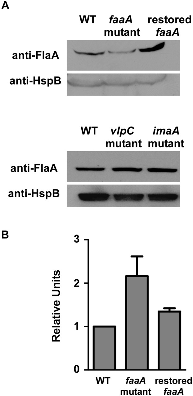
FaaA has a role in flagellar stability. (A) WT H. pylori strain J99, isogenic faaA, vlpC, and imaA mutant strains, and a faaA mutant with a restored intact faaA allele were cultured overnight in BB-FBS, and the presence of FlaA was assessed by Western blotting. The presence of HspB was monitored as a control. The level of FlaA was reduced in the faaA mutant compared to the other strains. The experiment was performed multiple times with similar results. (B) Transcription of flaA was analyzed by real-time RT-PCR, as described in Materials and Methods. RNA from WT bacteria served as the calibrator, and relative units are shown in comparison to the WT strain. Transcription of flaA was increased in the faaA mutant strain compared to the WT strain. Error bars represent mean plus standard error from combined results of three independent experiments performed in triplicate.
Decreased motility of faaA mutant strains.
Since FaaA is localized to flagella and various flagellar alterations were detected in faaA mutant strains, we hypothesized that FaaA might be required for optimal H. pylori motility. To test this hypothesis, we analyzed the motility of a WT strain, a faaA isogenic mutant, and a mutant with a restored intact copy of faaA. In comparison to the WT strain, the faaA mutant strain exhibited decreased motility (Fig. 7A). Restoration of an intact faaA gene reversed the defect in motility (Fig. 7A). There were no significant differences in the motility of imaA or vlpC mutants compared to the WT strain (Fig. 7B). These results indicate that FaaA is required not only for flagellar stability and proper flagellar localization but also for optimal motility.
FIG 7 .
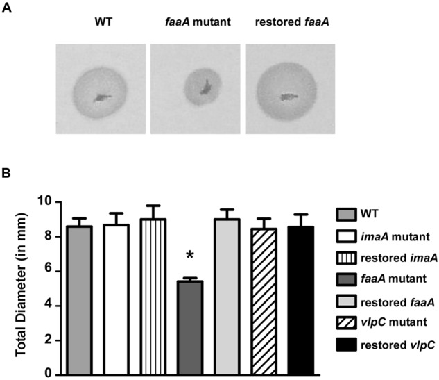
Mutation of faaA results in decreased motility. Motility of WT H. pylori strain J99, isogenic imaA, faaA, and vlpC mutant derivatives, and mutants with restored intact forms of imaA, faaA, and vlpC was assessed. Bacterial suspensions were inoculated into semisolid brucella medium, and the outward migration was measured over a period of 5 days. (A) Analysis of the motility of a faaA mutant compared to the WT strain and a mutant with a restored intact faaA. (B) Quantification of H. pylori motility. The motility of the faaA mutant was significantly decreased compared to that of the WT strain and the other strains tested. Data are means plus standard errors from combined results of three independent experiments, each performed in triplicate. The asterisk indicates a P value of <0.05 compared to all other strains (Student’s t test).
Role of FaaA in H. pylori colonization of the mouse stomach.
Competition experiments showed that a strain containing intact VacA-like proteins has a competitive advantage over a faaA mutant strain, based on analysis at two weeks postinfection (Fig. 2). To further investigate the role of faaA in vivo, we orogastrically infected C57BL/6 mice with either the WT strain alone or an isogenic faaA mutant strain alone. Mice were euthanized, and colonization of the stomach was analyzed at 4 days or 1 month postinfection. At 4 days postinfection, the faaA mutant strain demonstrated attenuated colonization compared to the WT strain (Fig. 8A) (P = 0.0172). In contrast, there was no significant difference at 1 month postinfection (Fig. 8B). These data, along with the competition experiments (Fig. 2), indicate that FaaA is required for optimal H. pylori colonization during early stages of infection, when flagella are required for bacterial entry into the gastric mucus layer (35, 36).
FIG 8 .
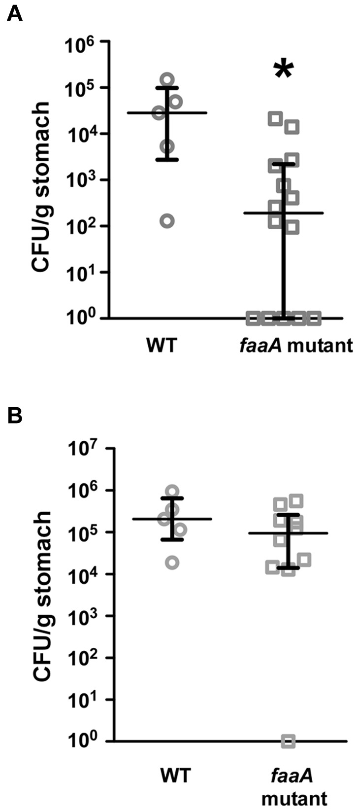
Role of FaaA in H. pylori colonization of the mouse stomach. C57BL/6 mice were infected with WT H. pylori strain X47 or an isogenic mutant for 4 days or 1 month. Mice were then euthanized and H. pylori colonization of the stomach was analyzed as described in Materials and Methods. (A) C57BL/6 mice were orogastrically infected with either the WT strain or an isogenic faaA mutant strain (WT, n = 5; faaA mutant, n = 14) for 4 days. The asterisk indicates a P value of 0.019 (Mann-Whitney U test). (B) Mice were infected with the WT strain or a faaA mutant strain (WT, n = 5; faaA mutant, n = 10) for 1 month. CFUs for individual mice are shown. Medians with interquartile ranges are shown.
DISCUSSION
The H. pylori genome contains three vacA-like genes that have C-terminal regions related to that of vacA, which encodes a secreted toxin. The C-terminal regions shared by VacA and the VacA-like proteins correspond to a predicted β-barrel domain that is required for protein secretion via the autotransporter pathway. The results of the present study show that these three VacA-like proteins share several common features. ImaA, FaaA, and VlpC are among the largest proteins produced by H. pylori (each >250 kDa in mass), and they are each localized on the bacterial surface. The three vacA-like genes are all upregulated during H. pylori colonization of the mouse stomach compared to H. pylori growth in vitro. Finally, imaA, faaA, and vlpC mutants each have a competitive disadvantage compared to the WT strain in mouse colonization experiments.
Unlike ImaA and VlpC, which are localized to a bacterial pole, FaaA is localized to the flagella. Correspondingly, faaA mutants exhibit multiple flagellar abnormalities, including absence of flagella, decreased numbers of flagella, increased flagellar fragility, and mislocalization of flagella to the lateral side of the bacteria instead of the pole. In addition, faaA mutant bacteria exhibit decreased motility compared to the WT strain. Thus, FaaA is required not only for flagellar stability and proper flagellar localization but also for optimal flagellar function. In an analysis of gastric colonization, a faaA mutant strain colonized the mouse stomach less efficiently than WT bacteria at an early time point postinfection, which is consistent with a known essential role of motility at early stages of H. pylori infection (36). FaaA might also have a role at later stages of infection, since flagella are likely to be required for continuous H. pylori colonization of the gastric mucus layer during the natural turnover of gastric mucus and exfoliation of gastric epithelial cells.
To the best of our knowledge, flagellar localization of autotransporter proteins has not previously been reported. The atypical localization of FaaA is probably attributable at least in part to unusual features of H. pylori flagella. H. pylori contains 2 to 6 polar flagella that are characterized by the presence of a sheath and a terminal bulb (30–34). Thus far, there has been relatively little analysis of the H. pylori flagellar sheath, and only one protein, HpaA, has been previously localized to this site (37–39). We speculate that the flagellar sheath contains multiple components that are derived from the H. pylori outer membrane. Therefore, we propose that FaaA is exported to the outer membrane and subsequently becomes a component of the flagellar sheath. Interestingly, we noted that levels of FlaA (the major component of flagella) were diminished in a faaA mutant, but there was no detectable reduction in flaA transcription in this mutant. We hypothesize that FaaA interacts directly or indirectly with multiple flagellar proteins; thus, the absence of FaaA may result in decreased protein stability of FlaA and possibly decreased stability of other flagellar components as well.
In summary, this study highlights important features of the H. pylori VacA-like proteins, including upregulation of the corresponding genes in vivo and a role for these proteins in colonization of the mammalian stomach. Unexpectedly, we show that one of these proteins, FaaA, localizes to flagella and that FaaA is required for proper flagellar localization and optimal flagellar function. This unusual localization and function of an autotransporter protein presumably reflects an adaptation designed to optimize H. pylori colonization of the gastric mucosal niche.
MATERIALS AND METHODS
Bacterial strains and culture conditions.
H. pylori strains 60190 (ATCC 49503), 60190 vacA::cat-rdxA (40), and J99 (ATCC 700824) and mouse-adapted versions of strain G27 and X47 were selected for use in this study. H. pylori strains were routinely grown at 37°C on Trypticase soy agar plates containing 5% sheep blood in ambient air containing 5% CO2. H. pylori mutant strains were selected based on resistance to chloramphenicol (2.5 µg ml−1) or resistance to metronidazole (7.5 µg ml−1) on brucella agar plates (brucella broth containing 1.35% agar and 10% fetal bovine serum [FBS]). H. pylori liquid cultures were grown in brucella broth (Sigma) supplemented with 5 to 10% fetal bovine serum (Atlanta Biologicals) or cholesterol (Gibco) (41). Prior to mouse infections, H. pylori strains were grown in brucella broth containing 10% FBS and 10 µg ml−1 vancomycin at 37°C under microaerobic conditions generated by a GasPakEZ Campy Container System (BD Biosciences).
Mutagenesis of imaA, faaA, and vlpC and production of c-Myc-tagged proteins.
In this present study, we designate HP0289/JHP0274 as ImaA (for immunomodulatory autotransporter protein A [17]), HP0609/JHP0556 as FaaA (for flagella-associated autotransporter A), and HP0922/JHP0856 as VlpC (for VacA-like protein C). To introduce mutations into these genes, we used a previously described mutagenesis method (42, 43). As a first step, metronidazole-resistant forms of strains 60190, X47, and J99 (60190ΔrdxA, X47ΔrdxA, and J99ΔrdxA) were generated by deleting the rdxA gene (43). Next, fragments of each gene (nucleotides 1,261 to 3,530 for imaA, 2,041 to 4,284 for faaA, and 1,390 to 3,639 for vlpC, with numbers based on the DNA sequences from H. pylori strain J99) were PCR amplified from H. pylori 60190, X47, and J99 genomic DNA and cloned into pGEM-T Easy (Promega). These plasmids were then used as templates for inverse PCR to generate modified plasmids containing a BamHI site after nucleotide 2199, 2997, and 2067 (numbers based on the sequences of genes in H. pylori strain J99) in imaA, faaA, and vlpC, respectively. The locations of the BamHI sites were selected based on the identification of regions that are predicted to be surface-exposed in a Hopp-Woods hydrophobicity analysis (http://www.vivo.colostate.edu/molkit/hydropathy/). A cat-rdxA cassette was cloned into the BamHI site, and cat-rdxA-containing plasmids, which are unable to replicate in H. pylori, were then transformed into H. pylori 60190ΔrdxA, X47ΔrdxA, and J99ΔrdxA, thereby allowing insertion of the cat-rdxA cassette into imaA, faaA, or vlpC. Single colonies were selected based on chloramphenicol resistance and metronidazole sensitivity. In each case, correct insertion of the cat-rdxA cassette was confirmed by PCR analysis.
To restore an intact copy of the relevant gene in strains that had been mutated and simultaneously insert a sequence encoding a c-Myc epitope tag into the gene of interest, we used a counterselection method, as described previously (42, 43). A DNA sequence encoding a c-Myc tag (5′ GAA CAA AAA CTT ATT AGT GAA GAA GAT CTT 3′) was inserted into the BamHI site in the plasmids described above using a QuikChange II XL site-directed mutagenesis kit (Agilent). Correct insertion of the c-Myc tag was confirmed by DNA sequencing. Plasmids containing the c-Myc-tagged versions of each VacA-like protein were then transformed into the appropriate H. pylori strains containing the cat-rdxA cassette, and metronidazole-resistant transformants were selected. This resulted in replacement of the cat-rdxA cassette with a sequence that contained the c-Myc tag.
Experiments in this study analyzed properties of metronidazole-resistant strains that contain WT copies of imaA, faaA, and vlpC, compared to isogenic mutant strains with disruptions of imaA, faaA, and vlpC and derivatives that contain restored intact forms of these genes with a c-Myc epitope tag. For convenience, the parental strains are designated WT here (despite the presence of the rdxA mutation) because they contain WT copies of all three vacA-like genes.
Detection of c-Myc-tagged proteins.
The presence of c-Myc-tagged VacA-like proteins was assessed by Western blot analysis using an anti-c-Myc antibody (4F6, 1:1,000; Vanderbilt Monoclonal Antibody Core) followed by a horseradish peroxidase (HRP)-conjugated secondary antibody (1:10,000; Promega). Proteins were visualized by incubation with chemiluminescent substrate solution (Pierce) and exposure to X-ray film.
Proteinase K susceptibility.
The susceptibility of the VacA-like proteins to proteinase K digestion was assessed using a modified version of previous protocols (43, 44). H. pylori strains were grown in liquid medium for 18 h and then harvested and washed with PBS. Bacteria were then resuspended in RPMI medium only or RPMI medium containing 50 µg/ml of proteinase K and incubated at 37°C for 30 min. Proteinase K activity was abrogated by the addition of phenylmethylsulfonyl fluoride (PMSF; 2 mM final concentration). The bacteria were washed in RPMI containing 2 mM PMSF, resuspended in SDS sample buffer, and analyzed by immunoblotting using an anti-c-Myc antibody. As controls, we monitored proteolysis of VacA, which is known to be localized to the bacterial surface (13), and heat shock protein B (HspB), which is localized within the cytoplasm, using antisera directed toward these proteins. Antibody concentrations used were 1:1,000 (anti-c-Myc), 1:10,000 (anti-VacA), and 1:20,000 (anti-HspB).
Immunofluorescence microscopy.
H. pylori strains were washed in PBS (pH 7.4), fixed in PBS containing 2.5% glutaraldehyde and 2.0% paraformaldehyde for 1 h at room temperature, washed twice with PBS, and blocked for 1 h in PBS containing 0.1% bovine serum albumin. Cells were incubated with anti-c-Myc antibody for 4 h at 4°C. Afterward, cells were washed three times with PBS and then incubated overnight at 4°C with an Alexa Fluor 488-conjugated secondary antibody (goat anti-mouse IgG; Invitrogen). Bacterial cells were washed three times with PBS before being counterstained with propidium iodide. As negative controls, replicate samples were processed by applying secondary antibodies alone. Samples were mounted using ProLong Gold antifade reagent (Invitrogen) and viewed using a Zeiss Axiophot wide-field microscope or a Zeiss LSM710 confocal laser scanning microscope.
Immunoelectron microscopy.
Immunoelectron microscopy was performed as described previously (43). Briefly, H. pylori strains were washed in 0.05 M sodium cacodylate buffer (pH 7.4) and then fixed in 2.5% glutaraldehyde and 2.0% paraformaldehyde in 0.05 M sodium cacodylate buffer for 1 h at room temperature. Cells were washed twice with sodium cacodylate buffer and blocked for 1 h in sodium cacodylate buffer containing 0.1% gelatin. Cells were incubated with primary antibody (mouse monoclonal anti-c-Myc) for 4 h at 4°C. Afterward, cells were washed three times with sodium cacodylate buffer before incubation overnight at 4°C with goat anti-mouse IgG conjugated to 10 nm gold particles (Ted Pella) or 25 nm gold particles (Electron Microscopy Sciences). The following day, bacterial cells were spotted onto Formvar-coated grids (Electron Microscopy Sciences) and negatively stained with 1% ammonium molybdate. As negative controls, replicate samples were processed by applying secondary antibodies alone. Samples were viewed with a FEI T-12 or a Philips C-12 transmission electron microscope. FaaA labeling and localization were quantified by counting the number of gold particles. Quantitation was done by one person (J.N.R.), who was blinded to the identity of the samples.
Electron microscopy analysis of flagella.
H. pylori strains were cultured on blood agar plates or in brucella broth (BB)-5% FBS overnight at 37°C in 5% CO2. Cells were harvested from the plate in 0.05 M sodium cacodylate buffer (pH 7.4) for an initial wash. Bacterial cells were spotted onto Formvar-coated grids and negatively stained with 1% ammonium molybdate. Samples were viewed with a FEI T-12 or a Philips C-12 transmission electron microscope. The number and localization of flagella were quantified by counting the number of flagella per bacterium. Quantification was done by one person (J.N.R.), who was blinded to the identity of the samples.
Motility assay.
H. pylori motility was analyzed as described previously (36, 45). Briefly, 1-µl aliquots of bacterial suspensions (overnight broth cultures back-diluted to an optical density at 600 nm [OD600] of 0.1) were stabbed into soft agar plates composed of BB–10% FBS and 0.35% agar. The plates were then incubated for 5 days at 37°C in 5% CO2, and the diameters of the bacterial halos were measured each day.
Enrichment of flagella.
A preparation enriched in flagella was prepared using a protocol adapted from previously described methods (46–48). Briefly, H. pylori strains were grown in liquid culture at 37°C for 18 h under microaerobic conditions. Bacteria were centrifuged at 6,000 rpm for 10 min, pellets were resuspended in sucrose solution (0.5 M sucrose, 10 mM Tris-HCl [pH 8.0]) containing lysozyme (0.02 mg/ml final concentration), EDTA (10 mM final concentration), and Zwittergent (2 mg/ml final concentration), and the suspensions were incubated at 4°C for 4 to 6 h with gentle shaking. Suspensions were then centrifuged at 25,000 × g for 30 min and pellets containing the flagella were resuspended in 5 ml PBS. Flagellar preparations were examined by Western blot analysis using anti-c-Myc (4F6; Vanderbilt Monoclonal Antibody Core) and anti-FlaA (Fisher) antibodies (each at a 1:1,000 dilution), followed by appropriate HRP-conjugated secondary antibodies.
RNA isolation and analysis of gene expression by real-time RT-PCR.
Total RNA was isolated from H. pylori using TRI reagent solution (Ambion) according to the manufacturer’s instructions, with slight modifications. Bacterial pellets were resuspended in TRI reagent solution, and one chloroform extraction was performed. The RNA was then mixed with 70% ethanol and purified using an RNeasy minikit (Qiagen). RNA samples were DNase treated using DNA-free (Ambion). RNA was then reverse transcribed using a high-capacity RNA-to-cDNA kit (Applied Biosystems). As a control, samples were processed without reverse transcriptase. The cDNA and control reactions were diluted 1:10 and used in real-time RT-PCR reactions. Real-time RT-PCR was performed using a Step One Plus real-time PCR machine (Applied Biosystems), with SYBR green as the fluorochrome. Abundance of transcript was calculated using the ΔΔCT method, with each transcript signal normalized to 16S rRNA. Transcript signals for each experimental sample were then compared to transcript signals from control bacteria grown in vitro. Primer sequences were as follows: imaA, 5′ GACACCAATAGCGCGGTTGT 3′ and 5′ TCAGCCGAGCTGGACTCTAAA 3′; faaA, 5′ GATAACGGCTTGACTTACTACATCAAA 3′ and 5′ CACGGTGTTACTGGCGTTGT 3′; vlpC, 5′ TGGGCGACAGGAGTTGGA 3′ and 5′ TTTGCATGAAACCCG CTATACC 3′; flaA, 5′ CATGGGGATTATCCAGGTTG 3′ and 5′ CGATACGAACCTGACCGATT 3′; 16S rRNA, 5′ CTAGCGGATTCTCTCAATGTCAA 3′ and 5′ GGAGTACGGTCGCAAGATTAAA 3′.
Infection of mice with H. pylori.
Eight-week-old Helicobacter-free male C57BL/6 mice (Jackson Laboratory) were used in all experiments, with a minimum of 5 to 10 mice per group. Prior to infection of mice, H. pylori was inoculated into liquid medium and cultured for 18 h under microaerobic conditions. Mice were orogastrically inoculated with a suspension of 5 × 10⁸ CFU of H. pylori in 0.5 ml of brucella broth without supplemental FBS (49). For competition experiments, mice were coinfected with a 1:1 ratio of the WT strain plus imaA, faaA, or vlpC mutant H. pylori strains, using a total input of approximately 5 × 10⁸ CFU in 0.5 ml of brucella broth. The inocula of the WT and mutant strains used for coinfection experiments were verified to contain equivalent CFU/ml, based on colony counting. Mice were orogastrically infected with two doses, administered two days apart. For in vivo gene expression studies, mice were euthanized at 2, 6, and 16 weeks postinfection. For competition experiments, mice were euthanized at 2 weeks postinfection. For colonization studies in which animals were infected with a single strain, mice were euthanized at 4 days or 1 month postinfection.
Processing of mouse stomachs and culturing of H. pylori from mouse stomachs.
Mouse stomachs were processed as described previously, with minor modifications (49). The stomach was removed from each mouse by excising between the esophagus and the duodenum. The forestomach (nonglandular stomach) was removed from the glandular stomach and discarded. The glandular stomach was opened and rinsed gently in PBS. For colonization studies, the glandular stomach was cut in half and placed into brucella broth for immediate processing. Gastric tissue was then homogenized using a Tissue Tearor (BioSpec Products), and serial dilutions of the homogenate were plated on Trypticase soy agar plates containing 5% sheep blood, 10 µg ml−1 nalidixic acid, 100 µg ml−1 vancomycin, 2 µg ml−1 amphotericin, and 200 µg ml−1 bacitracin. For coinfection experiments, plates also contained either chloramphenicol (2.5 µg ml−1) or metronidazole (7.5 µg ml−1), in order to permit selective isolation of mutant and WT strains, respectively (the WT strain X47 used in these experiments is metronidazole resistant, and the imaA, faaA, and vlpC mutants are chloramphenicol resistant). Plates were cultured under microaerobic conditions for 5 days before colonies were counted.
For experiments designed to analyze bacterial transcription in vivo, stomachs were incised and washed as described above, the gastric mucosa was scraped with cell scrapers (Fisher), and the scraped mucosa was placed in RNAprotect (Qiagen). Scrapings from 3 or 4 mouse stomachs were combined and analyzed as a single pooled sample. RNA was isolated and RT-PCR was performed as described above. Competitive index was determined by dividing the number of cultured mutant bacteria by the number of cultured WT bacteria, followed by corrections for any deviations from an input ratio of 1:1 (17).
Statistical analysis.
Gene transcription data, motility data, mouse colonization data, competition data, and flagellar localization data were analyzed using Student’s t test. Bacterial colonization densities were analyzed using the Mann-Whitney U test. Quantitative data pertaining to FaaA labeling, flagellar numbers, and flagellar localization were analyzed using the Kruskal-Wallis test. All statistical analyses were performed using the GraphPad Prism 5 program.
Ethics statement.
This study was carried out in strict accordance with the recommendations in the Guide for the Care and Use of Laboratory Animals of the National Institutes of Health. The protocol was approved by the Institutional Animal Care and Use Committee of Vanderbilt University School of Medicine and the VA Institutional Animal Care and Use Committee (V/10/157 and M/06/333).
SUPPLEMENTAL MATERIAL
Extracellular release of the VacA-like proteins. H. pylori strains 60190 ImaA c-Myc and 60190 FaaA c-Myc were cultured in BB-FBS broth for 48 h. After centrifugation at 14,000 × g to remove intact bacteria, the supernatants were ultracentrifuged at 200,000 × g yielding a fraction containing soluble proteins (S) and an insoluble fraction (INS). Western blot analysis using an anti-c-Myc antibody showed that FaaA and ImaA localize mainly to the insoluble fraction. Download
Analysis of localization of the VacA-like proteins by immunoelectron microscopy. H. pylori strains were immunolabeled with primary antibodies to either VacA or c-Myc, followed by secondary antibodies conjugated to 10-nm immunogold particles. In each case, the images represent visualization of the nonflagellar pole. (A) Labeling of VacA in strain 60190, (B) labeling of VacA in a 60190 vacA mutant strain (negative control), (C) labeling of ImaA in 60190 ImaA c-Myc, (D) labeling of VlpC in 60190 VlpC c-Myc, (E) labeling of FaaA in 60190 FaaA c-Myc, and (F) labeling of strain 60190 (no c-Myc tag, negative control) with an anti-c-Myc antibody. VacA, ImaA, and VlpC localize mainly to the nonflagellar pole, whereas FaaA does not. The experiment was performed three times in multiple strains with similar results. Arrows point to immunogold particles. Bars, 100 nm. Download
ACKNOWLEDGMENTS
This study is based on work supported by NIH AI039657, CA116087, AI068009, F32 AI102568, T32 AI007281, and the Department of Veterans Affairs, Veterans Health Administration, Office of Research and Development. Experiments in the Vanderbilt Cell Imaging Shared Resource were supported by the Vanderbilt University Digestive Disease Research Center (NIH grant P30DK058404) and the Vanderbilt University Ingram Cancer Center.
We thank Mark S. McClain for helpful discussions. We thank David Hendrixson and Deborah Ribardo at UT Southwestern Medical Center for advice on methods for flagellar enrichment. We thank Tatsuki Koyama for advice on statistical analyses.
Footnotes
Citation Radin JN, Gaddy JA, González-Rivera C, Loh JT, Scott Algood HM, and Cover TL. 2013. Flagellar localization of a Helicobacter pylori autotransporter protein. mBio 4(2):e00613-12. doi:10.1128/mBio.00613-12.
REFERENCES
- 1. Amieva MR, El-Omar EM. 2008. Host-bacterial interactions in Helicobacter pylori infection. Gastroenterology 134:306–323 [DOI] [PubMed] [Google Scholar]
- 2. Atherton JC, Blaser MJ. 2009. Coadaptation of Helicobacter pylori and humans: ancient history, modern implications. J. Clin. Invest. 119:2475–2487 [DOI] [PMC free article] [PubMed] [Google Scholar]
- 3. Cover TL, Blaser MJ. 2009. Helicobacter pylori in health and disease. Gastroenterology 136:1863–1873 [DOI] [PMC free article] [PubMed] [Google Scholar]
- 4. Suerbaum S, Michetti P. 2002. Helicobacter pylori infection. N Engl J. Med. 347:1175–1186 [DOI] [PubMed] [Google Scholar]
- 5. Blaser MJ, Berg DE. 2001. Helicobacter pylori genetic diversity and risk of human disease. J. Clin. Invest. 107:767–773 [DOI] [PMC free article] [PubMed] [Google Scholar]
- 6. Castillo AR, Woodruff AJ, Connolly LE, Sause WE, Ottemann KM. 2008. Recombination-based in vivo expression technology identifies Helicobacter pylori genes important for host colonization. Infect. Immun. 76:5632–5644 [DOI] [PMC free article] [PubMed] [Google Scholar]
- 7. de Bernard M, Cappon A, Del Giudice G, Rappuoli R, Montecucco C. 2004. The multiple cellular activities of the VacA cytotoxin of Helicobacter pylori. Int. J. Med. Microbiol. 293:589–597 [DOI] [PubMed] [Google Scholar]
- 8. Fischer W, Prassl S, Haas R. 2009. Virulence mechanisms and persistence strategies of the human gastric pathogen Helicobacter pylori. Curr. Top. Microbiol. Immunol. 337:129–171 [DOI] [PubMed] [Google Scholar]
- 9. Kim IJ, Blanke SR. 2012. Remodeling the host environment: modulation of the gastric epithelium by the Helicobacter pylori vacuolating toxin (VacA). Front. Cell. Infect. Microbiol. 2:37. [DOI] [PMC free article] [PubMed] [Google Scholar]
- 10. Cover TL, Blaser MJ. 1992. Purification and characterization of the vacuolating toxin from Helicobacter pylori. J. Biol. Chem. 267:10570–10575 [PubMed] [Google Scholar]
- 11. Cover TL, Tummuru MK, Cao P, Thompson SA, Blaser MJ. 1994. Divergence of genetic sequences for the vacuolating cytotoxin among Helicobacter pylori strains. J. Biol. Chem. 269:10566–10573 [PubMed] [Google Scholar]
- 12. Cover TL, Blanke SR. 2005. Helicobacter pylori VacA, a paradigm for toxin multifunctionality. Nat. Rev. Microbiol. 3:320–332 [DOI] [PubMed] [Google Scholar]
- 13. Ilver D, Barone S, Mercati D, Lupetti P, Telford JL. 2004. Helicobacter pylori toxin VacA is transferred to host cells via a novel contact-dependent mechanism. Cell. Microbiol. 6:167–174 [DOI] [PubMed] [Google Scholar]
- 14. Alm RA, Ling LS, Moir DT, King BL, Brown ED, Doig PC, Smith DR, Noonan B, Guild BC, deJonge BL, Carmel G, Tummino PJ, Caruso A, Uria-Nickelsen M, Mills DM, Ives C, Gibson R, Merberg D, Mills SD, Jiang Q, Taylor DE, Vovis GF, Trust TJ. 1999. Genomic-sequence comparison of two unrelated isolates of the human gastric pathogen Helicobacter pylori. Nature 397:176–180 [DOI] [PubMed] [Google Scholar]
- 15. Tomb JF, White O, Kerlavage AR, Clayton RA, Sutton GG, Fleischmann RD, Ketchum KA, Klenk HP, Gill S, Dougherty BA, Nelson K, Quackenbush J, Zhou L, Kirkness EF, Peterson S, Loftus B, Richardson D, Dodson R, Khalak HG, Glodek A, McKenney K, Fitzegerald LM, Lee N, Adams MD, Hickey EK, Berg DE, Gocayne JD, Utterback TR, Peterson JD, Kelley JM, Cotton MD, Weidman JM, Fujii C, Bowman C, Watthey L, Wallin E, Hayes WS, Borodovsky M, Karp PD, Smith HO, Fraser CM, Venter JC. 1997. The complete genome sequence of the gastric pathogen Helicobacter pylori. Nature 388:539–547 [DOI] [PubMed] [Google Scholar]
- 16. Kavermann H, Burns BP, Angermuller K, Odenbreit S, Fischer W, Melchers K, Haas R. 2003. Identification and characterization of Helicobacter pylori genes essential for gastric colonization. J. Exp. Med. 197:813–822 [DOI] [PMC free article] [PubMed] [Google Scholar]
- 17. Sause WE, Castillo AR, Ottemann KM. 2012. The Helicobacter pylori autotransporter ImaA (HP0289) modulates the immune response and contributes to host colonization. Infect. Immun. 80:2286–2296 [DOI] [PMC free article] [PubMed] [Google Scholar]
- 18. Baldwin DN, Shepherd B, Kraemer P, Hall MK, Sycuro LK, Pinto-Santini DM, Salama NR. 2007. Identification of Helicobacter pylori genes that contribute to stomach colonization. Infect. Immun. 75:1005–1016 [DOI] [PMC free article] [PubMed] [Google Scholar]
- 19. Fischer W, Buhrdorf R, Gerland E, Haas R. 2001. Outer membrane targeting of passenger proteins by the vacuolating cytotoxin autotransporter of Helicobacter pylori. Infect. Immun. 69:6769–6775 [DOI] [PMC free article] [PubMed] [Google Scholar]
- 20. Schmitt W, Haas R. 1994. Genetic analysis of the Helicobacter pylori vacuolating cytotoxin: structural similarities with the IgA protease type of exported protein. Mol. Microbiol. 12:307–319 [DOI] [PubMed] [Google Scholar]
- 21. Telford JL, Ghiara P, Dell’Orco M, Comanducci M, Burroni D, Bugnoli M, Tecce MF, Censini S, Covacci A, Xiang Z, et al. 1994. Gene structure of the Helicobacter pylori cytotoxin and evidence of its key role in gastric disease. J. Exp. Med. 179:1653–1658 [DOI] [PMC free article] [PubMed] [Google Scholar]
- 22. Dautin N, Bernstein HD. 2007. Protein secretion in gram-negative bacteria via the autotransporter pathway. Annu. Rev. Microbiol. 61:89–112 [DOI] [PubMed] [Google Scholar]
- 23. Henderson IR, Navarro-Garcia F, Desvaux M, Fernandez RC, Ala’Aldeen D. 2004. Type V protein secretion pathway: the autotransporter story. Microbiol. Mol. Biol. Rev. 68:692–744 [DOI] [PMC free article] [PubMed] [Google Scholar]
- 24. Leyton DL, Rossiter AE, Henderson IR. 2012. From self sufficiency to dependence: mechanisms and factors important for autotransporter biogenesis. Nat. Rev. Microbiol. 10:213–225 [DOI] [PubMed] [Google Scholar]
- 25. Gangwer KA, Mushrush DJ, Stauff DL, Spiller B, McClain MS, Cover TL, Lacy DB. 2007. Crystal structure of the Helicobacter pylori vacuolating toxin p55 domain. Proc. Natl. Acad. Sci. U. S. A. 104:16293–16298 [DOI] [PMC free article] [PubMed] [Google Scholar]
- 26. Junker M, Schuster CC, McDonnell AV, Sorg KA, Finn MC, Berger B, Clark PL. 2006. Pertactin beta-helix folding mechanism suggests common themes for the secretion and folding of autotransporter proteins. Proc. Natl. Acad. Sci. U. S. A. 103:4918–4923 [DOI] [PMC free article] [PubMed] [Google Scholar]
- 27. Renn JP, Clark PL. 2008. A conserved stable core structure in the passenger domain beta-helix of autotransporter virulence proteins. Biopolymers 89:420–427 [DOI] [PubMed] [Google Scholar]
- 28. Olofsson A, Vallström A, Petzold K, Tegtmeyer N, Schleucher J, Carlsson S, Haas R, Backert S, Wai SN, Gröbner G, Arnqvist A. 2010. Biochemical and functional characterization of Helicobacter pylori vesicles. Mol. Microbiol. 77:1539–1555 [DOI] [PMC free article] [PubMed] [Google Scholar]
- 29. Salama NR, Otto G, Tompkins L, Falkow S. 2001. Vacuolating cytotoxin of Helicobacter pylori plays a role during colonization in a mouse model of infection. Infect. Immun. 69:730–736 [DOI] [PMC free article] [PubMed] [Google Scholar]
- 30. Lertsethtakarn P, Ottemann KM, Hendrixson DR. 2011. Motility and chemotaxis in campylobacter and Helicobacter. Annu. Rev. Microbiol. 65:389–410 [DOI] [PMC free article] [PubMed] [Google Scholar]
- 31. O’Toole PW, Lane MC, Porwollik S. 2000. Helicobacter pylori motility. Microbes Infect. 2:1207–1214 [DOI] [PubMed] [Google Scholar]
- 32. Kostrzynska M, Betts JD, Austin JW, Trust TJ. 1991. Identification, characterization, and spatial localization of two flagellin species in Helicobacter pylori flagella. J. Bacteriol. 173:937–946 [DOI] [PMC free article] [PubMed] [Google Scholar]
- 33. Schirm M, Soo EC, Aubry AJ, Austin J, Thibault P, Logan SM. 2003. Structural, genetic and functional characterization of the flagellin glycosylation process in Helicobacter pylori. Mol. Microbiol. 48:1579–1592 [DOI] [PubMed] [Google Scholar]
- 34. Suerbaum S, Josenhans C, Labigne A. 1993. Cloning and genetic characterization of the Helicobacter pylori and Helicobacter mustelae flaB flagellin genes and construction of H. pylori flaA- and flaB-negative mutants by electroporation-mediated allelic exchange. J. Bacteriol. 175:3278–3288 [DOI] [PMC free article] [PubMed] [Google Scholar]
- 35. Eaton KA, Morgan DR, Krakowka S. 1992. Motility as a factor in the colonisation of gnotobiotic piglets by Helicobacter pylori. J. Med. Microbiol. 37:123–127 [DOI] [PubMed] [Google Scholar]
- 36. Ottemann KM, Lowenthal AC. 2002. Helicobacter pylori uses motility for initial colonization and to attain robust infection. Infect. Immun. 70:1984–1990 [DOI] [PMC free article] [PubMed] [Google Scholar]
- 37. Jones AC, Logan RP, Foynes S, Cockayne A, Wren BW, Penn CW. 1997. A flagellar sheath protein of Helicobacter pylori is identical to HpaA, a putative N-acetylneuraminyllactose-binding hemagglutinin, but is not an adhesin for AGS cells. J. Bacteriol. 179:5643–5647 [DOI] [PMC free article] [PubMed] [Google Scholar]
- 38. Luke CJ, Penn CW. 1995. Identification of a 29 kDa flagellator sheath protein in Helicobacter pylori using a murine monoclonal antibody. Microbiology 141:597–604 [DOI] [PubMed] [Google Scholar]
- 39. Lundström AM, Blom K, Sundaeus V, Bölin I. 2001. HpaA shows variable surface localization but the gene expression is similar in different Helicobacter pylori strains. Microb. Pathog. 31:243–253 [DOI] [PubMed] [Google Scholar]
- 40. González-Rivera C, Algood HM, Radin JN, McClain MS, Cover TL. 2012. The intermediate region of Helicobacter pylori VacA is a determinant of toxin potency in a Jurkat T cell assay. Infect. Immun. 80:2578–2588 [DOI] [PMC free article] [PubMed] [Google Scholar]
- 41. Jiménez-Soto LF, Rohrer S, Jain U, Ertl C, Sewald X, Haas R. 2012. Effects of cholesterol on Helicobacter pylori growth and virulence properties in vitro. Helicobacter 17:133–139 [DOI] [PubMed] [Google Scholar]
- 42. Loh JT, Shaffer CL, Piazuelo MB, Bravo LE, McClain MS, Correa P, Cover TL. 2011. Analysis of cagA in Helicobacter pylori strains from Colombian populations with contrasting gastric cancer risk reveals a biomarker for disease severity. Cancer Epidemiol. Biomarkers Prev. 20:2237–2249 [DOI] [PMC free article] [PubMed] [Google Scholar]
- 43. Shaffer CL, Gaddy JA, Loh JT, Johnson EM, Hill S, Hennig EE, McClain MS, McDonald WH, Cover TL. 2011. Helicobacter pylori exploits a unique repertoire of type IV secretion system components for pilus assembly at the bacteria-host cell interface. PLoS Pathog. 7:e1002237 http://dx.doi.org/10.1371/journal.ppat.1002237 [DOI] [PMC free article] [PubMed] [Google Scholar]
- 44. Ivie SE, McClain MS, Algood HM, Lacy DB, Cover TL. 2010. Analysis of a beta-helical region in the p55 domain of Helicobacter pylori vacuolating toxin. BMC Microbiol. 10:60 [DOI] [PMC free article] [PubMed] [Google Scholar]
- 45. Croxen MA, Sisson G, Melano R, Hoffman PS. 2006. The Helicobacter pylori chemotaxis receptor TlpB (HP0103) is required for pH taxis and for colonization of the gastric mucosa. J. Bacteriol. 188:2656–2665 [DOI] [PMC free article] [PubMed] [Google Scholar]
- 46. Aizawa SI, Dean GE, Jones CJ, Macnab RM, Yamaguchi S. 1985. Purification and characterization of the flagellar hook-basal body complex of Salmonella typhimurium. J. Bacteriol. 161:836–849 [DOI] [PMC free article] [PubMed] [Google Scholar]
- 47. Akiba T, Yoshimura H, Namba K. 1991. Monolayer crystallization of flagellar L-P rings by sequential addition and depletion of lipid. Science 252:1544–1546 [DOI] [PubMed] [Google Scholar]
- 48. Schuster SC, Baeuerlein E. 1992. Location of the basal disk and a ringlike cytoplasmic structure, two additional structures of the flagellar apparatus of Wolinella succinogenes. J. Bacteriol. 174:263–268 [DOI] [PMC free article] [PubMed] [Google Scholar]
- 49. Algood HM, Allen SS, Washington MK, Peek RM, Jr, Miller GG, Cover TL. 2009. Regulation of gastric B cell recruitment is dependent on IL-17 receptor A signaling in a model of chronic bacterial infection. J. Immunol. 183:5837–5846 [DOI] [PMC free article] [PubMed] [Google Scholar]
Associated Data
This section collects any data citations, data availability statements, or supplementary materials included in this article.
Supplementary Materials
Extracellular release of the VacA-like proteins. H. pylori strains 60190 ImaA c-Myc and 60190 FaaA c-Myc were cultured in BB-FBS broth for 48 h. After centrifugation at 14,000 × g to remove intact bacteria, the supernatants were ultracentrifuged at 200,000 × g yielding a fraction containing soluble proteins (S) and an insoluble fraction (INS). Western blot analysis using an anti-c-Myc antibody showed that FaaA and ImaA localize mainly to the insoluble fraction. Download
Analysis of localization of the VacA-like proteins by immunoelectron microscopy. H. pylori strains were immunolabeled with primary antibodies to either VacA or c-Myc, followed by secondary antibodies conjugated to 10-nm immunogold particles. In each case, the images represent visualization of the nonflagellar pole. (A) Labeling of VacA in strain 60190, (B) labeling of VacA in a 60190 vacA mutant strain (negative control), (C) labeling of ImaA in 60190 ImaA c-Myc, (D) labeling of VlpC in 60190 VlpC c-Myc, (E) labeling of FaaA in 60190 FaaA c-Myc, and (F) labeling of strain 60190 (no c-Myc tag, negative control) with an anti-c-Myc antibody. VacA, ImaA, and VlpC localize mainly to the nonflagellar pole, whereas FaaA does not. The experiment was performed three times in multiple strains with similar results. Arrows point to immunogold particles. Bars, 100 nm. Download