Abstract
Here, we show that a small-molecule activator of telomerase (TA-65) purified from the root of Astragalus membranaceus is capable of increasing average telomere length and decreasing the percentage of critically short telomeres and of DNA damage in haploinsufficient mouse embryonic fibroblasts (MEFs) that harbor critically short telomeres and a single copy of the telomerase RNA Terc gene (G3 Terc+/− MEFs). Importantly, TA-65 does not cause telomere elongation or rescues DNA damage in similarly treated telomerase-deficient G3 Terc−/− littermate MEFs. These results indicate that TA-65 treatment results in telomerase-dependent elongation of short telomeres and rescue of associated DNA damage, thus demonstrating that TA-65 mechanism of action is through the telomerase pathway. In addition, we demonstrate that TA-65 is capable of increasing mTERT levels in some mouse tissues and elongating critically short telomeres when supplemented as part of a standard diet in mice. Finally, TA-65 dietary supplementation in female mice leads to an improvement of certain health-span indicators including glucose tolerance, osteoporosis and skin fitness, without significantly increasing global cancer incidence.
Keywords: telomerase activation, TA-65, telomere length, aging, mouse
Introduction
Progressive attrition of telomeres is one of the best understood molecular changes associated with organismal aging in humans (Harley et al. 1990) and in mice (Flores et al. 2008). Telomeres are specialized structures at the ends of chromosomes, with an essential role in protecting the chromosome ends from fusions and degradation (Blackburn 2001; de Lange 2005). Mammalian telomeres consist of TTAGGG repeats bound by a six-protein complex known as shelterin (de Lange 2005). A minimum length of TTAGGG repeats and the integrity of the shelterin complex are necessary for telomere protection (Blackburn 2001; de Lange 2005). Telomerase is a cellular reverse transcriptase (TERT, telomerase reverse transcriptase) capable of compensating telomere attrition through de novo addition of TTAGGG repeats onto the chromosome ends by using an associated RNA component as template (Terc, telomerase RNA component) (Greider & Blackburn 1985). Telomerase expression can be detected in a number of adult cell types, including peripheral lymphocytes and adult stem cell compartments, however, this is not sufficient to maintain telomere length with age in most human and mouse tissues (Blasco et al. 1997; Harley 2005; Flores et al. 2008). Further supporting the notion that telomerase levels may be rate-limiting for organismal aging, some diseases characterized by premature loss of tissue renewal and premature death, such as dyskeratosis congenita, aplastic anemia and idiopathic pulmonary fibrosis, are linked to germline mutations in Tert and Terc genes, which result in decreased telomerase activity and accelerated telomere shortening (Mitchell et al. 1999; Vulliamy et al. 2001; Yamaguchi et al. 2005; Armanios et al. 2007; Tsakiri et al. 2007). A role for telomerase in tissue renewal and organismal life-span is also supported by telomerase-deficient (Terc−/−) mice (Blasco et al. 1997). These mice show progressive telomere shortening from the first generation (G1) until the third (G3) or fourth (G4) generation when in a pure C57BL6 background by which stage they present critically short telomeres, defective stem cell proliferative capacity, infertility due to germ cell apoptosis and increased genomic instability (Blasco et al. 1997; Herrera et al. 1999; Flores et al. 2005). We and others have shown that restoration of telomerase activity in these mice, by re-introduction of one copy of the Terc gene, rescues critically short telomeres and reverses chromosomal instability and cell and tissue defects associated with late-generation telomerase deficiency, including rescue of stem cell dysfunction and of organismal life-span (Hemann et al. 2001a; Samper et al. 2001a; Siegl-Cachedenier et al. 2007). These studies illustrate that telomerase is preferentially recruited to the shortest telomeres, similarly to that shown for budding yeast (Teixeira et al. 2004), thereby ensuring chromosomal stability and tissue fitness. Importantly, these findings also suggested that therapies aimed to re-activate telomerase and elongate short telomeres with age, could have significant anti-aging effects. In this regard, we recently demonstrated that enhanced telomerase activity in mice over-expressing TERT is able to delay aging and extent the median life span by 40%, when combined with increased cancer resistance (Tomas-Loba et al. 2008).
Here we report on our initial findings on the mechanism of action of TA-65, a small molecule telomerase activator derived from an extract of a plant commonly used in traditional Chinese medicine, Astragalus membranaceous. TA-65 was identified in an empirical screen based on its ability to upregulate basal telomerase activity levels in neonatal human keratinocytes, and has been studied in humans as a dietary supplement (Harley et al. 2010) leading to a decline of senescent and natural killer cells together with a significant reduction of the percentage of cells with short telomeres. Telomerase activation data and functional studies on a related molecule from this plant has been recently reported (Fauce et al. 2008). In that study, human immune cells were exposed ex vivo to the activator and this resulted in significant telomere elongation and enhanced proliferative capacity of these cells. Here, we demonstrate that TA-65 is capable of increasing telomerase activity and elongating critically short telomeres in a telomerase-dependent manner in MEFs haploinsufficient for the telomerase RNA component and in vivo, when supplemented as part of a standard diet in mice. We also report initial findings on the outcomes of a TA-65 dietary supplementation in female mice, and how it leads to an improvement of certain health-span indicators. Importantly, treatment with TA-65 did not show any detectable negative secondary effects, including no increase in the incidence of cancer.
Results
TA-65 stimulates telomerase activity and leads to telomerase-dependent elongation of short telomeres in MEF haploinsufficient for the telomerase RNA component
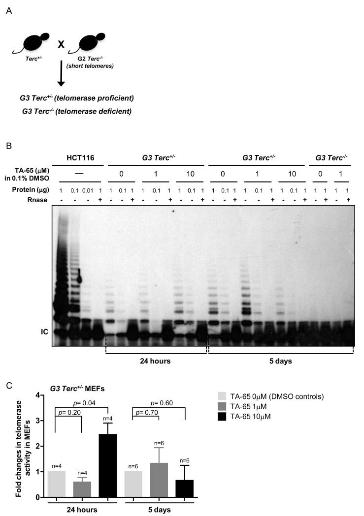
TA-65 has been identified as an effective telomerase activator in human immune cells, and neonatal keratinocytes and fibroblasts (Fauce et al. 2008; Harley et al. 2010). To delineate whether TA-65 could also have an impact on telomerase-dependent telomere extension we tested its capacity to affect the length of the shortest telomeres, which are the known preferred substrates of telomerase (Bianchi & Shore 2007; Sabourin et al. 2007), in an ex vivo model haploinsufficient for telomerase. To this end, we crossed Terc+/− females mice with G2 Terc−/− males mice to generate littermate populations of mouse embryonic fibroblasts (MEFs) that were either G3 Terc−/− or G3 Terc+/− (hereafter referred to as G3 Terc−/− or G3 Terc+/− MEFs, respectively). The G3 progeny of these crosses inherit a set of chromosomes with short telomeres from the male Terc−/− parent and a set of chromosomes with normal telomeres from the female Terc+/− parent (Fig. 1a, scheme).
Figure 1. TA-65 rescues short telomeres in a telomerase-dependent manner.
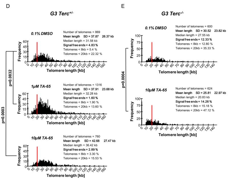
A. Scheme of mouse crosses. B. Telomerase activity was measured in MEFs extracts grown in the presence or absence of TA-65 for 24h and 5 days. Two different concentrations of TA-65 were tested. A cellular extract from HCT116 cells was included as a positive control, and RNase was added in all experimental settings as negative control. IC, PCR efficiency control. C. Fold changes in telomerase activity were quantified from the TRAP experiments, comparing to the values obtained for the control situation (without TA-65). At least 4 independent experiments were used per condition. The mean amounts of telomerase activity and error bars (standard deviation) in the different experimental settings are shown. One-way ANOVA was used for statistical analysis. D, E. Telomere fluorescence determined by Q-FISH on MEFs from the indicated genotypes grown in different concentrations of TA-65. Histograms represent the frequency of telomere fluorescence in Kb per telomere dot from a total of 50 nuclei, representing at least 650 telomeric dots per genotype. The red line indicates telomeres presenting <20 Kb of size. F, G. Comparison of the percentage of “signal-free” ends and short telomeres (< 8Kb) between MEFs of different genotypes, growth in the presence or absence of TA-65. Student’s t-test was used for statistical comparisons. H. Comparison of the percentage of “signal-free” ends and short telomeres (< 8Kb) between MEFs of different genotypes. Student’s t-test was used for statistical comparisons. I. Mean γ-H2AX immunofluorescence (green) per nucleus of MEF of the indicated genotype, in the presence or absence of 5 days incubation with TA-65 (DAPI in blue; at least 200 nucleus were scored per condition). Quantitative image analysis was performed using the Definiens Developer Cell software (version XD 1.2; Definiens AG). J. Representative immuno images showing the γ-H2AX (green) and nucleus stainings (DAPI - Blue), in the indicated genotypes.
Noteworthy that only the G3 Terc+/− progeny will inherit a copy of the Terc gene and, thereafter, is telomerase proficient (Fig. 1a). Using a telomere repeat amplification protocol assay (TRAP) we confirmed that reintroduction of the Terc allele successfully reconstituted telomerase activity in G3 Terc+/− cells, while G3 Terc−/− littermates persisted telomerase negative (Fig. 1b and Sup Fig. 1). Importantly, when we cultured G3 Terc−/− or G3 Terc+/− MEFs in the presence of 1 μM or 10 μM of TA-65 we observed its capacity to increase telomerase activity in telomerase positive (G3 Terc+/−) cells, with a significantly higher effect 24 hours post-treatment for the 10 μm dose treatment and later for the 1 μm dose treatment demonstrating a concentration dependent kinetics of telomerase activation (Fig. 1b-c and Sup Fig. 1). The observed telomerase stimulation is consistent with previous results with human keratinocytes (Harley et al. 2010). The absence of telomerase in MEFs leads, among other phenotypes, to an increase in the percentage of short telomeres and “signal-free ends” and the correlated appearance of chromosomal instability (Blasco et al. 1997; Herrera et al. 1999; Samper et al. 2001a; Cayuela et al. 2005). By using the quantitative telomere FISH (Q-FISH) assay we could observe that control telomerase reconstituted G3 Terc+/− cells treated with 0.1% DMSO present a significant decrease of “signal-free” ends (12.3% to 4.8%; Fig 1d-h) and critically short telomeres (telomeres with <8Kb length) compared to similarly treated G3 Terc−/− littermates (12.8% to 5.4% Fig. 1d-h). Treatment of telomerase-proficient G3 Terc+/− cells with TA-65 during 5 days at concentrations of 1 μM or 10 μM resulted in an additional decrease of the percentage of “signal-free ends” to 1.6% and 2.9%, and short telomeres to 1.9% and 3.3%, respectively, compared to their control situation (0.1% DMSO) (Fig. 1d,f). In marked contrast, TA-65 treated G3 telomerase-deficient Terc−/− littermate MEFs presented similar number of short telomeres or “signal-free ends” confirming the telomerase-dependent mechanism of action of TA-65 (Fig. 1e,g). Additionally, a significantly increased of average telomere length was observed in G3 Terc+/− cells when treated with 10 μM of TA-65 (37.87 Kb to 42.68 Kb, Fig. 1d). The presence of short or uncapped telomeres results in higher cellular levels of gamma-H2AX, an indicator of DNA double strand breaks and damage response (Martinez et al. 2010). We observed that TERC−/− MEFs present higher levels of nuclear gamma-H2AX comparing to TERC proficient cells (Fig. 1i and representative image at Fig 1j). The presence of TA-65 could additionally decrease gamma-H2AX signal in TERC proficient cells but not in TERC deficient background (Fig. 1i).
Although TA-65 can significantly rescue the percentage of short telomeres in telomerase proficient cells, TA-65 per se cannot mimic the positive effects of re-introducing telomerase in a telomerase deficient background (Hemann et al. 2001b; Samper et al. 2001a; Samper et al. 2001b; Siegl-Cachedenier et al. 2007). These results delineate the mechanism of action of TA-65, demonstrating its ability to decrease the percentage of short telomeres and “signal-free ends”, only when in the presence of an active telomerase complex.
Dietary supplementation of TA-65 increases TERT expression in some tissues and rescues short telomeres in mice
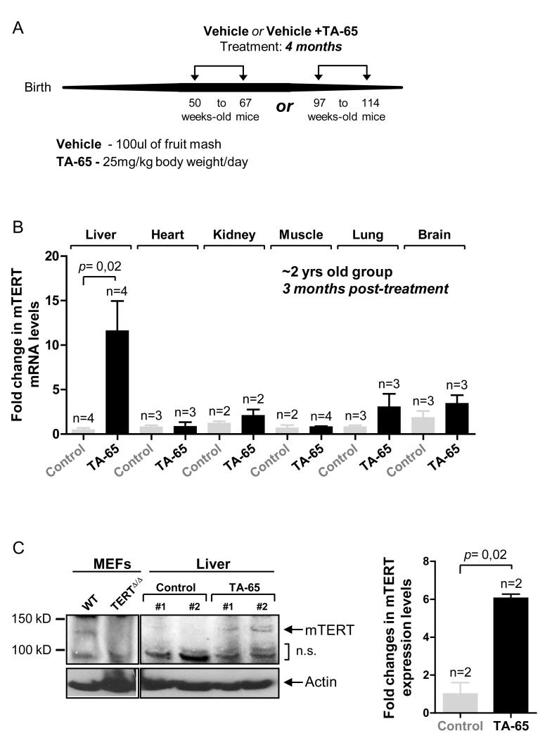
Telomere attrition and the accumulation of short telomeres are parallel to the ageing process in humans and mice, being a probable cause of the appearance of age-related pathologies (Harley et al. 1990; Canela et al. 2007; Jiang et al. 2008; Calado & Young 2009). Short telomeres are therefore, both an indicator and a possible cause of healthspan decay with age either in humans and mice. Following the above-described results with MEFs, we set to address whether a dietary supplementation with TA-65 could similarly lead to detectable changes on telomere dynamics in vivo. To this end, we fed two cohorts of mature or old female mice (1 yr or 2 yrs old, respectively) with control vehicle (fruit mash) or vehicle plus TA-65 (final concentration of 25mg/kg body weight/day) for 4 months (scheme in Fig 2a, and detailed information in Materials and Methods). Treated mice demonstrated a complete tolerance to the administration of the vehicle or vehicle+TA-65 as no deaths or other overt pathologies related to treatment were observed during this period. In line with this, body weight was maintained and comparable between the different mouse cohorts throughout the treatment period (Sup Fig. 2).
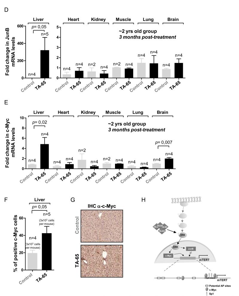
Figure 2. TA-65 leads to an increased TERT expression in vivo.
A. Scheme of the TA-65 treatment plan. B. Fold change in mTERT mRNA levels 3 months post treatment in different tissues of 2 years old TA-65 treated mice compared to age-matched controls. mTERT mRNA values were normalized to actin. Student’s t-test was used for statistical comparisons. C. mTERT protein expression in liver extracts from control or 2 years old TA-65 treated mice (NS: non-specific band) using a mTERT antibody previously validated by us (Martinez et al. 2010)). As controls, mTERT protein expression is detected in wild-type MEFs but not in TERT−/− MEFs (Liu et al. 2000). Quantification of mTERT expression is presented in the right panel (fold changes in telomerase activity are plotted, comparing to the values obtained for the control situation (without TA-65)). The quantification was made with Scion Image Software. Student’s t-test was used for statistical comparisons. D. Fold change in JunB mRNA levels 3 months post treatment in different tissues of 2 years old TA-65 treated mice compared to age-matched controls. JunB mRNA values were normalized to actin. E. Fold change in c-Myc mRNA levels 3 months post treatment in different tissues of 2 years old TA-65 treated mice compared to age-matched controls. c-Myc mRNA values were normalized to actin. Student’s t-test was used for statistical comparisons. F. Percentage of c-Myc positive cells, quantified from at least 3 independent mice. Measures were realized post mortem in cancer-free mice. Student’s t-test was used for statistical comparisons. At least 5 high power fields (HPF, x100) were used per independent mouse (around 3000 cells scored per mouse, AxioVision was used for image analysis). G. Representative IHC of c-Myc staining in either control or TA-65 treated 2 years old female mice. H. Hypothetical model of action of TA-65 based in ours and previous results.
It has been previously described that treatment of CD8+ T lymphocytes with a similar telomerase activator compound (TAT2) leads to an 8-fold increase in hTERT mRNA levels (Fauce et al. 2008). To follow the effects of TA-65 dietary supplementation in vivo, we examined mTERT mRNA levels in different tissues from the 2-years old TA-65 treated or control cohorts at 3 months post-treatment. Notably, TA-65 treatment resulted in a significant 10-fold increase of mTERT mRNA (Fig. 2b) and protein (Fig. 2c) levels in the liver. mTERT mRNA levels were also modestly increased in other mouse tissues including kidney, lung and brain, although these increases did not reach statistical significance (Fig. 2b). Of note, the same patterns were observed in the 1 year old group (Sup Fig. 3a). Interestingly, increased levels of mTERT in the liver of 2 yrs old TA-65 treated mice correlated with an increase in mRNA levels of JunB (Fig. 2d) and c-Myc (Fig 2e), two transcription factors regulated by the MAPK pathway (Gupta et al. 1993; Lefloch et al. 2008), which is a known potential mediator of TA-65 action (Fauce et al. 2008) The same activation was observed in the 1 yr old group (Sup Fig. 3b,c). Higher levels of c-Myc in the liver were confirmed by IHC in the 2-year-old group of mice, at time of death (Fig 2f,g). Of note, TA-65 did not altered significantly the levels of targets of the Wnt (CD44, CyclinD1) or TGFβ (Fibronectin, Klf4 or p16) pathways (Sup Fig 4), further supporting that TA-65-dependent telomerase activation occurs through transcription factors regulated by the MAPK pathway, which may directly or indirectly regulate the mTERT promoter (hypothetical mechanism in Fig. 2h, based in our current findings and previous results (Wang et al. 1998; Greenberg et al. 1999; Chang & Karin 2001; Inui et al. 2001; Takakura et al. 2005; Pericuesta et al. 2006)).
To address the in vivo effect of TA-65 treatment on telomerase-mediated telomere elongation, we measured telomere length in blood samples from the 1 yr and 2 yrs old controls and TA-65 treated groups, 3 months post the dietary supplementation period ending. Telomere length was assessed from peripheral blood leukocytes by using a high-throughput (HT) Q-FISH technique optimized for blood samples (Canela et al. 2007). Average telomere length was not significantly increased in the 1 yr or 2 yrs TA-65 treated groups compared to the untreated controls (Fig. 3a,c). Interestingly, we observed a significant decrease of very short telomeres (telomeres below 2, 3 and 4 Kb, Fig. 3b,d) in the 1 yr and 2 yrs old TA-65 treated groups at 3 months post-treatment, indicating a significant and consistent capacity of TA-65 to promote rescue of short telomeres both in vitro (MEFs) and in vivo (mice). A similar scenario has been previously described in a study assessing the role of TA-65 in humans, where a reduced percentage of cells with short telomeres appeared concomitant with minimal effects in the mean telomere length (Harley et al. 2010).
Figure 3. TA-65 rescues short telomeres in vivo.
A, C. HT-QFISH telomere length analysis of the 1 year old (A) or 2 year old (C), control or TA-65 treated group, 3 month post-treatment. B, D. Percentage of short telomeres (fraction of telomeres presenting a size below 2, 3 or 4 Kb), 3 month post-treatment, in the 1 year old (B) or 2 year old (D) group of mice. Data are given as mean±SEM.
TA-65 treatment enhances health-span in female mice
Previous studies have demonstrated that the shortest telomeres are causal of reduced cell viability (Hemann et al. 2001b; Hao et al. 2005) and that a stable and enforced expression of telomerase leads to an improved health-span, accompanied by an extension of lifespan, possibly through the rescue of short telomeres in mice (Tomas-Loba et al. 2008). Since TA-65 influences the percentage of cellular short telomeres through the activation of telomerase, we were interested in addressing if this compound could have an impact on health and/or life-span in the treated mice.
The aging process can be evaluated by studying some of the so-called biomarkers of aging. One established indicator of ageing in mice is glucose intolerance and insulin resistance, which increase with age (Bailey & Flatt 1982; Guarente 2006). Importantly, TA-65 administration during 4 months significantly improved the capacity to uptake glucose after a glucose pulse (Area Under the Curve, AUC) in the 1yr old treated mice compared to the control groups at 6 and 12 months post-treatment (Fig. 4a,d; Materials and Methods). Furthermore, TA-65 treated mice presented a tendency to show lower levels of blood insulin 6 months-post treatment which, together with the glucose levels at fasting, resulted in a tendency to have a better HOMA-IR score at 6 months post-treatment (Fig. 4b,c) (Heikkinen et al. 2007). Of note, although we could observed a better glucose uptake after 12 months in the 1 yr old group post TA-65 treatment (Fig 4d) we did not find major differences at the basal insulin levels or HOMA-IR score; the same situation was observed in the 2 yrs old group of mice (Fig 4e-i) which present a minor amelioration on the glucose tolerance test, insulin and HOMA-IR parameters post treatment. This situation could indicate that a discontinued TA-65 intake could result in an attenuation of some health-span improvements (a situation in agreement with previous observations (Fauce et al. 2008)), or that old mice are less refractory to TA-65 treatment.
Figure 4. TA-65 treatment increases metabolic fitness.
A, D, G. Glucose tolerance measured as fold changes to the area under the curve (AUC) at the indicated time post-treatment in the 1 yr or 2 yrs old cohort of mice. B, E, H. Insulin levels at the indicated times post-treatment in the 1 yr or 2 yrs old cohort of mice. C, F, I. Homeostatic model assessment scores (HOMA) at the indicated time post-treatment in the 1 yr or 2 yrs old cohort of mice. TA-65 supplemented mice present a better score at 6 months post treatment, indicating a better metabolic rate. J. Oil red O staining of lipid droplets in frozen liver sections from either controls or TA-65 treated mice on a standard diet. Measures were realized post mortem in cancer-free mice. Quantification of Oil Red O area is presented in the right panel (5 images per mice; Photoshop CS3 and Scion Image software were used for image analysis).
Liver steatosis is caused by lipid accumulation within hepatocytes both in humans and mice. Despite being a relatively benign condition, when combined with inflammation it may progress to serious liver disease (Feldstein et al. 2003; Higuchi & Gores 2003). Although mouse liver steatosis models involve usually intake of a high-fat diet (and subsequent diet-induced obesity) (Pfluger et al. 2008), this condition may also occur in aged mice under standard diet conditions (Kelder et al. 2007). As expected, lipid droplets were found in liver sections of both control and TA-65 treated mice at time of death (Fig 4j). Interestingly, control mice presented the strongest accumulation of fat in the liver at older ages comparing to the TA-65 treated cohorts, which, together with the previous results, could indicate a liver protective action of TA-65 (Fig 4j).
Other well-established biomarkers of ageing are the loss of the epidermal and subcutaneous adipose skin layers (Shimokata et al. 1989; Tomas-Loba et al. 2008). The weakness of the skin barrier associates with infections and deficient water exchange. TA-65 treatment resulted in an increase of subcutaneous fat content and epidermal layer in the 1 yr old group of mice (Fig. 5a,c and representative image in Fig. 5b), but did not significantly change these parameters in the 2 yrs old groups (Fig. 5a,c), comparing to the untreated controls. In line with an improved epithelial fitness, TA-65 treatment increased the in vitro wound-healing capacity of keratinocytes (Fig 5d) as well as significantly accelerated hair re-grow in vivo upon plucking of old mice (Fig 5e and representative image in Fig 5f); moreover, TA-65 treated mice present higher levels of positive Ki67 cells at the epidermis (Fig. 5g and representative image at Fig. 5h) together with a significant protection of TUNEL positive cells (Fig. 5i and representative image at Fig. 5j), partially explaining the previous results and demonstrating TA-65 capacity to favor cell proliferation in vivo.
Figure 5. TA-65 treatment delays some age-associated pathologies.
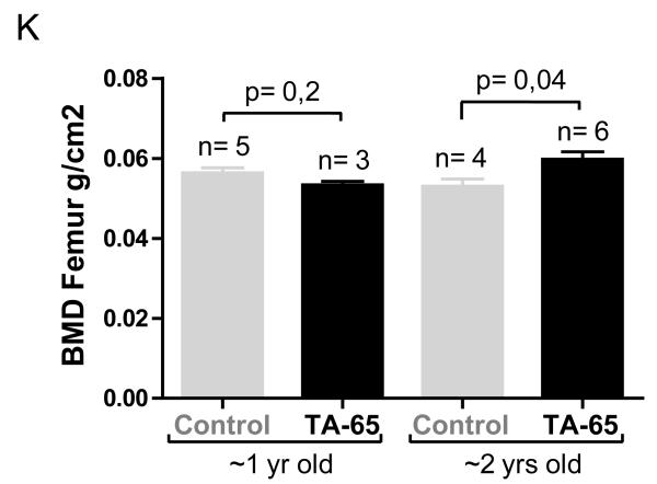
A. Thickness of the subcutaneous fat layer at time of death in the different mice cohorts. B. Representative images of subcutaneous fat and epidermal layers in mice feed with vehicle or vehicle plus TA-65. C. Thickness of the skin epidermal layer, at time of death, in the different mice cohorts. D. Stimulation of wound closure in HEKn cells incubated in the presence of TA-65. Images were taken after incubating for 24, 48 and 72 hours. Wound closure was calculated and is presented in the right panel. E. Hair re-growth capacity was quantified in arbitrary units (a.u., see Materials and Methods) 14 d after plucking. Fisher’s Exact test was used for statistical analysis. Experiments were carried 12 months post the ending to the TA-65 supplementation period in the 1 yr old cohort of mice. Six independent mouse were used. F. Representative images of hair-regrowth. Images were acquired in anesthetized female mice of the 1 yr old group before and 14 days after hair plucking. G. Percentage of Ki67-positive cells in the epidermis (tail skin) from mice non-treated (control) or treated with TA-65. Student’s t-test was used for statistical assessments. At least 6 high-power fields (HPF, x100) were used per independent mouse (around 2000 skin epidermis cells scored per mouse). H. Representative Ki67 immunohistochemistry images of skin epidermis (tail) from the indicated mice cohorts. I. Percentage of TUNEL-positive (Apoptag detection kit) cells in the epidermis (tail skin) from mice non-treated (control) or treated with TA-65. Student’s t-test was used for statistical assessments. At least 6 HPF (x100) were used per independent mouse (around 2000 skin epidermis cells scored per mouse). J. Representative TUNEL stained images of skin from the indicated mice cohorts. K. Femur bone mineral density (BMD femur) measured at the time of death in the 2 yrs old cohorts.
Bone loss, which results from the imbalance between osteoclast and osteoblasts, is also a common finding with increasing age both in mice and humans, leading to age-related bone fragility (Ferguson et al. 2003). Bone density was improved in the 2 yrs old TA-65 treated group compared to their controls without TA-65 supplementation (measurements at time of death, Fig. 5k).
Previous findings demonstrated that TA-65 uptake leads to significant changes in blood or immune parameters in humans (Fauce et al. 2008; Harley et al. 2010). Table 1 summarizes the hematological analysis of either 1 yr and 2 yrs control mice compared to TA-65 treated groups. Although no significant differences were observed in the 1 yr old group, we could observe significant differences in the level of red blood cells (RBC), hemoglobin, and platelets in the TA-65 treated 2 yrs old group compared to age-matched cohorts. The absence of differences in the 1 yr old group could be due to the fact that the blood analysis was performed at time of death, which occurred much after the treatment period in the 1 yr old group, where the effect of TA-65 has been already partially lost.
Table 1.
Values for relevant blood/immune variables among TA-65 treated and controls, in both aged cohorts.
| Blood/Immune variables |
Control 1 yr old (time of death) |
TA-65 1 yr old (time of death) |
T- test |
Control 2 yrs old (time of death) |
TA-65 2 yrs old (time of death) |
T- test |
|
|---|---|---|---|---|---|---|---|
| WBC | 10^9/ l |
7,4 | 5,4 | 0,1 | 8,1 | 4,5 | * |
| LYM | % | 49,6 | 50,0 | n.s. | 41,4 | 36,9 | n.s. |
| MI | % | 2,5 | 4,7 | n.s. | 6,8 | 4,2 | n.s. |
| GR | % | 47,9 | 44,9 | n.s. | 51,7 | 62,2 | n.s. |
| RBC | 10^12/ l |
7,1 | 7,1 | n.s. | 4,8 | 6,3 | ** |
| HGB | g/dl | 10,1 | 10,0 | n.s. | 7,6 | 9,2 | ** |
| HCT | % | 24,3 | 23,9 | n.s. | 21,5 | 26,6 | ** |
| PLT | 10^9/ l |
354 | 289 | n.s. | 292 | 587 | * |
These results demonstrate that TA-65 per se can modestly but significantly enhance overall organ fitness of both mature (1 yr old) and old (2 yrs old) female mice.
TA-65 intake does not impact on mean or maximum longevity of female mice
The known impact of telomerase in ageing and cancer lead us to address whether TA-65 supplementation could have any beneficial or undesirable effects on mouse survival and cancer incidence, respectively. Recent evidences demonstrate that enforced expression of telomerase either from the germline (together with enforced expression of tumor suppressors), or ectopically in adult/old mice leads to a significant extension of both mean and maximum lifespan ((Tomas-Loba et al. 2008) and unpublished results).
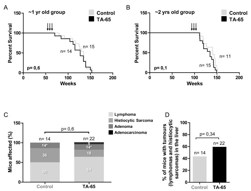
Analysis of the Kaplan-Meier survival curves of control versus TA-65 treated female mice demonstrated no significant effects of TA-65 intake on survival (Fig. 6a,b). Accordingly, TA-65 administration for 4 months did not change statistically the mean or maximal lifespan of female mice under our experimental conditions.
Figure 6. Dietary supplementation of TA-65 has no effect on lifespan in female mice.
A, B. Survival curves of the indicated control of TA-65 treated 1 yr and 2 yrs old mouse cohorts. Alive mice are plotted as a vertical line. The Log rank test was used for statistical analysis. C. Percentage of mice with the indicated tumour at their time of death. (* Of note that 100% [2 mice] and 33% [1 mice] of the mice presenting adenomas also present lymphomas in the control and TA-65 situations, respectively). D. Percentage of cancer penetrance (histiocytic sarcomas and lymphomas) in the liver of the indicated mice cohorts.
TA-65 intake does not increase cancer incidence
A disadvantage of mTERT potentiation could be associated to its capacity to favor proliferation of cancerous cells in murine models (Gonzalez-Suarez et al. 2001; Artandi et al. 2002; Canela et al. 2004; McKay et al. 2008; Tomas-Loba et al. 2008; Rafnar et al. 2009). To address whether TA-65 supplemented diet had undesirable long-term side effects, we performed a pathological analysis of all female mice under treatment and controls at time of death. We observed that TA-65 treated mice presented a similar incidence of malignant cancers at time of death, with a tendency to show decreased sarcomas and slightly increased lymphomas (Fig. 6c,d). Moreover, although we observed an increased incidence of cancer in the liver of TA-65 treated mice (were TA-65 treatment resulted in a 10-fold increase of TERT expression), these values were not significant comparing to the untreated situation (Fig. 6d).
Discussion
Strategies to extend heath span conditions with lifetime extension have been the target of scientific investigation for decades. From the preliminary studies describing the effects of caloric restriction, which evolve to detailed characterizations of pathways and potential targets resulting in recent conceivable treatments, the outcome has been continually focused in a healthier organismal living (Guarente & Kenyon 2000; Hayflick 2000; Kirkwood & Austad 2000; Kenyon 2010).
Here, we characterize a small molecule compound (TA-65) extracted from the roots of a medicinal Chinese plant used, among other things, to “protect” the immune system or “preserve” the liver (Shen et al. 2006; Clement-Kruzel et al. 2008; Mao et al. 2009). We demonstrate here that TA-65 leads to a significant rescue of short telomeres through telomerase activation in MEFs haploinsufficient for the telomerase RNA component. Indeed, TA-65 treatment increases proliferation and mobilization potential of mouse keratinocytes in vitro, a situation mimicking telomerase overexpression (Greider 1998; Cerezo et al. 2003). Recently, Fauce and colleagues demonstrated that TAT2, a similar molecule, have beneficial effects in the activation of CD8+ T lymphocytes from HIV-infected patients where they observe an increase of the proliferative potential and enhancement of cytokine/chemokine production (Fauce et al. 2008).
The use of TA-65 as a treatment to improve health-span in humans has been tested in past few years, where subjects took part in an open label comprehensive dietary supplementation program, which included a TA-65 dose of 10-50mg daily (Harley et al. 2010). Report analysis of the first treatment year has been recently released, demonstrating high tolerability and some beneficial effects in humans (Harley et al. 2010). We have now assessed the specific effects of TA-65 in a blinded study of two cohorts of 1 year old (mature) or 2 years old (old) female mice where an adjusted dose of TA-65 was administrated daily for 4 months. TA-65 resulted in a similar rescue of short telomeres in leukocytes post-treatment as observed with humans, most likely through an activation of telomerase. Actually, when we measured mRNA transcripts we observe that TA-65 lead to 10 fold increase of telomerase RNA levels in the liver of treated mice comparing to the non-treated same-age cohorts. These results enforce the previous idea that TA-65 regulates telomerase at the transcription level, probably through the regulation of the MAPK pathway (Fauce et al. 2008). Whether or not TA-65 could regulate post-transcriptionally the levels of telomerase in the liver or other tissues has not been assessed here or elsewhere, although this is improbable in the case of mouse telomerase since some mTERT post-translational modifications such as phosphorylation is mediated through the AKT pathway, which seems not to be influenced by TA-65 (Kang et al. 1999; Gomez-Garcia et al. 2005; Fauce et al. 2008).
We further show here that TA-65 dependent telomerase activation results in a better organ fitness as demonstrated by the improved scores at the glucose tolerance test and insulin levels at fasting. These enhancements could be related to the TA-65 dependent ERK pathway activation in the liver and subsequent telomerase expression, which have been shown to mediate the glucose tolerance and insulin response (Tomas-Loba et al. 2008). Moreover, ERK signal is silenced by the activation of the p38 MAPK, which responds to external damage signals arising usually with ageing progression and oxidative damage accumulation, and result in age-related insulin insensitivity and glucose intolerance (Kyriakis & Avruch 2001; Au et al. 2003; Li et al. 2007; Bluher et al. 2009). TA-65 supplemented mice also present modest enhancement of the subcutaneous and epidermal thickness, as well as higher bone density, representative of an overall fitness status improvement. A similar situation has been previously observed in mice overexpressing the telomerase catalytic subunit (Tomas-Loba et al. 2008). Analysis of the blood parameters also demonstrated that 2 yrs old TA-65 treated mice present higher levels of RBC and hemoglobin comparing to the control cohorts. The decrease of such parameters is a regular situation experienced during aging progression in healthy old mice and could be related to RBC fragility and deficient regulation of the stem cell pools (Ewing & Tauber 1964; Finch & Foster 1973; Tyan 1980; Tyan 1982a; Tyan 1982b; Xing et al. 2006). Importantly, improved health-span of TA-65 treated mice is not accompanied by increased cancer incidence, which may be related to the fact that TERT levels are very modestly increased in all tissues tested except for the liver. Alternatively, telomerase enhancement in adult (1 yr old) or very old mice (2 yr old) may have beneficial effects without increasing cancer incidence owe to the fact that older organisms are more refractory to proliferative stimuli. In conclusion, the TA-65-dependent mechanisms mediating healthspan improvements could involve, and be mostly mediated, by the increased levels of telomerase expression. Indeed, it has been recently observed that systemic telomerase overexpression from the germline leads to protection from aging associated pathologies (Tomas-Loba et al. 2008), and a similar situation could be mimicked expressing telomerase late in life in a telomerase deficient background (Jaskelioff et al. 2010). Telomerase was also shown to support stem cell mobilization which could explain a better tissue regeneration capacity through time (Flores et al. 2005). In line with this, we observed a higher proliferation rate and a partial protection from cell death in some tissues of TA65 treated mice (Fig. 5g-j and Sup. Fig. 5) comparing to the control situation although other tissues did not present significant alterations (Sup Fig 5). Even if novel roles have been described for telomerase (for instance, in activating the Wnt pathway (Park et al. 2009)), the lack of scientific support did not permit to unveil so far which of its functions could further contribute to the observed effects.
Here, we provide evidence on the role of TA-65 in a healthy ageing improvement but not survival (either mean or maximum longevity) in female mice. The balance between health enhancement and longevity do not always correlate. Indeed, if a long-lived mammalian organism presents regularly health benefits (Berryman et al. 2008; Tomas-Loba et al. 2008) the opposite as shown to be potentially untrue (Herranz & Serrano 2010). Of importance is the fact that we feed our mice cohorts for a limited period of time (4 months), being the impact of the compound shown to be partially lost during the absence period (Fauce et al. 2008); even though, we show that some phenotypic rescues (such as the hair re-growth) can be maintained longer after TA-65 uptake conclusion. We cannot discard that a longer or constant dietary supplementation of TA-65 could also impact on lifespan or cancer. In conclusion, here we described new findings on the potential therapeutic use of TA-65, delineating for the first time its mechanism of action and organismal response.
Methods
Formulation of TA-65
TA-65 was obtained from TA Sciences. TA-65 is a >95% pure single chemical entity as judged by HPLC and NMR (data not shown), which was isolated from a proprietary extract of the dried root of Astragalus membranaceus (Harley et al. 2010). TA-65 was dissolved in DMSO at 1mM or 10mM stock concentration (1000x final concentration).
Mice, cells and treatments
All G2 Terc−/− and Terc+/− mice used in this study were of the same identical genetic background (C57BL6). Third generation G3 Terc−/− and G3 Terc+/− littermate mouse embryos were derived from crossing G2 Terc−/− with Terc+/− mice (Fig. 1a). Genotyping was performed by Southern blotting as described (Blasco et al. 1997). Mice were maintained at the Spanish National Cancer Centre (CNIO) under specific pathogen-free conditions in accordance with the recommendations of the Federation of European Laboratory Animal Science Associations.
Mouse embryonic fibroblasts (MEF) were prepared from day 13.5 embryos as described (Blasco et al. 1997). Two independent TA-65 treatments were performed using independent MEF cultures. G3 Terc−/− and G3 Terc+/− primary MEF (passage 1) were cultured for 6 days in Dulbecco’s modified Eagle’s medium (DMEM; Gibco, Paisley, UK) supplemented with 10% fetal bovine serum (FBS) and antibiotic/antimycotic at 37°C / 5% CO2. Fresh TA-65 was added to the media at a final concentration of 1μM or 10μM every 24 to 48 hours, along with change of media, and cells were passaged at day 2 and day 5. Control cells were treated with 0.1% DMSO.
Aged cohorts of female mice were obtained directly from Harlan Laboratories. Both 1 yr and 2 yrs old groups were aged under standard conditions and present 100% C57BL/6JOlaHsd background. Mice were conditioned in a pathogen-free area of CNIO after arrival, in accordance with the recommendations of the Federation of European Laboratory Animal Science Associations. Half of the mice were supplemented with 25mg of TA-65 per kg of mouse body weight per day, mixed in 100μl of fruit mash, for 4 months. Fruit mash alone was supplemented to the control cohorts (100μl, with the same periodicity as treatment groups). Mice were daily inspected by an authorized animal facility supervisor, before and after TA-65 treatment. Mice were sacrificed when presenting signs of illness or tumors in accordance to the Guidelines for Humane Endpoints for Animals Used in Biomedical Research.
Telomere repeat amplification protocol (TRAP)
Telomerase activity in G3 Terc−/− and G3 Terc+/− MEF was measured by Telomeric Repeat Amplification Protocol (TRAP) as described previously (Blasco et al. 1997). Protein extracts of HCT116 cells served as a positive control. As negative controls, protein extracts were treated with 5 ug RNase for 10 min at 30°C before performing the TRAP assay. Radioactive gels were exposed to BioMax MS films (Kodak) for at least 12 hours at -80°C. Quantification was done on digitalized films, using ImageJ software (NIH). The mean intensity of each lane after background subtraction was calculated, normalized by the dilution factor of the applied protein extract and expressed as folds relative to the corresponding DMSO treated cells.
Quantitative telomere fluorescence in situ hybridization (Q-FISH) and High throughput (HT) QFISH
At 70% confluence, primary MEFs (passage 3) were incubated with 0.1mg/mL Colcemide (Gibco, Paisley, UK) at 37°C for 4 hours to induce metaphases. Cells were then harvested and fixed in methanol:acetic acid (3:1). Metaphases were spread on glass slides and dried overnight, and Q-FISH was performed using a PNA-telomeric probe as described previously (Herrera et al. 1999; Samper et al. 2000).
Metaphases were analyzed with a fluorescence microscope (DMRA2, Leica, Wetzlar, Germany) and images were captured with a COHU CCD camera (San Diego, CA, USA) using the Leica Q-FISH software. Fluorescence intensity of telomeres was quantified by spot IOD analysis using the TFL-TELO software (provided by P.Landsdorp, Vancouver, Canada) and normalized to the fluorescence intensity of fluorescent beads (Molecular Probes, Carlsbad, CA, USA).
HT-QFISH was performed as previously described (Canela et al. 2007). Briefly, blood was extracted at the indicated time points after TA-65 treatment was finished. Peripheral blood leukocytes were plated on a clear bottom black-walled 96-well plate. Telomere length values were analyzed using individual telomere spots (> 5000 per sample) corresponding to the specific binding of a Cy3 labeled telomeric probe. Fluorescence intensities were converted into Kb as previously described (McIlrath et al. 2001; Canela et al. 2007).
Quantitative real-time RT–PCR
Total RNA from liver was extracted with Trizol (Life Technologies). Samples were treated with DNase I before reverse transcription using random primers and Superscript Reverse Transcriptase (Life Technologies), following manufacturer’s guidelines. The primers used were:
Actin-For: GGCACCACACCTTCTACAATG;
Actin-Rev: GTGGTGGTGAAGCTGTAG;
TERT-For: GGATTGCCACTGGCTCCG;
TERT-Rev: TGCCTGACCTCCTCTTGTGAC;
JunB-For: AGC CGC CTC CCG TCT ACA CCA;
JunB-Rev GCC GCT TTC GCTCCA CTT TGA TG;
c-Myc-For: ATGCCCCTCAACGTGAACTTC;
c-Myc-Rev: CGCAACATAGGATGGAGAGCA;
CyclinD1-For: TGCGCCCTCCGTATCTTAC;
CyclinD1-Rev: ATCTTAGAGGCCACGAACATGC;
CD44-For: CAGCCTACTGGAGATCAGGATGA;
CD44-Rev: GGAGTCCTTGGATGAGTCTCGA;
p16-For: CGTACCCCGATTCAGGTGAT;
p16-Rev: TTGAGCAGAAGAGCTGCTACGT;
Klf4-For: GCGAACTCACACAGGCGAGAAACC;
Klf4-Rev: TCGCTTCCTCTTCCTCCGACACA;
Fibronectin-For: TACCAAGGTCAATCCACACCCC;
Fibronectin-Rev: CAGATGGCAAAAGAAAGCAGAGG;
Student’s t-test was performed on the ΔΔCt as previously recommended (Yuan et al. 2006; Matheu et al. 2007).
Histological analyses
We performed c-Myc (sc-764, Santa Cruz biotechnology) Ki67 (M7249, clone TEC-3) and TUNEL (ApopTag Detection Kit) stainings on paraformaldehyde-fixed, paraffin-embedded sections (AxioVision and Scion Image software were used for image analysis; quantitation of immunostainings was determined by counting the number of peroxidase stained nuclei over the total number of hematoxylin-stained nuclei) and oil red O staining (fat cells) on liver frozen sections. Samples were processed and acquired under identical conditions.
For complete blood cell counts, we collected blood samples just before euthanasia. Data represents information collected from, at least, 5 mice. Counts were done using an Abacus Junior Vet System.
Glucose Tolerance Tests, Insulin Levels and HOMA-IR
Glucose tolerance test and analysis was performed as described elsewhere (Moynihan et al. 2005). Mice were fasted for at least 6 hr, and injected intraperitoneally with a 50% dextrose solution (2 g/kg body weight).
Blood insulin levels were measured with an Ultra-Sensitive Mouse Insulin ELISA Kit (Crystal Chem Inc. #90080), following manufacturers protocol. Mice were fasted for at least 6 h prior to blood insulin analysis. HOMA-IR was calculated as previously described (Matthews et al. 1985; Heikkinen et al. 2007).
Bone density, skin measurements and cell migration
Bone mineral density was measured in the femur of female mice post-mortem using a Dual Energy X-ray Absorptiometry (DEXA) scan device. Subcutaneous fat and epidermal layers measurements were performed as previously described (Tomas-Loba et al. 2008). ImageJ software was used for skin length measurements.
Cell migration assays were performed on neonatal human epidermal keratinocytes (HEKn). Briefly, a confluent monolayer of HEKn was scraped with a sterile pipette tip. Migration into the wound was scored after 24h, 48h or 72h in the presence of DMSO or TA-65 (10 μM). ImageJ software was used for length measurements.
Hair re-growth assay was performed and quantified as previously described (Matheu et al. 2007). Briefly, dorsal hair was removed by plucking from a square of approximately 1.5 cm × 1.5 cm. Hair re-growth was scored two weeks later and a semi-quantitative assessment using an arbitrary scale from one to four (where four represents complete hair regeneration).
Statistical analyses
Statistical significances were calculated using GraphPad Prism 5 software. An unpaired student’s t-test was used to calculate statistical significances of telomerase activity (TRAP), mRNA expression levels, protein levels, IHC quantification, glucose tolerance, insulin levels, HOMA-IR, skin measurements and BMD. Statistical analysis of hair regrowth experiments was assessed with Fisher’s exact test. Statistical significances of differences in overall telomere length were determined by Wilcoxon-Mann-Whitney rank sum test. For statistical analyses of percentage of short telomeres and pathological analysis at time of death a Chi-square test was used. Finally, a Log-Rank test was used for survival comparisons between cohorts.
Supplementary Material
Supplementary Figure 1. A. Telomerase activity was measured in MEFs extracts grown in the presence or absence of TA-65 for 24h and 5 days. Two different concentrations of TA-65 were tested. A cellular extract from HCT116 cells was included as a positive control, and RNase was added in all experimental settings as negative control. IC, PCR efficiency control.
Supplementary Figure 2. A Body weight of the indicated mice before or after treatment is given as mean±SEM.
Supplementary Figure 3. A. Fold change in mTERT mRNA levels 3 months post treatment in different tissues of 1 year old TA-65 treated mice compared to age-matched controls. mTERT mRNA values were normalized to actin. B. Fold change in JunB mRNA levels 3 months post treatment in different tissues of 1 year old TA-65 treated mice compared to age-matched controls. JunB mRNA values were normalized to actin. C. Fold change in c-Myc mRNA levels 3 months post treatment in different tissues of 1 year old TA-65 treated mice compared to age-matched controls. c-Myc mRNA values were normalized to actin.
Supplementary Figure 4. A. Fold changes in CD44, CyclinD1, Klf4, Fibronectin and p16 mRNA levels in liver extracts of TA-65 treated mice compared to age-matched controls. mRNA values were normalized to actin.
Supplementary Figure 5. A. Percentage of Ki67-positive cells in the lung from mice non-treated (control) or treated with TA-65. Student’s t-test was used for statistical assessments. At least 4 HPF (x100) were used per independent mouse (around 24000 lung cells scored per mouse). B. Representative Ki67 immunohistochemistry images of lung from the indicated mice cohorts. C. Percentage of TUNEL-positive (Apoptag detection kit) cells in the lung from mice non-treated (control) or treated with TA-65. Student’s t-test was used for statistical assessments. At least 4 HPF (x100) were used per independent mouse (around 24000 cells scored per mouse). D. Representative TUNEL stained images of lung from the indicated mice cohorts. E. Percentage of Ki67-positive cells in the liver from mice non-treated (control) or treated with TA-65. Student’s t-test was used for statistical assessments. At least 5 HPF (x100) were used per independent mouse (around 3000 hepatocytes scored per mouse). F. Representative Ki67 immunohistochemistry images of liver from the indicated mice cohorts. G. Percentage of TUNEL-positive (Apoptag detection kit) cells in the liver from mice non-treated (control) or treated with TA-65. Student’s t-test was used for statistical assessments. At least 5 HPF (x100) were used per independent mouse (around 3000 hepatocytes scored per mouse). H. Representative TUNEL stained images of liver from the indicated mice cohorts.
Acknowledgements
We thank R. Serrano for mouse care and J. Flores for pathology analyses. We thank Comparative Pathology Unit at CNIO for technical assistance. M.A. Blasco’s laboratory is funded by the Spanish Ministry of Science and Innovation (MICINN), the European Union (GENICA, TELOMARKER), the European Research Council (ERC Advance Grants), the Körber European Science Award and the Fundacion Lilly Pre-clinical Award to M.A. Blasco.
Footnotes
Author contributions M.A.B conceived the idea. B.B. performed most of the experiments of the paper. K.S. performed the TA-65 administration, and performed Fig 1d-g and Fig. 5d. E.V. performed telomere length determinations (Fig. 3). A.T. performed the TRAP assays (Fig 1b and Sup. Fig. 1). M.A.B and B.B. wrote the paper.
References
- Armanios MY, Chen JJ, Cogan JD, Alder JK, Ingersoll RG, Markin C, Lawson WE, Xie M, Vulto I, Phillips JA, 3rd, Lansdorp PM, Greider CW, Loyd JE. Telomerase mutations in families with idiopathic pulmonary fibrosis. N Engl J Med. 2007;356:1317–1326. doi: 10.1056/NEJMoa066157. [DOI] [PubMed] [Google Scholar]
- Artandi SE, Alson S, Tietze MK, Sharpless NE, Ye S, Greenberg RA, Castrillon DH, Horner JW, Weiler SR, Carrasco RD, DePinho RA. Constitutive telomerase expression promotes mammary carcinomas in aging mice. Proc Natl Acad Sci U S A. 2002;99:8191–8196. doi: 10.1073/pnas.112515399. [DOI] [PMC free article] [PubMed] [Google Scholar]
- Au WS, Kung HF, Lin MC. Regulation of microsomal triglyceride transfer protein gene by insulin in HepG2 cells: roles of MAPKerk and MAPKp38. Diabetes. 2003;52:1073–1080. doi: 10.2337/diabetes.52.5.1073. [DOI] [PubMed] [Google Scholar]
- Bailey CJ, Flatt PR. Hormonal control of glucose homeostasis during development and ageing in mice. Metabolism. 1982;31:238–246. doi: 10.1016/0026-0495(82)90059-2. [DOI] [PubMed] [Google Scholar]
- Berryman DE, Christiansen JS, Johannsson G, Thorner MO, Kopchick JJ. Role of the GH/IGF-1 axis in lifespan and healthspan: lessons from animal models. Growth Horm IGF Res. 2008;18:455–471. doi: 10.1016/j.ghir.2008.05.005. [DOI] [PMC free article] [PubMed] [Google Scholar]
- Bianchi A, Shore D. Increased association of telomerase with short telomeres in yeast. Genes Dev. 2007;21:1726–1730. doi: 10.1101/gad.438907. [DOI] [PMC free article] [PubMed] [Google Scholar]
- Blackburn EH. Switching and signaling at the telomere. Cell. 2001;106:661–673. doi: 10.1016/s0092-8674(01)00492-5. [DOI] [PubMed] [Google Scholar]
- Blasco MA, Lee HW, Hande MP, Samper E, Lansdorp PM, DePinho RA, Greider CW. Telomere shortening and tumor formation by mouse cells lacking telomerase RNA. Cell. 1997;91:25–34. doi: 10.1016/s0092-8674(01)80006-4. [DOI] [PubMed] [Google Scholar]
- Bluher M, Bashan N, Shai I, Harman-Boehm I, Tarnovscki T, Avinaoch E, Stumvoll M, Dietrich A, Kloting N, Rudich A. Activated Ask1-MKK4-p38MAPK/JNK stress signaling pathway in human omental fat tissue may link macrophage infiltration to whole-body Insulin sensitivity. J Clin Endocrinol Metab. 2009;94:2507–2515. doi: 10.1210/jc.2009-0002. [DOI] [PubMed] [Google Scholar]
- Calado RT, Young NS. Telomere diseases. N Engl J Med. 2009;361:2353–2365. doi: 10.1056/NEJMra0903373. [DOI] [PMC free article] [PubMed] [Google Scholar]
- Canela A, Martin-Caballero J, Flores JM, Blasco MA. Constitutive expression of tert in thymocytes leads to increased incidence and dissemination of T-cell lymphoma in Lck-Tert mice. Mol Cell Biol. 2004;24:4275–4293. doi: 10.1128/MCB.24.10.4275-4293.2004. [DOI] [PMC free article] [PubMed] [Google Scholar]
- Canela A, Vera E, Klatt P, Blasco MA. High-throughput telomere length quantification by FISH and its application to human population studies. Proc Natl Acad Sci U S A. 2007;104:5300–5305. doi: 10.1073/pnas.0609367104. [DOI] [PMC free article] [PubMed] [Google Scholar]
- Cayuela ML, Flores JM, Blasco MA. The telomerase RNA component Terc is required for the tumour-promoting effects of Tert overexpression. EMBO Rep. 2005;6:268–274. doi: 10.1038/sj.embor.7400359. [DOI] [PMC free article] [PubMed] [Google Scholar]
- Cerezo A, Stark HJ, Moshir S, Boukamp P. Constitutive overexpression of human telomerase reverse transcriptase but not c-myc blocks terminal differentiation in human HaCaT skin keratinocytes. J Invest Dermatol. 2003;121:110–119. doi: 10.1046/j.1523-1747.2003.12304.x. [DOI] [PubMed] [Google Scholar]
- Clement-Kruzel S, Hwang SA, Kruzel MC, Dasgupta A, Actor JK. Immune modulation of macrophage pro-inflammatory response by goldenseal and Astragalus extracts. J Med Food. 2008;11:493–498. doi: 10.1089/jmf.2008.0044. [DOI] [PubMed] [Google Scholar]
- Chang L, Karin M. Mammalian MAP kinase signalling cascades. Nature. 2001;410:37–40. doi: 10.1038/35065000. [DOI] [PubMed] [Google Scholar]
- de Lange T. Shelterin: the protein complex that shapes and safeguards human telomeres. Genes Dev. 2005;19:2100–2110. doi: 10.1101/gad.1346005. [DOI] [PubMed] [Google Scholar]
- Ewing KL, Tauber OE. Hematological Changes in Aging Male C57bl/6 Jax Mice. J Gerontol. 1964;19:165–167. doi: 10.1093/geronj/19.2.165. [DOI] [PubMed] [Google Scholar]
- Fauce SR, Jamieson BD, Chin AC, Mitsuyasu RT, Parish ST, Ng HL, Kitchen CM, Yang OO, Harley CB, Effros RB. Telomerase-based pharmacologic enhancement of antiviral function of human CD8+ T lymphocytes. J Immunol. 2008;181:7400–7406. doi: 10.4049/jimmunol.181.10.7400. [DOI] [PMC free article] [PubMed] [Google Scholar]
- Feldstein AE, Canbay A, Angulo P, Taniai M, Burgart LJ, Lindor KD, Gores GJ. Hepatocyte apoptosis and fas expression are prominent features of human nonalcoholic steatohepatitis. Gastroenterology. 2003;125:437–443. doi: 10.1016/s0016-5085(03)00907-7. [DOI] [PubMed] [Google Scholar]
- Ferguson VL, Ayers RA, Bateman TA, Simske SJ. Bone development and age-related bone loss in male C57BL/6J mice. Bone. 2003;33:387–398. doi: 10.1016/s8756-3282(03)00199-6. [DOI] [PubMed] [Google Scholar]
- Finch CE, Foster JR. Hematologic and serum electrolyte values of the C57BL-6J male mouse in maturity and senescence. Lab Anim Sci. 1973;23:339–349. [PubMed] [Google Scholar]
- Flores I, Canela A, Vera E, Tejera A, Cotsarelis G, Blasco MA. The longest telomeres: a general signature of adult stem cell compartments. Genes Dev. 2008;22:654–667. doi: 10.1101/gad.451008. [DOI] [PMC free article] [PubMed] [Google Scholar]
- Flores I, Cayuela ML, Blasco MA. Effects of telomerase and telomere length on epidermal stem cell behavior. Science. 2005;309:1253–1256. doi: 10.1126/science.1115025. [DOI] [PubMed] [Google Scholar]
- Gomez-Garcia L, Sanchez FM, Vallejo-Cremades MT, de Segura IA, del Campo Ede M. Direct activation of telomerase by GH via phosphatidylinositol 3′-kinase. J Endocrinol. 2005;185:421–428. doi: 10.1677/joe.1.05766. [DOI] [PubMed] [Google Scholar]
- Gonzalez-Suarez E, Samper E, Ramirez A, Flores JM, Martin-Caballero J, Jorcano JL, Blasco MA. Increased epidermal tumors and increased skin wound healing in transgenic mice overexpressing the catalytic subunit of telomerase, mTERT, in basal keratinocytes. EMBO J. 2001;20:2619–2630. doi: 10.1093/emboj/20.11.2619. [DOI] [PMC free article] [PubMed] [Google Scholar]
- Greenberg RA, O’Hagan RC, Deng H, Xiao Q, Hann SR, Adams RR, Lichtsteiner S, Chin L, Morin GB, DePinho RA. Telomerase reverse transcriptase gene is a direct target of c-Myc but is not functionally equivalent in cellular transformation. Oncogene. 1999;18:1219–1226. doi: 10.1038/sj.onc.1202669. [DOI] [PubMed] [Google Scholar]
- Greider CW. Telomerase activity, cell proliferation, and cancer. Proc Natl Acad Sci U S A. 1998;95:90–92. doi: 10.1073/pnas.95.1.90. [DOI] [PMC free article] [PubMed] [Google Scholar]
- Greider CW, Blackburn EH. Identification of a specific telomere terminal transferase activity in Tetrahymena extracts. Cell. 1985;43:405–413. doi: 10.1016/0092-8674(85)90170-9. [DOI] [PubMed] [Google Scholar]
- Guarente L. Sirtuins as potential targets for metabolic syndrome. Nature. 2006;444:868–874. doi: 10.1038/nature05486. [DOI] [PubMed] [Google Scholar]
- Guarente L, Kenyon C. Genetic pathways that regulate ageing in model organisms. Nature. 2000;408:255–262. doi: 10.1038/35041700. [DOI] [PubMed] [Google Scholar]
- Gupta S, Seth A, Davis RJ. Transactivation of gene expression by Myc is inhibited by mutation at the phosphorylation sites Thr-58 and Ser-62. Proc Natl Acad Sci U S A. 1993;90:3216–3220. doi: 10.1073/pnas.90.8.3216. [DOI] [PMC free article] [PubMed] [Google Scholar]
- Hao LY, Armanios M, Strong MA, Karim B, Feldser DM, Huso D, Greider CW. Short telomeres, even in the presence of telomerase, limit tissue renewal capacity. Cell. 2005;123:1121–1131. doi: 10.1016/j.cell.2005.11.020. [DOI] [PubMed] [Google Scholar]
- Harley CB. Telomerase therapeutics for degenerative diseases. Curr Mol Med. 2005;5:205–211. doi: 10.2174/1566524053586671. [DOI] [PubMed] [Google Scholar]
- Harley CB, Futcher AB, Greider CW. Telomeres shorten during ageing of human fibroblasts. Nature. 1990;345:458–460. doi: 10.1038/345458a0. [DOI] [PubMed] [Google Scholar]
- Harley CB, Liu W, Blasco M, Vera E, Andrews WH, Briggs LA, Raffaele JM. A Natural Product Telomerase Activator As Part of a Health Maintenance Program. Rejuvenation Res. 2010 doi: 10.1089/rej.2010.1085. [DOI] [PMC free article] [PubMed] [Google Scholar]
- Hayflick L. The future of ageing. Nature. 2000;408:267–269. doi: 10.1038/35041709. [DOI] [PubMed] [Google Scholar]
- Heikkinen S, Argmann CA, Champy MF, Auwerx J. Evaluation of glucose homeostasis. Curr Protoc Mol Biol. 2007 doi: 10.1002/0471142727.mb29b03s77. Chapter 29, Unit 29B 23. [DOI] [PubMed] [Google Scholar]
- Hemann MT, Rudolph KL, Strong MA, DePinho RA, Chin L, Greider CW. Telomere dysfunction triggers developmentally regulated germ cell apoptosis. Mol Biol Cell. 2001a;12:2023–2030. doi: 10.1091/mbc.12.7.2023. [DOI] [PMC free article] [PubMed] [Google Scholar]
- Hemann MT, Strong MA, Hao LY, Greider CW. The shortest telomere, not average telomere length, is critical for cell viability and chromosome stability. Cell. 2001b;107:67–77. doi: 10.1016/s0092-8674(01)00504-9. [DOI] [PubMed] [Google Scholar]
- Herranz D, Serrano M. Impact of Sirt1 on mammalian aging. Aging (Albany NY) 2010;2:315–316. doi: 10.18632/aging.100156. [DOI] [PMC free article] [PubMed] [Google Scholar]
- Herrera E, Samper E, Martin-Caballero J, Flores JM, Lee HW, Blasco MA. Disease states associated with telomerase deficiency appear earlier in mice with short telomeres. EMBO J. 1999;18:2950–2960. doi: 10.1093/emboj/18.11.2950. [DOI] [PMC free article] [PubMed] [Google Scholar]
- Higuchi H, Gores GJ. Mechanisms of liver injury: an overview. Curr Mol Med. 2003;3:483–490. doi: 10.2174/1566524033479528. [DOI] [PubMed] [Google Scholar]
- Inui T, Shinomiya N, Fukasawa M, Kuranaga N, Ohkura S, Seki S. Telomerase activation and MAPK pathways in regenerating hepatocytes. Hum Cell. 2001;14:275–282. [PubMed] [Google Scholar]
- Jaskelioff M, Muller FL, Paik JH, Thomas E, Jiang S, Adams AC, Sahin E, Kost-Alimova M, Protopopov A, Cadinanos J, Horner JW, Maratos-Flier E, Depinho RA. Telomerase reactivation reverses tissue degeneration in aged telomerase-deficient mice. Nature. 2010 doi: 10.1038/nature09603. [DOI] [PMC free article] [PubMed] [Google Scholar]
- Jiang H, Schiffer E, Song Z, Wang J, Zurbig P, Thedieck K, Moes S, Bantel H, Saal N, Jantos J, Brecht M, Jeno P, Hall MN, Hager K, Manns MP, Hecker H, Ganser A, Dohner K, Bartke A, Meissner C, Mischak H, Ju Z, Rudolph KL. Proteins induced by telomere dysfunction and DNA damage represent biomarkers of human aging and disease. Proc Natl Acad Sci U S A. 2008;105:11299–11304. doi: 10.1073/pnas.0801457105. [DOI] [PMC free article] [PubMed] [Google Scholar]
- Kang SS, Kwon T, Kwon DY, Do SI. Akt protein kinase enhances human telomerase activity through phosphorylation of telomerase reverse transcriptase subunit. J Biol Chem. 1999;274:13085–13090. doi: 10.1074/jbc.274.19.13085. [DOI] [PubMed] [Google Scholar]
- Kelder B, Boyce K, Kriete A, Clark R, Berryman DE, Nagatomi S, List EO, Braughler M, Kopchick JJ. CIDE-A is expressed in liver of old mice and in type 2 diabetic mouse liver exhibiting steatosis. Comp Hepatol. 2007;6:4. doi: 10.1186/1476-5926-6-4. [DOI] [PMC free article] [PubMed] [Google Scholar]
- Kenyon CJ. The genetics of ageing. Nature. 2010;464:504–512. doi: 10.1038/nature08980. [DOI] [PubMed] [Google Scholar]
- Kirkwood TB, Austad SN. Why do we age? Nature. 2000;408:233–238. doi: 10.1038/35041682. [DOI] [PubMed] [Google Scholar]
- Kyriakis JM, Avruch J. Mammalian mitogen-activated protein kinase signal transduction pathways activated by stress and inflammation. Physiol Rev. 2001;81:807–869. doi: 10.1152/physrev.2001.81.2.807. [DOI] [PubMed] [Google Scholar]
- Lefloch R, Pouyssegur J, Lenormand P. Single and combined silencing of ERK1 and ERK2 reveals their positive contribution to growth signaling depending on their expression levels. Mol Cell Biol. 2008;28:511–527. doi: 10.1128/MCB.00800-07. [DOI] [PMC free article] [PubMed] [Google Scholar]
- Li G, Barrett EJ, Barrett MO, Cao W, Liu Z. Tumor necrosis factor-alpha induces insulin resistance in endothelial cells via a p38 mitogen-activated protein kinase-dependent pathway. Endocrinology. 2007;148:3356–3363. doi: 10.1210/en.2006-1441. [DOI] [PubMed] [Google Scholar]
- Liu Y, Snow BE, Hande MP, Yeung D, Erdmann NJ, Wakeham A, Itie A, Siderovski DP, Lansdorp PM, Robinson MO, Harrington L. The telomerase reverse transcriptase is limiting and necessary for telomerase function in vivo. Curr Biol. 2000;10:1459–1462. doi: 10.1016/s0960-9822(00)00805-8. [DOI] [PubMed] [Google Scholar]
- Mao XQ, Yu F, Wang N, Wu Y, Zou F, Wu K, Liu M, Ouyang JP. Hypoglycemic effect of polysaccharide enriched extract of Astragalus membranaceus in diet induced insulin resistant C57BL/6J mice and its potential mechanism. Phytomedicine. 2009;16:416–425. doi: 10.1016/j.phymed.2008.12.011. [DOI] [PubMed] [Google Scholar]
- Martinez P, Thanasoula M, Carlos AR, Gomez-Lopez G, Tejera AM, Schoeftner S, Dominguez O, Pisano DG, Tarsounas M, Blasco MA. Mammalian Rap1 controls telomere function and gene expression through binding to telomeric and extratelomeric sites. Nat Cell Biol. 2010;12:768–780. doi: 10.1038/ncb2081. [DOI] [PMC free article] [PubMed] [Google Scholar]
- Matheu A, Maraver A, Klatt P, Flores I, Garcia-Cao I, Borras C, Flores JM, Vina J, Blasco MA, Serrano M. Delayed ageing through damage protection by the Arf/p53 pathway. Nature. 2007;448:375–379. doi: 10.1038/nature05949. [DOI] [PubMed] [Google Scholar]
- Matthews DR, Hosker JP, Rudenski AS, Naylor BA, Treacher DF, Turner RC. Homeostasis model assessment: insulin resistance and beta-cell function from fasting plasma glucose and insulin concentrations in man. Diabetologia. 1985;28:412–419. doi: 10.1007/BF00280883. [DOI] [PubMed] [Google Scholar]
- McIlrath J, Bouffler SD, Samper E, Cuthbert A, Wojcik A, Szumiel I, Bryant PE, Riches AC, Thompson A, Blasco MA, Newbold RF, Slijepcevic P. Telomere length abnormalities in mammalian radiosensitive cells. Cancer Res. 2001;61:912–915. [PubMed] [Google Scholar]
- McKay JD, Hung RJ, Gaborieau V, Boffetta P, Chabrier A, Byrnes G, Zaridze D, Mukeria A, Szeszenia-Dabrowska N, Lissowska J, Rudnai P, Fabianova E, Mates D, Bencko V, Foretova L, Janout V, McLaughlin J, Shepherd F, Montpetit A, Narod S, Krokan HE, Skorpen F, Elvestad MB, Vatten L, Njolstad I, Axelsson T, Chen C, Goodman G, Barnett M, Loomis MM, Lubinski J, Matyjasik J, Lener M, Oszutowska D, Field J, Liloglou T, Xinarianos G, Cassidy A, Vineis P, Clavel-Chapelon F, Palli D, Tumino R, Krogh V, Panico S, Gonzalez CA, Ramon Quiros J, Martinez C, Navarro C, Ardanaz E, Larranaga N, Kham KT, Key T, Bueno-de-Mesquita HB, Peeters PH, Trichopoulou A, Linseisen J, Boeing H, Hallmans G, Overvad K, Tjonneland A, Kumle M, Riboli E, Zelenika D, Boland A, Delepine M, Foglio M, Lechner D, Matsuda F, Blanche H, Gut I, Heath S, Lathrop M, Brennan P. Lung cancer susceptibility locus at 5p15.33. Nat Genet. 2008;40:1404–1406. doi: 10.1038/ng.254. [DOI] [PMC free article] [PubMed] [Google Scholar]
- Mitchell JR, Wood E, Collins K. A telomerase component is defective in the human disease dyskeratosis congenita. Nature. 1999;402:551–555. doi: 10.1038/990141. [DOI] [PubMed] [Google Scholar]
- Moynihan KA, Grimm AA, Plueger MM, Bernal-Mizrachi E, Ford E, Cras-Meneur C, Permutt MA, Imai S. Increased dosage of mammalian Sir2 in pancreatic beta cells enhances glucose-stimulated insulin secretion in mice. Cell Metab. 2005;2:105–117. doi: 10.1016/j.cmet.2005.07.001. [DOI] [PubMed] [Google Scholar]
- Park JI, Venteicher AS, Hong JY, Choi J, Jun S, Shkreli M, Chang W, Meng Z, Cheung P, Ji H, McLaughlin M, Veenstra TD, Nusse R, McCrea PD, Artandi SE. Telomerase modulates Wnt signalling by association with target gene chromatin. Nature. 2009;460:66–72. doi: 10.1038/nature08137. [DOI] [PMC free article] [PubMed] [Google Scholar]
- Pericuesta E, Ramirez MA, Villa-Diaz A, Relano-Gines A, Torres JM, Nieto M, Pintado B, Gutierrez-Adan A. The proximal promoter region of mTert is sufficient to regulate telomerase activity in ES cells and transgenic animals. Reprod Biol Endocrinol. 2006;4:5. doi: 10.1186/1477-7827-4-5. [DOI] [PMC free article] [PubMed] [Google Scholar]
- Pfluger PT, Herranz D, Velasco-Miguel S, Serrano M, Tschop MH. Sirt1 protects against high-fat diet-induced metabolic damage. Proc Natl Acad Sci U S A. 2008;105:9793–9798. doi: 10.1073/pnas.0802917105. [DOI] [PMC free article] [PubMed] [Google Scholar]
- Rafnar T, Sulem P, Stacey SN, Geller F, Gudmundsson J, Sigurdsson A, Jakobsdottir M, Helgadottir H, Thorlacius S, Aben KK, Blondal T, Thorgeirsson TE, Thorleifsson G, Kristjansson K, Thorisdottir K, Ragnarsson R, Sigurgeirsson B, Skuladottir H, Gudbjartsson T, Isaksson HJ, Einarsson GV, Benediktsdottir KR, Agnarsson BA, Olafsson K, Salvarsdottir A, Bjarnason H, Asgeirsdottir M, Kristinsson KT, Matthiasdottir S, Sveinsdottir SG, Polidoro S, Hoiom V, Botella-Estrada R, Hemminki K, Rudnai P, Bishop DT, Campagna M, Kellen E, Zeegers MP, de Verdier P, Ferrer A, Isla D, Vidal MJ, Andres R, Saez B, Juberias P, Banzo J, Navarrete S, Tres A, Kan D, Lindblom A, Gurzau E, Koppova K, de Vegt F, Schalken JA, van der Heijden HF, Smit HJ, Termeer RA, Oosterwijk E, van Hooij O, Nagore E, Porru S, Steineck G, Hansson J, Buntinx F, Catalona WJ, Matullo G, Vineis P, Kiltie AE, Mayordomo JI, Kumar R, Kiemeney LA, Frigge ML, Jonsson T, Saemundsson H, Barkardottir RB, Jonsson E, Jonsson S, Olafsson JH, Gulcher JR, Masson G, Gudbjartsson DF, Kong A, Thorsteinsdottir U, Stefansson K. Sequence variants at the TERT-CLPTM1L locus associate with many cancer types. Nat Genet. 2009;41:221–227. doi: 10.1038/ng.296. [DOI] [PMC free article] [PubMed] [Google Scholar]
- Sabourin M, Tuzon CT, Zakian VA. Telomerase and Tel1p preferentially associate with short telomeres in S. cerevisiae. Mol Cell. 2007;27:550–561. doi: 10.1016/j.molcel.2007.07.016. [DOI] [PMC free article] [PubMed] [Google Scholar]
- Samper E, Flores JM, Blasco MA. Restoration of telomerase activity rescues chromosomal instability and premature aging in Terc-/- mice with short telomeres. EMBO Rep. 2001a;2:800–807. doi: 10.1093/embo-reports/kve174. [DOI] [PMC free article] [PubMed] [Google Scholar]
- Samper E, Goytisolo FA, Menissier-de Murcia J, Gonzalez-Suarez E, Cigudosa JC, de Murcia G, Blasco MA. Normal telomere length and chromosomal end capping in poly(ADP-ribose) polymerase-deficient mice and primary cells despite increased chromosomal instability. J Cell Biol. 2001b;154:49–60. doi: 10.1083/jcb.200103049. [DOI] [PMC free article] [PubMed] [Google Scholar]
- Samper E, Goytisolo FA, Slijepcevic P, van Buul PP, Blasco MA. Mammalian Ku86 protein prevents telomeric fusions independently of the length of TTAGGG repeats and the G-strand overhang. EMBO Rep. 2000;1:244–252. doi: 10.1093/embo-reports/kvd051. [DOI] [PMC free article] [PubMed] [Google Scholar]
- Shen P, Liu MH, Ng TY, Chan YH, Yong EL. Differential effects of isoflavones, from Astragalus membranaceus and Pueraria thomsonii, on the activation of PPARalpha, PPARgamma, and adipocyte differentiation in vitro. J Nutr. 2006;136:899–905. doi: 10.1093/jn/136.4.899. [DOI] [PubMed] [Google Scholar]
- Shimokata H, Tobin JD, Muller DC, Elahi D, Coon PJ, Andres R. Studies in the distribution of body fat: I. Effects of age, sex, and obesity. J Gerontol. 1989;44:M66–73. doi: 10.1093/geronj/44.2.m66. [DOI] [PubMed] [Google Scholar]
- Siegl-Cachedenier I, Flores I, Klatt P, Blasco MA. Telomerase reverses epidermal hair follicle stem cell defects and loss of long-term survival associated with critically short telomeres. J Cell Biol. 2007;179:277–290. doi: 10.1083/jcb.200704141. [DOI] [PMC free article] [PubMed] [Google Scholar]
- Takakura M, Kyo S, Inoue M, Wright WE, Shay JW. Function of AP-1 in transcription of the telomerase reverse transcriptase gene (TERT) in human and mouse cells. Mol Cell Biol. 2005;25:8037–8043. doi: 10.1128/MCB.25.18.8037-8043.2005. [DOI] [PMC free article] [PubMed] [Google Scholar]
- Teixeira MT, Arneric M, Sperisen P, Lingner J. Telomere length homeostasis is achieved via a switch between telomerase-extendible and -nonextendible states. Cell. 2004;117:323–335. doi: 10.1016/s0092-8674(04)00334-4. [DOI] [PubMed] [Google Scholar]
- Tomas-Loba A, Flores I, Fernandez-Marcos PJ, Cayuela ML, Maraver A, Tejera A, Borras C, Matheu A, Klatt P, Flores JM, Vina J, Serrano M, Blasco MA. Telomerase reverse transcriptase delays aging in cancer-resistant mice. Cell. 2008;135:609–622. doi: 10.1016/j.cell.2008.09.034. [DOI] [PubMed] [Google Scholar]
- Tsakiri KD, Cronkhite JT, Kuan PJ, Xing C, Raghu G, Weissler JC, Rosenblatt RL, Shay JW, Garcia CK. Adult-onset pulmonary fibrosis caused by mutations in telomerase. Proc Natl Acad Sci U S A. 2007;104:7552–7557. doi: 10.1073/pnas.0701009104. [DOI] [PMC free article] [PubMed] [Google Scholar]
- Tyan ML. Marrow colony-forming units: age-related changes in responses to anti-theta-sensitive helper/suppressor stimuli. Proc Soc Exp Biol Med. 1980;165:354–360. doi: 10.3181/00379727-165-40985. [DOI] [PubMed] [Google Scholar]
- Tyan ML. Age-related increase in erythrocyte oxidant sensitivity. Mech Ageing Dev. 1982a;20:25–32. doi: 10.1016/0047-6374(82)90071-9. [DOI] [PubMed] [Google Scholar]
- Tyan ML. Effect of age on the intrinsic regulation of murine hemopoiesis. Mech Ageing Dev. 1982b;19:15–20. doi: 10.1016/0047-6374(82)90045-8. [DOI] [PubMed] [Google Scholar]
- Vulliamy T, Marrone A, Goldman F, Dearlove A, Bessler M, Mason PJ, Dokal I. The RNA component of telomerase is mutated in autosomal dominant dyskeratosis congenita. Nature. 2001;413:432–435. doi: 10.1038/35096585. [DOI] [PubMed] [Google Scholar]
- Wang J, Xie LY, Allan S, Beach D, Hannon GJ. Myc activates telomerase. Genes Dev. 1998;12:1769–1774. doi: 10.1101/gad.12.12.1769. [DOI] [PMC free article] [PubMed] [Google Scholar]
- Xing Z, Ryan MA, Daria D, Nattamai KJ, Van Zant G, Wang L, Zheng Y, Geiger H. Increased hematopoietic stem cell mobilization in aged mice. Blood. 2006;108:2190–2197. doi: 10.1182/blood-2005-12-010272. [DOI] [PMC free article] [PubMed] [Google Scholar]
- Yamaguchi H, Calado RT, Ly H, Kajigaya S, Baerlocher GM, Chanock SJ, Lansdorp PM, Young NS. Mutations in TERT, the gene for telomerase reverse transcriptase, in aplastic anemia. N Engl J Med. 2005;352:1413–1424. doi: 10.1056/NEJMoa042980. [DOI] [PubMed] [Google Scholar]
- Yuan JS, Reed A, Chen F, Stewart CN., Jr. Statistical analysis of real-time PCR data. BMC Bioinformatics. 2006;7:85. doi: 10.1186/1471-2105-7-85. [DOI] [PMC free article] [PubMed] [Google Scholar]
Associated Data
This section collects any data citations, data availability statements, or supplementary materials included in this article.
Supplementary Materials
Supplementary Figure 1. A. Telomerase activity was measured in MEFs extracts grown in the presence or absence of TA-65 for 24h and 5 days. Two different concentrations of TA-65 were tested. A cellular extract from HCT116 cells was included as a positive control, and RNase was added in all experimental settings as negative control. IC, PCR efficiency control.
Supplementary Figure 2. A Body weight of the indicated mice before or after treatment is given as mean±SEM.
Supplementary Figure 3. A. Fold change in mTERT mRNA levels 3 months post treatment in different tissues of 1 year old TA-65 treated mice compared to age-matched controls. mTERT mRNA values were normalized to actin. B. Fold change in JunB mRNA levels 3 months post treatment in different tissues of 1 year old TA-65 treated mice compared to age-matched controls. JunB mRNA values were normalized to actin. C. Fold change in c-Myc mRNA levels 3 months post treatment in different tissues of 1 year old TA-65 treated mice compared to age-matched controls. c-Myc mRNA values were normalized to actin.
Supplementary Figure 4. A. Fold changes in CD44, CyclinD1, Klf4, Fibronectin and p16 mRNA levels in liver extracts of TA-65 treated mice compared to age-matched controls. mRNA values were normalized to actin.
Supplementary Figure 5. A. Percentage of Ki67-positive cells in the lung from mice non-treated (control) or treated with TA-65. Student’s t-test was used for statistical assessments. At least 4 HPF (x100) were used per independent mouse (around 24000 lung cells scored per mouse). B. Representative Ki67 immunohistochemistry images of lung from the indicated mice cohorts. C. Percentage of TUNEL-positive (Apoptag detection kit) cells in the lung from mice non-treated (control) or treated with TA-65. Student’s t-test was used for statistical assessments. At least 4 HPF (x100) were used per independent mouse (around 24000 cells scored per mouse). D. Representative TUNEL stained images of lung from the indicated mice cohorts. E. Percentage of Ki67-positive cells in the liver from mice non-treated (control) or treated with TA-65. Student’s t-test was used for statistical assessments. At least 5 HPF (x100) were used per independent mouse (around 3000 hepatocytes scored per mouse). F. Representative Ki67 immunohistochemistry images of liver from the indicated mice cohorts. G. Percentage of TUNEL-positive (Apoptag detection kit) cells in the liver from mice non-treated (control) or treated with TA-65. Student’s t-test was used for statistical assessments. At least 5 HPF (x100) were used per independent mouse (around 3000 hepatocytes scored per mouse). H. Representative TUNEL stained images of liver from the indicated mice cohorts.


