Abstract
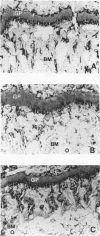
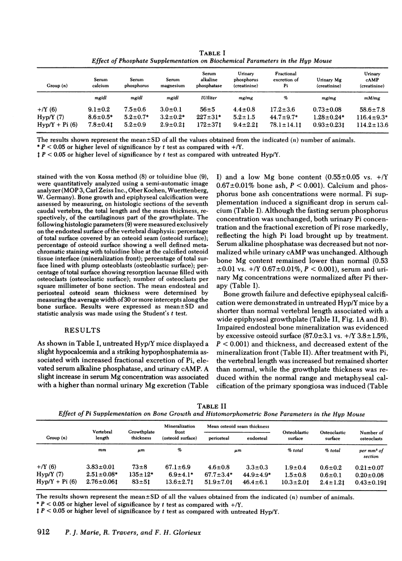
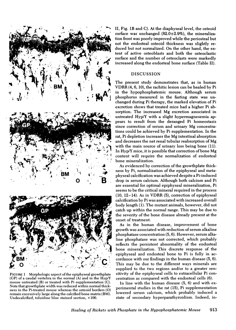
The hypophosphatemic male mouse, an animal model for human vitamin D-resistant rickets, is characterized by low serum phosphorus concentration due to increased urinary phosphate excretion, rickets, osteomalacia, and dwarfism. Because phosphate administration can heal rickets but not osteomalacia in the human disease, we have compared the effect of phosphate supplementation on the epiphyseal and endosteal bone mineralization in the mutant animal. Phosphate was given in drinking water for 137 d and the biochemical and bone responses were assessed by analytical and histomorphometric methods. Treatment with phosphate normalized the endochondral calcification (vertebral growthplate thickness: 83 +/- 5 SD vs. controls [+/Y] 73 +/- 8 micrometers, NS), but did not correct the endosteal bone mineralization (mineralization front: 13.6 +/- 2.7 vs. +/Y 67.1 +/- 6.9% osteoid surface, P less than 0.001, endosteal mean osteoid seam thickness: 46.4 +/- 6.1 vs. +/Y 3.3 +/- 0.3 micrometers, P less than 0.001). In addition, both osteoblastic and osteoclastic recruitment and activity were stimulated, as a result of a probable increase in parathyroid hormone secretion following the phosphate induced fall in serum calcium. Our results show that in the hypophosphatemic mouse, phosphate supplementation can heal the epiphyseal, but not the endosteal defective bone mineralization. Then, the biochemical and skeletal response to phosphate therapy appear to be similar to what we have observed in the human disease, further stressing the interest of the animal model.
Full text
PDF
Images in this article
Selected References
These references are in PubMed. This may not be the complete list of references from this article.
- Brighton C. T., Hunt R. M. The role of mitochondria in growth plate calcification as demonstrated in a rachitic model. J Bone Joint Surg Am. 1978 Jul;60(5):630–639. [PubMed] [Google Scholar]
- Cowgill L. D., Goldfarb S., Lau K., Slatopolsky E., Agus Z. S. Evidence for an intrinsic renal tubular defect in mice with genetic hypophosphatemic rickets. J Clin Invest. 1979 Jun;63(6):1203–1210. doi: 10.1172/JCI109415. [DOI] [PMC free article] [PubMed] [Google Scholar]
- Eicher E. M., Southard J. L., Scriver C. R., Glorieux F. H. Hypophosphatemia: mouse model for human familial hypophosphatemic (vitamin D-resistant) rickets. Proc Natl Acad Sci U S A. 1976 Dec;73(12):4667–4671. doi: 10.1073/pnas.73.12.4667. [DOI] [PMC free article] [PubMed] [Google Scholar]
- Glorieux F. H., Marie P. J., Pettifor J. M., Delvin E. E. Bone response to phosphate salts, ergocalciferol, and calcitriol in hypophosphatemic vitamin D-resistant rickets. N Engl J Med. 1980 Oct 30;303(18):1023–1031. doi: 10.1056/NEJM198010303031802. [DOI] [PubMed] [Google Scholar]
- Glorieux F. H., Scriver C. R., Reade T. M., Goldman H., Roseborough A. Use of phosphate and vitamin D to prevent dwarfism and rickets in X-linked hypophosphatemia. N Engl J Med. 1972 Sep 7;287(10):481–487. doi: 10.1056/NEJM197209072871003. [DOI] [PubMed] [Google Scholar]
- Harris W. H., Heaney R. P., Davis L. A., Weinberg E. H., Coutts R. D., Schiller A. L. Stimulation of bone formation in vivo by phosphate supplementation. Calcif Tissue Res. 1976 Nov 24;22(1):85–98. doi: 10.1007/BF02010349. [DOI] [PubMed] [Google Scholar]
- Kreusser W. J., Kurokawa K., Aznar E., Sachtjen E., Massry S. G. Effect of phosphate depletion on magnesium homeostasis in rats. J Clin Invest. 1978 Mar;61(3):573–581. doi: 10.1172/JCI108968. [DOI] [PMC free article] [PubMed] [Google Scholar]
- Meyer R. A., Jr, Jowsey J., Meyer M. H. Osteomalacia and altered magnesium metabolism in the X-linked hypophosphatemic mouse. Calcif Tissue Int. 1979 Mar 13;27(1):19–26. doi: 10.1007/BF02441156. [DOI] [PubMed] [Google Scholar]
- Simmons D. J., Kunin A. S. Development and healing of rickets in rats. I. Studies with tritiated thymidine and nutritional considerations. Clin Orthop Relat Res. 1970 Jan-Feb;68:251–260. [PubMed] [Google Scholar]
- Tenenhouse H. S., Scriver C. R., McInnes R. R., Glorieux F. H. Renal handling of phosphate in vivo and in vitro by the X-linked hypophosphatemic male mouse: evidence for a defect in the brush border membrane. Kidney Int. 1978 Sep;14(3):236–244. doi: 10.1038/ki.1978.115. [DOI] [PubMed] [Google Scholar]


