Abstract
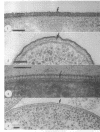
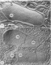
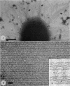
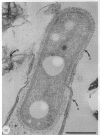
Since bacteria are so small, microscopy has traditionally been used to study them as individual cells. To this end, electron microscopy has been a most powerful tool for studying bacterial surfaces; the viewing of macromolecular arrangements of some surfaces is now possible. This review compares older conventional electron-microscopic methods with new cryotechniques currently available and the results each has produced. Emphasis is not placed on the methodology but, rather, on the importance of the results in terms of our perception of the makeup and function of bacterial surfaces and their interaction with the surrounding environment.
Full text
PDF










Images in this article
Selected References
These references are in PubMed. This may not be the complete list of references from this article.
- Acker G., Bitter-Suermann D., Meier-Dieter U., Peters H., Mayer H. Immunocytochemical localization of enterobacterial common antigen in Escherichia coli and Yersinia enterocolitica cells. J Bacteriol. 1986 Oct;168(1):348–356. doi: 10.1128/jb.168.1.348-356.1986. [DOI] [PMC free article] [PubMed] [Google Scholar]
- Acker G., Kammerer C. Localization of enterobacterial common antigen immunoreactivity in the ribosomal cytoplasm of Escherichia coli cells cryosubstituted and embedded at low temperature. J Bacteriol. 1990 Feb;172(2):1106–1113. doi: 10.1128/jb.172.2.1106-1113.1990. [DOI] [PMC free article] [PubMed] [Google Scholar]
- Adams L. F., Ghiorse W. C. Characterization of extracellular Mn2+-oxidizing activity and isolation of an Mn2+-oxidizing protein from Leptothrix discophora SS-1. J Bacteriol. 1987 Mar;169(3):1279–1285. doi: 10.1128/jb.169.3.1279-1285.1987. [DOI] [PMC free article] [PubMed] [Google Scholar]
- Adrian M., Dubochet J., Lepault J., McDowall A. W. Cryo-electron microscopy of viruses. Nature. 1984 Mar 1;308(5954):32–36. doi: 10.1038/308032a0. [DOI] [PubMed] [Google Scholar]
- Amako K., Meno Y., Takade A. Fine structures of the capsules of Klebsiella pneumoniae and Escherichia coli K1. J Bacteriol. 1988 Oct;170(10):4960–4962. doi: 10.1128/jb.170.10.4960-4962.1988. [DOI] [PMC free article] [PubMed] [Google Scholar]
- Amako K., Murata K., Umeda A. Structure of the envelope of Escherichia coli observed by the rapid-freezing and substitution fixation method. Microbiol Immunol. 1983;27(1):95–99. doi: 10.1111/j.1348-0421.1983.tb03571.x. [DOI] [PubMed] [Google Scholar]
- Amako K., Okada K., Miake S. Evidence for the presence of a capsule in Vibrio vulnificus. J Gen Microbiol. 1984 Oct;130(10):2741–2743. doi: 10.1099/00221287-130-10-2741. [DOI] [PubMed] [Google Scholar]
- Amako K., Takade A. The fine structure of Bacillus subtilis revealed by the rapid-freezing and substitution-fixation method. J Electron Microsc (Tokyo) 1985;34(1):13–17. [PubMed] [Google Scholar]
- Armbruster B. L., Carlemalm E., Chiovetti R., Garavito R. M., Hobot J. A., Kellenberger E., Villiger W. Specimen preparation for electron microscopy using low temperature embedding resins. J Microsc. 1982 Apr;126(Pt 1):77–85. doi: 10.1111/j.1365-2818.1982.tb00358.x. [DOI] [PubMed] [Google Scholar]
- Bar-Ness R., Avrahamy N., Matsuyama T., Rosenberg M. Increased cell surface hydrophobicity of a Serratia marcescens NS 38 mutant lacking wetting activity. J Bacteriol. 1988 Sep;170(9):4361–4364. doi: 10.1128/jb.170.9.4361-4364.1988. [DOI] [PMC free article] [PubMed] [Google Scholar]
- Bar-Ness R., Rosenberg M. Putative role of a 70 kDa outer-surface protein in promoting cell-surface hydrophobicity of Serratia marcescens RZ. J Gen Microbiol. 1989 Aug;135(8):2277–2281. doi: 10.1099/00221287-135-8-2277. [DOI] [PubMed] [Google Scholar]
- Bayer M. E., Thurow H. Polysaccharide capsule of Escherichia coli: microscope study of its size, structure, and sites of synthesis. J Bacteriol. 1977 May;130(2):911–936. doi: 10.1128/jb.130.2.911-936.1977. [DOI] [PMC free article] [PubMed] [Google Scholar]
- Beveridge T. J., Davies J. A. Cellular responses of Bacillus subtilis and Escherichia coli to the Gram stain. J Bacteriol. 1983 Nov;156(2):846–858. doi: 10.1128/jb.156.2.846-858.1983. [DOI] [PMC free article] [PubMed] [Google Scholar]
- Beveridge T. J. Mechanism of gram variability in select bacteria. J Bacteriol. 1990 Mar;172(3):1609–1620. doi: 10.1128/jb.172.3.1609-1620.1990. [DOI] [PMC free article] [PubMed] [Google Scholar]
- Beveridge T. J., Meloche J. D., Fyfe W. S., Murray R. G. Diagenesis of metals chemically complexed to bacteria: laboratory formation of metal phosphates, sulfides, and organic condensates in artificial sediments. Appl Environ Microbiol. 1983 Mar;45(3):1094–1108. doi: 10.1128/aem.45.3.1094-1108.1983. [DOI] [PMC free article] [PubMed] [Google Scholar]
- Beveridge T. J., Murray R. G. Sites of metal deposition in the cell wall of Bacillus subtilis. J Bacteriol. 1980 Feb;141(2):876–887. doi: 10.1128/jb.141.2.876-887.1980. [DOI] [PMC free article] [PubMed] [Google Scholar]
- Beveridge T. J., Murray R. G. Uptake and retention of metals by cell walls of Bacillus subtilis. J Bacteriol. 1976 Sep;127(3):1502–1518. doi: 10.1128/jb.127.3.1502-1518.1976. [DOI] [PMC free article] [PubMed] [Google Scholar]
- Beveridge T. J. Role of cellular design in bacterial metal accumulation and mineralization. Annu Rev Microbiol. 1989;43:147–171. doi: 10.1146/annurev.mi.43.100189.001051. [DOI] [PubMed] [Google Scholar]
- Beveridge T. J., Southam G., Jericho M. H., Blackford B. L. High-resolution topography of the S-layer sheath of the archaebacterium Methanospirillum hungatei provided by scanning tunneling microscopy. J Bacteriol. 1990 Nov;172(11):6589–6595. doi: 10.1128/jb.172.11.6589-6595.1990. [DOI] [PMC free article] [PubMed] [Google Scholar]
- Beveridge T. J., Sprott G. D., Whippey P. Ultrastructure, inferred porosity, and gram-staining character of Methanospirillum hungatei filament termini describe a unique cell permeability for this archaeobacterium. J Bacteriol. 1991 Jan;173(1):130–140. doi: 10.1128/jb.173.1.130-140.1991. [DOI] [PMC free article] [PubMed] [Google Scholar]
- Beveridge T. J., Stewart M., Doyle R. J., Sprott G. D. Unusual stability of the Methanospirillum hungatei sheath. J Bacteriol. 1985 May;162(2):728–737. doi: 10.1128/jb.162.2.728-737.1985. [DOI] [PMC free article] [PubMed] [Google Scholar]
- Beveridge T. J. The bacterial surface: general considerations towards design and function. Can J Microbiol. 1988 Apr;34(4):363–372. doi: 10.1139/m88-067. [DOI] [PubMed] [Google Scholar]
- Beveridge T. J. Ultrastructure, chemistry, and function of the bacterial wall. Int Rev Cytol. 1981;72:229–317. doi: 10.1016/s0074-7696(08)61198-5. [DOI] [PubMed] [Google Scholar]
- Boogerd F. C., de Vrind J. P. Manganese oxidation by Leptothrix discophora. J Bacteriol. 1987 Feb;169(2):489–494. doi: 10.1128/jb.169.2.489-494.1987. [DOI] [PMC free article] [PubMed] [Google Scholar]
- CHAPMAN G. B., HILLIER J. Electron microscopy of ultra-thin sections of bacteria I. Cellular division in Bacillus cereus. J Bacteriol. 1953 Sep;66(3):362–373. doi: 10.1128/jb.66.3.362-373.1953. [DOI] [PMC free article] [PubMed] [Google Scholar]
- Carlemalm E., Villiger W., Hobot J. A., Acetarin J. D., Kellenberger E. Low temperature embedding with Lowicryl resins: two new formulations and some applications. J Microsc. 1985 Oct;140(Pt 1):55–63. doi: 10.1111/j.1365-2818.1985.tb02660.x. [DOI] [PubMed] [Google Scholar]
- Chang C. F., Shuman H., Somlyo A. P. Electron probe analysis, X-ray mapping, and electron energy-loss spectroscopy of calcium, magnesium, and monovalent ions in log-phase and in dividing Escherichia coli B cells. J Bacteriol. 1986 Sep;167(3):935–939. doi: 10.1128/jb.167.3.935-939.1986. [DOI] [PMC free article] [PubMed] [Google Scholar]
- Costerton J. W., Irvin R. T., Cheng K. J. The bacterial glycocalyx in nature and disease. Annu Rev Microbiol. 1981;35:299–324. doi: 10.1146/annurev.mi.35.100181.001503. [DOI] [PubMed] [Google Scholar]
- Costerton J. W. The role of electron microscopy in the elucidation of bacterial structure and function. Annu Rev Microbiol. 1979;33:459–479. doi: 10.1146/annurev.mi.33.100179.002331. [DOI] [PubMed] [Google Scholar]
- Crepeau R. H., Fram E. K. Reconstruction of imperfectly ordered zinc-induced tubulin sheets using cross-correlation and real space averaging. Ultramicroscopy. 1981;6(1):7–17. doi: 10.1016/s0304-3991(81)80173-8. [DOI] [PubMed] [Google Scholar]
- Davies J. A., Anderson G. K., Beveridge T. J., Clark H. C. Chemical mechanism of the Gram stain and synthesis of a new electron-opaque marker for electron microscopy which replaces the iodine mordant of the stain. J Bacteriol. 1983 Nov;156(2):837–845. doi: 10.1128/jb.156.2.837-845.1983. [DOI] [PMC free article] [PubMed] [Google Scholar]
- Doyle R. J., Koch A. L. The functions of autolysins in the growth and division of Bacillus subtilis. Crit Rev Microbiol. 1987;15(2):169–222. doi: 10.3109/10408418709104457. [DOI] [PubMed] [Google Scholar]
- Doyle R. J., Matthews T. H., Streips U. N. Chemical basis for selectivity of metal ions by the Bacillus subtilis cell wall. J Bacteriol. 1980 Jul;143(1):471–480. doi: 10.1128/jb.143.1.471-480.1980. [DOI] [PMC free article] [PubMed] [Google Scholar]
- Dubochet J., McDowall A. W., Menge B., Schmid E. N., Lickfeld K. G. Electron microscopy of frozen-hydrated bacteria. J Bacteriol. 1983 Jul;155(1):381–390. doi: 10.1128/jb.155.1.381-390.1983. [DOI] [PMC free article] [PubMed] [Google Scholar]
- Dubreuil J. D., Kostrzynska M., Austin J. W., Trust T. J. Antigenic differences among Campylobacter fetus S-layer proteins. J Bacteriol. 1990 Sep;172(9):5035–5043. doi: 10.1128/jb.172.9.5035-5043.1990. [DOI] [PMC free article] [PubMed] [Google Scholar]
- Dubreuil J. D., Logan S. M., Cubbage S., Eidhin D. N., McCubbin W. D., Kay C. M., Beveridge T. J., Ferris F. G., Trust T. J. Structural and biochemical analyses of a surface array protein of Campylobacter fetus. J Bacteriol. 1988 Sep;170(9):4165–4173. doi: 10.1128/jb.170.9.4165-4173.1988. [DOI] [PMC free article] [PubMed] [Google Scholar]
- FEDER N., SIDMAN R. L. Methods and principles of fixation by freeze-substitution. J Biophys Biochem Cytol. 1958 Sep 25;4(5):593–600. doi: 10.1083/jcb.4.5.593. [DOI] [PMC free article] [PubMed] [Google Scholar]
- Ferris F. G., Beveridge T. J. Physicochemical roles of soluble metal cations in the outer membrane of Escherichia coli K-12. Can J Microbiol. 1986 Jul;32(7):594–601. doi: 10.1139/m86-110. [DOI] [PubMed] [Google Scholar]
- Ferris F. G., Beveridge T. J. Site specificity of metallic ion binding in Escherichia coli K-12 lipopolysaccharide. Can J Microbiol. 1986 Jan;32(1):52–55. doi: 10.1139/m86-010. [DOI] [PubMed] [Google Scholar]
- Ferris F. G., Schultze S., Witten T. C., Fyfe W. S., Beveridge T. J. Metal Interactions with Microbial Biofilms in Acidic and Neutral pH Environments. Appl Environ Microbiol. 1989 May;55(5):1249–1257. doi: 10.1128/aem.55.5.1249-1257.1989. [DOI] [PMC free article] [PubMed] [Google Scholar]
- Flemming C. A., Ferris F. G., Beveridge T. J., Bailey G. W. Remobilization of toxic heavy metals adsorbed to bacterial wall-clay composites. Appl Environ Microbiol. 1990 Oct;56(10):3191–3203. doi: 10.1128/aem.56.10.3191-3203.1990. [DOI] [PMC free article] [PubMed] [Google Scholar]
- Funahara Y., Nikaido H. Asymmetric localization of lipopolysaccharides on the outer membrane of Salmonella typhimurium. J Bacteriol. 1980 Mar;141(3):1463–1465. doi: 10.1128/jb.141.3.1463-1465.1980. [DOI] [PMC free article] [PubMed] [Google Scholar]
- Ghiorse W. C. Biology of iron- and manganese-depositing bacteria. Annu Rev Microbiol. 1984;38:515–550. doi: 10.1146/annurev.mi.38.100184.002503. [DOI] [PubMed] [Google Scholar]
- Glaeser R. M., Chiu W., Grano D. Structure of the surface layer protein of the outer membrane of Spirillum serpens. J Ultrastruct Res. 1979 Mar;66(3):235–242. doi: 10.1016/s0022-5320(79)90121-7. [DOI] [PubMed] [Google Scholar]
- Golovchenko J. A. The tunneling microscope: a new look at the atomic world. Science. 1986 Apr 4;232(4746):48–53. doi: 10.1126/science.232.4746.48. [DOI] [PubMed] [Google Scholar]
- Graham L. L., Beveridge T. J. Effect of chemical fixatives on accurate preservation of Escherichia coli and Bacillus subtilis structure in cells prepared by freeze-substitution. J Bacteriol. 1990 Apr;172(4):2150–2159. doi: 10.1128/jb.172.4.2150-2159.1990. [DOI] [PMC free article] [PubMed] [Google Scholar]
- Graham L. L., Beveridge T. J. Evaluation of freeze-substitution and conventional embedding protocols for routine electron microscopic processing of eubacteria. J Bacteriol. 1990 Apr;172(4):2141–2149. doi: 10.1128/jb.172.4.2141-2149.1990. [DOI] [PMC free article] [PubMed] [Google Scholar]
- Graham L. L., Beveridge T. J., Nanninga N. Periplasmic space and the concept of the periplasm. Trends Biochem Sci. 1991 Sep;16(9):328–329. doi: 10.1016/0968-0004(91)90135-i. [DOI] [PubMed] [Google Scholar]
- Graham L. L., Harris R., Villiger W., Beveridge T. J. Freeze-substitution of gram-negative eubacteria: general cell morphology and envelope profiles. J Bacteriol. 1991 Mar;173(5):1623–1633. doi: 10.1128/jb.173.5.1623-1633.1991. [DOI] [PMC free article] [PubMed] [Google Scholar]
- HEARST J. E., VINOGRAD J. The net hydration of T-4 bacteriophage deoxyribonuecleic acid and the effect of hydration on buoyant behavior in a density gradient at equilibrium in the ultracentrifuge. Proc Natl Acad Sci U S A. 1961 Jul 15;47:1005–1014. doi: 10.1073/pnas.47.7.1005. [DOI] [PMC free article] [PubMed] [Google Scholar]
- Hancock R. E. Alterations in outer membrane permeability. Annu Rev Microbiol. 1984;38:237–264. doi: 10.1146/annurev.mi.38.100184.001321. [DOI] [PubMed] [Google Scholar]
- Hobot J. A., Carlemalm E., Villiger W., Kellenberger E. Periplasmic gel: new concept resulting from the reinvestigation of bacterial cell envelope ultrastructure by new methods. J Bacteriol. 1984 Oct;160(1):143–152. doi: 10.1128/jb.160.1.143-152.1984. [DOI] [PMC free article] [PubMed] [Google Scholar]
- Hobot J. A., Villiger W., Escaig J., Maeder M., Ryter A., Kellenberger E. Shape and fine structure of nucleoids observed on sections of ultrarapidly frozen and cryosubstituted bacteria. J Bacteriol. 1985 Jun;162(3):960–971. doi: 10.1128/jb.162.3.960-971.1985. [DOI] [PMC free article] [PubMed] [Google Scholar]
- Holt S. C., Beveridge T. J. Electron microscopy: its development and application to microbiology. Can J Microbiol. 1982 Jan;28(1):1–53. doi: 10.1139/m82-001. [DOI] [PubMed] [Google Scholar]
- Hovmöller S., Sjögren A., Wang D. N. The structure of crystalline bacterial surface layers. Prog Biophys Mol Biol. 1988;51(2):131–163. doi: 10.1016/0079-6107(88)90012-0. [DOI] [PubMed] [Google Scholar]
- Howard R. J., Aist J. R. Hyphal tip cell ultrastructure of the fungus Fusarium: improved preservation by freeze-substitution. J Ultrastruct Res. 1979 Mar;66(3):224–234. doi: 10.1016/s0022-5320(79)90120-5. [DOI] [PubMed] [Google Scholar]
- Hoyle B. D., Beveridge T. J. Metal binding by the peptidoglycan sacculus of Escherichia coli K-12. Can J Microbiol. 1984 Feb;30(2):204–211. doi: 10.1139/m84-031. [DOI] [PubMed] [Google Scholar]
- Ishiguro E. E., Kay W. W., Ainsworth T., Chamberlain J. B., Austen R. A., Buckley J. T., Trust T. J. Loss of virulence during culture of Aeromonas salmonicida at high temperature. J Bacteriol. 1981 Oct;148(1):333–340. doi: 10.1128/jb.148.1.333-340.1981. [DOI] [PMC free article] [PubMed] [Google Scholar]
- Jacques M., Gottschalk M., Foiry B., Higgins R. Ultrastructural study of surface components of Streptococcus suis. J Bacteriol. 1990 Jun;172(6):2833–2838. doi: 10.1128/jb.172.6.2833-2838.1990. [DOI] [PMC free article] [PubMed] [Google Scholar]
- Jacques M., Graham L. Improved preservation of bacterial capsule for electron microscopy. J Electron Microsc Tech. 1989 Feb;11(2):167–169. doi: 10.1002/jemt.1060110212. [DOI] [PubMed] [Google Scholar]
- Jaffe J. S., Glaeser R. M. Difference Fourier analysis of "surface features" of bacteriorhodopsin using glucose-embedded and frozen-hydrated purple membrane. Ultramicroscopy. 1987;23(1):17–28. doi: 10.1016/0304-3991(87)90223-3. [DOI] [PubMed] [Google Scholar]
- Jap B. K., Downing K. H., Walian P. J. Structure of PhoE porin in projection at 3.5 A resolution. J Struct Biol. 1990 Mar;103(1):57–63. doi: 10.1016/1047-8477(90)90086-r. [DOI] [PubMed] [Google Scholar]
- Jarrell K. F., Koval S. F. Ultrastructure and biochemistry of Methanococcus voltae. Crit Rev Microbiol. 1989;17(1):53–87. doi: 10.3109/10408418909105722. [DOI] [PubMed] [Google Scholar]
- Kellenberger E., Dürrenberger M., Villiger W., Carlemalm E., Wurtz M. The efficiency of immunolabel on Lowicryl sections compared to theoretical predictions. J Histochem Cytochem. 1987 Sep;35(9):959–969. doi: 10.1177/35.9.3302020. [DOI] [PubMed] [Google Scholar]
- Koch A. L., Doyle R. J. Inside-to-outside growth and turnover of the wall of gram-positive rods. J Theor Biol. 1985 Nov 7;117(1):137–157. doi: 10.1016/s0022-5193(85)80169-7. [DOI] [PubMed] [Google Scholar]
- Koch A. L., Pinette M. F. Nephelometric determination of turgor pressure in growing gram-negative bacteria. J Bacteriol. 1987 Aug;169(8):3654–3663. doi: 10.1128/jb.169.8.3654-3663.1987. [DOI] [PMC free article] [PubMed] [Google Scholar]
- Koch A. L. The surface stress theory of microbial morphogenesis. Adv Microb Physiol. 1983;24:301–366. doi: 10.1016/s0065-2911(08)60388-4. [DOI] [PubMed] [Google Scholar]
- Koval S. F., Murray R. G. The isolation of surface array proteins from bacteria. Can J Biochem Cell Biol. 1984 Nov;62(11):1181–1189. doi: 10.1139/o84-152. [DOI] [PubMed] [Google Scholar]
- Koval S. F., Murray R. The superficial protein arrays on bacteria. Microbiol Sci. 1986 Dec;3(12):357–361. [PubMed] [Google Scholar]
- Labischinski H., Goodell E. W., Goodell A., Hochberg M. L. Direct proof of a "more-than-single-layered" peptidoglycan architecture of Escherichia coli W7: a neutron small-angle scattering study. J Bacteriol. 1991 Jan;173(2):751–756. doi: 10.1128/jb.173.2.751-756.1991. [DOI] [PMC free article] [PubMed] [Google Scholar]
- Larkin J. M., Strohl W. R. Beggiatoa, Thiothrix, and Thioploca. Annu Rev Microbiol. 1983;37:341–367. doi: 10.1146/annurev.mi.37.100183.002013. [DOI] [PubMed] [Google Scholar]
- Leduc M., Frehel C. Characterization of adhesion zones in E. coli cells. FEMS Microbiol Lett. 1990 Jan 15;55(1-2):39–43. doi: 10.1016/0378-1097(90)90164-l. [DOI] [PubMed] [Google Scholar]
- Leduc M., Fréhel C., Siegel E., Van Heijenoort J. Multilayered distribution of peptidoglycan in the periplasmic space of Escherichia coli. J Gen Microbiol. 1989 May;135(5):1243–1254. doi: 10.1099/00221287-135-5-1243. [DOI] [PubMed] [Google Scholar]
- Lepault J., Pitt T. Projected structure of unstained, frozen-hydrated T-layer of Bacillus brevis. EMBO J. 1984 Jan;3(1):101–105. doi: 10.1002/j.1460-2075.1984.tb01768.x. [DOI] [PMC free article] [PubMed] [Google Scholar]
- Luckevich M. D., Beveridge T. J. Characterization of a dynamic S layer on Bacillus thuringiensis. J Bacteriol. 1989 Dec;171(12):6656–6667. doi: 10.1128/jb.171.12.6656-6667.1989. [DOI] [PMC free article] [PubMed] [Google Scholar]
- Martin N. L., Beveridge T. J. Gentamicin interaction with Pseudomonas aeruginosa cell envelope. Antimicrob Agents Chemother. 1986 Jun;29(6):1079–1087. doi: 10.1128/aac.29.6.1079. [DOI] [PMC free article] [PubMed] [Google Scholar]
- Matsuyama T., Kaneda K., Ishizuka I., Toida T., Yano I. Surface-active novel glycolipid and linked 3-hydroxy fatty acids produced by Serratia rubidaea. J Bacteriol. 1990 Jun;172(6):3015–3022. doi: 10.1128/jb.172.6.3015-3022.1990. [DOI] [PMC free article] [PubMed] [Google Scholar]
- McLean R. J., Beauchemin D., Clapham L., Beveridge T. J. Metal-Binding Characteristics of the Gamma-Glutamyl Capsular Polymer of Bacillus licheniformis ATCC 9945. Appl Environ Microbiol. 1990 Dec;56(12):3671–3677. doi: 10.1128/aem.56.12.3671-3677.1990. [DOI] [PMC free article] [PubMed] [Google Scholar]
- Mendelson N. H., Thwaites J. J. Cell wall mechanical properties as measured with bacterial thread made from Bacillus subtilis. J Bacteriol. 1989 Feb;171(2):1055–1062. doi: 10.1128/jb.171.2.1055-1062.1989. [DOI] [PMC free article] [PubMed] [Google Scholar]
- Meng K. E., Pfister R. M. Intracellular structures of Mycoplasma pneumoniae revealed after membrane removal. J Bacteriol. 1980 Oct;144(1):390–399. doi: 10.1128/jb.144.1.390-399.1980. [DOI] [PMC free article] [PubMed] [Google Scholar]
- Meno Y., Amako K. Morphological evidence for penetration of anti-O antibody through the capsule of Klebsiella pneumoniae. Infect Immun. 1990 May;58(5):1421–1428. doi: 10.1128/iai.58.5.1421-1428.1990. [DOI] [PMC free article] [PubMed] [Google Scholar]
- Messner P., Bock K., Christian R., Schulz G., Sleytr U. B. Characterization of the surface layer glycoprotein of Clostridium symbiosum HB25. J Bacteriol. 1990 May;172(5):2576–2583. doi: 10.1128/jb.172.5.2576-2583.1990. [DOI] [PMC free article] [PubMed] [Google Scholar]
- Messner P., Pum D., Sára M., Stetter K. O., Sleytr U. B. Ultrastructure of the cell envelope of the archaebacteria Thermoproteus tenax and Thermoproteus neutrophilus. J Bacteriol. 1986 Jun;166(3):1046–1054. doi: 10.1128/jb.166.3.1046-1054.1986. [DOI] [PMC free article] [PubMed] [Google Scholar]
- Mühlradt P. F., Golecki J. R. Asymmetrical distribution and artifactual reorientation of lipopolysaccharide in the outer membrane bilayer of Salmonella typhimurium. Eur J Biochem. 1975 Feb 21;51(2):343–352. doi: 10.1111/j.1432-1033.1975.tb03934.x. [DOI] [PubMed] [Google Scholar]
- Nanninga N. Growth and form in microorganisms: morphogenesis of Escherichia coli. Can J Microbiol. 1988 Apr;34(4):381–389. doi: 10.1139/m88-069. [DOI] [PubMed] [Google Scholar]
- Phipps B. M., Kay W. W. Immunoglobulin binding by the regular surface array of Aeromonas salmonicida. J Biol Chem. 1988 Jul 5;263(19):9298–9303. [PubMed] [Google Scholar]
- Rivera M., McGroarty E. J. Analysis of a common-antigen lipopolysaccharide from Pseudomonas aeruginosa. J Bacteriol. 1989 Apr;171(4):2244–2248. doi: 10.1128/jb.171.4.2244-2248.1989. [DOI] [PMC free article] [PubMed] [Google Scholar]
- SALTON M. R. The relationship between the nature of the cell wall and the Gram stain. J Gen Microbiol. 1963 Feb;30:223–235. doi: 10.1099/00221287-30-2-223. [DOI] [PubMed] [Google Scholar]
- Schwarz H., Humbel B. M. Influence of fixatives and embedding media on immunolabelling of freeze-substituted cells. Scanning Microsc Suppl. 1989;3:57–64. [PubMed] [Google Scholar]
- Sleytr U. B., Messner P. Crystalline surface layers in procaryotes. J Bacteriol. 1988 Jul;170(7):2891–2897. doi: 10.1128/jb.170.7.2891-2897.1988. [DOI] [PMC free article] [PubMed] [Google Scholar]
- Sleytr U. B., Messner P. Crystalline surface layers on bacteria. Annu Rev Microbiol. 1983;37:311–339. doi: 10.1146/annurev.mi.37.100183.001523. [DOI] [PubMed] [Google Scholar]
- Sonnenfeld E. M., Beveridge T. J., Doyle R. J. Discontinuity of charge on cell wall poles of Bacillus subtilis. Can J Microbiol. 1985 Sep;31(9):875–877. doi: 10.1139/m85-163. [DOI] [PubMed] [Google Scholar]
- Sonnenfeld E. M., Beveridge T. J., Koch A. L., Doyle R. J. Asymmetric distribution of charge on the cell wall of Bacillus subtilis. J Bacteriol. 1985 Sep;163(3):1167–1171. doi: 10.1128/jb.163.3.1167-1171.1985. [DOI] [PMC free article] [PubMed] [Google Scholar]
- Stanier R. Y., Cohen-Bazire G. Phototrophic prokaryotes: the cyanobacteria. Annu Rev Microbiol. 1977;31:225–274. doi: 10.1146/annurev.mi.31.100177.001301. [DOI] [PubMed] [Google Scholar]
- Stewart M., Beveridge T. J., Sprott G. D. Crystalline order to high resolution in the sheath of Methanospirillum hungatei: a cross-beta structure. J Mol Biol. 1985 Jun 5;183(3):509–515. doi: 10.1016/0022-2836(85)90019-1. [DOI] [PubMed] [Google Scholar]
- Stewart M., Beveridge T. J., Trust T. J. Two patterns in the Aeromonas salmonicida A-layer may reflect a structural transformation that alters permeability. J Bacteriol. 1986 Apr;166(1):120–127. doi: 10.1128/jb.166.1.120-127.1986. [DOI] [PMC free article] [PubMed] [Google Scholar]
- Stewart M. Computer image processing of electron micrographs of biological structures with helical symmetry. J Electron Microsc Tech. 1988 Aug;9(4):325–358. doi: 10.1002/jemt.1060090404. [DOI] [PubMed] [Google Scholar]
- Stewart M., Somlyo A. P., Somlyo A. V., Shuman H., Lindsay J. A., Murrell W. G. Distribution of calcium and other elements in cryosectioned Bacillus cereus T spores, determined by high-resolution scanning electron probe x-ray microanalysis. J Bacteriol. 1980 Jul;143(1):481–491. doi: 10.1128/jb.143.1.481-491.1980. [DOI] [PMC free article] [PubMed] [Google Scholar]
- Stewart M., Somlyo A. P., Somlyo A. V., Shuman H., Lindsay J. A., Murrell W. G. Scanning electron probe x-ray microanalysis of elemental distributions in freeze-dried cryosections of Bacillus coagulans spores. J Bacteriol. 1981 Aug;147(2):670–674. doi: 10.1128/jb.147.2.670-674.1981. [DOI] [PMC free article] [PubMed] [Google Scholar]
- Stewart M., Vigers G. Electron microscopy of frozen-hydrated biological material. Nature. 1986 Feb 20;319(6055):631–636. doi: 10.1038/319631a0. [DOI] [PubMed] [Google Scholar]
- Sára M., Kalsner I., Sleytr U. B. Surface properties from the S-layer of Clostridium thermosaccharolyticum D120-70 and Clostridium thermohydrosulfuricum L111-69. Arch Microbiol. 1988;149(6):527–533. doi: 10.1007/BF00446756. [DOI] [PubMed] [Google Scholar]
- Sára M., Sleytr U. B. Charge distribution on the S layer of Bacillus stearothermophilus NRS 1536/3c and importance of charged groups for morphogenesis and function. J Bacteriol. 1987 Jun;169(6):2804–2809. doi: 10.1128/jb.169.6.2804-2809.1987. [DOI] [PMC free article] [PubMed] [Google Scholar]
- Sára M., Sleytr U. B. Molecular sieving through S layers of Bacillus stearothermophilus strains. J Bacteriol. 1987 Sep;169(9):4092–4098. doi: 10.1128/jb.169.9.4092-4098.1987. [DOI] [PMC free article] [PubMed] [Google Scholar]
- Taylor K. A., Glaeser R. M. Electron microscopy of frozen hydrated biological specimens. J Ultrastruct Res. 1976 Jun;55(3):448–456. doi: 10.1016/s0022-5320(76)80099-8. [DOI] [PubMed] [Google Scholar]
- Trachtenberg S., DeRosier D. J. Three-dimensional structure of the frozen-hydrated flagellar filament. The left-handed filament of Salmonella typhimurium. J Mol Biol. 1987 Jun 5;195(3):581–601. doi: 10.1016/0022-2836(87)90184-7. [DOI] [PubMed] [Google Scholar]
- Umeda A., Ueki Y., Amako K. Structure of the Staphylococcus aureus cell wall determined by the freeze-substitution method. J Bacteriol. 1987 Jun;169(6):2482–2487. doi: 10.1128/jb.169.6.2482-2487.1987. [DOI] [PMC free article] [PubMed] [Google Scholar]
- VANHARREVELD A., CROWELL J. ELECTRON MICROSCOPY AFTER RAPID FREEZING ON A METAL SURFACE AND SUBSTITUTION FIXATION. Anat Rec. 1964 Jul;149:381–385. doi: 10.1002/ar.1091490307. [DOI] [PubMed] [Google Scholar]
- Walker S. G., Beveridge T. J. Amikacin disrupts the cell envelope of Pseudomonas aeruginosa ATCC 9027. Can J Microbiol. 1988 Jan;34(1):12–18. doi: 10.1139/m88-003. [DOI] [PubMed] [Google Scholar]
- Walker S. G., Flemming C. A., Ferris F. G., Beveridge T. J., Bailey G. W. Physicochemical interaction of Escherichia coli cell envelopes and Bacillus subtilis cell walls with two clays and ability of the composite to immobilize heavy metals from solution. Appl Environ Microbiol. 1989 Nov;55(11):2976–2984. doi: 10.1128/aem.55.11.2976-2984.1989. [DOI] [PMC free article] [PubMed] [Google Scholar]
- Weel J. F., Hopman C. T., van Putten J. P. Stable expression of lipooligosaccharide antigens during attachment, internalization, and intracellular processing of Neisseria gonorrhoeae in infected epithelial cells. Infect Immun. 1989 Nov;57(11):3395–3402. doi: 10.1128/iai.57.11.3395-3402.1989. [DOI] [PMC free article] [PubMed] [Google Scholar]
- Whitfield C. Bacterial extracellular polysaccharides. Can J Microbiol. 1988 Apr;34(4):415–420. doi: 10.1139/m88-073. [DOI] [PubMed] [Google Scholar]
- Zalokar M. A simple freeze-substitution method for electron microscopy. J Ultrastruct Res. 1966 Aug;15(5):469–479. doi: 10.1016/s0022-5320(66)80119-3. [DOI] [PubMed] [Google Scholar]


