Abstract
Signaling between cells in the anterior (A) and posterior (P) compartments directs Drosophila wing disc development and is dependent on expression of the homeodomain transcription factor Engrailed (En) in P cells. Downstream of en, posteriorly expressed Hedgehog (Hh) protein signals across the A/P border to establish a developmental organizer that directs pattern formation and growth throughout the wing primordium. Here we extend investigations of the processes downstream of en by using expression array analysis to compare A and P cells. A total of 102 candidate genes were identified that express differentially in the A and P compartments; four were characterized: Stubble (Sb) expression is restricted to A cells due to repression by en. CG15905, CG16884; CG10200/hase und igel (hui) are expressed in A cells downstream of Hh signaling; and RNA interference for hui, Stubble, and CG16884 revealed that each is essential to wing development.
Keywords: anteroposterior compartment border, pattern formation, expression microarray, engrailed, hedgehog
The subdivision of the wing imaginal disc into anterior (A) and posterior (P) compartments has several remarkable features. First, each compartment represents a defined and contiguous geographical area, and after the compartments are established in the early embryo, all descendents of the constituent cells retain the compartment identity of their ancestors, even to the adult stage (Garcia-Bellido et al. 1973). Second, in the wing blade primordium, the A and P compartments meet to form a remarkably straight boundary line. Third, the compartments are domains of gene expression for engrailed (en), invected (inv), and hedgehog (hh), which are expressed by all P compartment cells (Kornberg et al. 1985; Coleman et al. 1987; Tabata et al. 1992), and for cubitus interruptus (ci) and patched (ptc), which are expressed by all A compartment cells (Eaton and Kornberg 1990; Phillips et al. 1990). Other genes such as vein (Schnepp et al. 1996; Amin et al. 1999) and knot/collier (Vervoort et al. 1999) are expressed in a stripe that abuts the A/P compartment border’s anterior side.
Maintenance of the A/P compartment border depends upon en. In its absence, P cells transform into A type—they express ci and ptc, they are not confined by the A/P border and can join A cells across the border, and they make structures and patterns characteristic of the A compartment. In addition to this role as a selector gene in P cells that establishes P compartment identity and inhibits A compartment identity, en also creates a developmental organizer at the A/P compartment border by positively regulating hh. Hh made in P cells signals in a paracrine manner to A cells at the border, endowing them with organizer functionality (Basler and Struhl 1994; Tabata et al. 1995). Anterior cells at the border express proteins such as Decapentaplegic (Dpp) in response to Hh signaling, and the function of the organizer, which is dependent upon Dpp, regulates growth and patterning of both A and P cells (reviewed in Lawrence and Struhl 1996).
Despite our detailed understanding of these key signaling processes in wing development, many questions remain about the nature of the mechanisms that act downstream of A/P signaling. Among these are the processes that keep A and P cells separate and that define the position and shape of the border. The work described here was undertaken to identify additional genes that function at the A/P border. It sought target genes downstream of en and hh by searching for and characterizing genes with patterns of expression specific to either the A or P compartments.
We performed a global screen for genes with compartment-specific expression using expression array hybridization to compare transcript levels in A and P wing disc cells. In a previous expression microarray screen, we characterized transcripts isolated from single imaginal discs and identified and analyzed transcriptional differences between different types of discs from individual larvae (Klebes et al. 2002). These experiments were made possible by the application of linear RNA amplification protocols (Klebes and Kornberg 2008). In a second study, we applied this strategy to the analysis of microdissected imaginal disc cell populations in the state of transdetermination (Klebes et al. 2005). This investigation demonstrated that the direct microarray comparison of small cell populations that originate from the same imaginal discs is feasible. Here, we apply this strategy to a microarray comparison of sets of A and P compartment cells that had been microdissected from wing discs. This expression pattern-based approach identified 102 differentially expressed genes, of which approximately half had not been previously characterized by genetic or molecular studies. We show that Sb expression is downstream of En; that CG15905, CG16884, and hui are activated by ectopic Hh; and by using RNA interference (RNAi) knockdown, that Sb, hui, and CG16884 are required for wing development.
Materials and Methods
Fly stocks
The following fly stocks were used: w1118 or Oregon R for in situ detection experiments; hs-flp; P{ry,neoFRT43D, y+} and w; P{w, FRT}43D, enE/CyO for the generation of en/inv mutant cell clones [Df(2R)enE removes most of the en and inv transcription units (Gustavson et al. 1996)]; ptc-Gal4 [a hypomorphic enhancer trap allele (Speicher et al. 1994)], hh-Gal4 [an enhancer trap allele (Tanimoto et al. 2000)], C765-Gal4 (Nellen et al. 1996), en-Gal4 (generated by Andrea Brand, FlyBase ID FBrf0098595), and UAS-GFP (Bloomington stock #4775) for in vivo labeling and RNAi expression; vestigial boundary enhancer Gal4 (vgBE-Gal4; gift from G. Schubiger) and UAS-hh (Ingham and Fietz 1995), and UAS-dpp (Bloomington stock #1486) for overexpression experiments. UAS-RNAi transgenic stocks were obtained from the Vienna Drosophila RNAi Center (http://stockcenter.vdrc.at), for CG10200: 13321, 47612, 47613, 103328; for Sb: 1613; for CG15905: 13865, 13866; for CG16884: 51362, 51363; from the Kyoto National Institute of Genetics Stock Center, for CG15905: 15905R-1; for CG16884: 16884R-2 and for Sb: 4316R-1; and from the Bloomington Drosophila Stock Center, CG10200: 28759.
Immunolabeling
Imaginal discs were dissected and fixed (4% formaldehyde) following standard procedures (Sullivan et al. 2000). Antibodies were α-Twist (Thisse et al. 1988), α-Ci (Motzny and Holmgren 1995), α-Hh (Tabata and Kornberg 1994).
RNA amplification, microarray hybridization, and data analysis
Green fluorescent protein (GFP)-labeled wing imaginal discs were microdissected under a fluorescence dissecting microscope. RNA isolation, amplification, and microarray procedures were previously described (Klebes et al. 2002, 2005; Klebes and Kornberg 2008). Detailed information about the microarray platform (accession number: GPL2581) and the array data from this study (accession number: GSE46601) are accessible on the Gene Expression Omnibus database, http://www.ncbi.nlm.nih.gov/geo/. In brief, hybridization probes were generated by two rounds of T7-catalyzed linear RNA amplification and labeled with Cy3 and Cy5 dyes. Reciprocally labeled probes (“dye flip”) were hybridized to custom-produced glass microarrays that contained approximately 14,000 100- to 600-bp exon sequences that were generated by polymerase chain reaction (PCR). Signal intensities were collected with a GenePix 4000A Scanner and processed with GenePix software (Molecular Devices) and global median normalized with NOMAD (http://ucsf-nomad.sourceforge.net/). We performed two kinds of data analysis. First ,we used the significance analysis of microarrays software package (SAM; Tusher et al. 2001) to identify 203 and 76 transcripts that are enriched in the A or P compartment, respectively (Supporting Information, Table S1). A higher stringency analysis was performed by combining the SAM statistical tools with cluster analysis (Eisen et al. 1998) with stringent filter settings. Expression ratios were evaluated with SAM using a Delta setting of 0.733 (9.2% false discovery rate). For the cluster analysis, we eliminated spots with a sum of median intensities <300 and without data in more than 20% of the experiments. Only those spots that show ratios greater than 1 (log2-transformed) in at least 6 of 12 experiments were considered for hierarchical clustering and generation of self organizing maps. Genes of those sub-clusters that showed predominant enrichment in one channel were further used (109 in the A group and 17 in the P group). To generate the list of 102 A- or P-enriched genes, 11 duplicate spots and 13 genes that were not identified as significant by the SAM analysis were eliminated.
In situ hybridization experiments
In situ labelings with DIG-labeled RNA probes were performed as described (Klebes et al. 2002). RNA probes were generated by T7 or SP6 polymerase reaction on TOPO-TA (Invitrogen Inc.) subcloned PCR products. Primer sequences are available upon request. For the in situ detection experiments we selected genes from both lists, that is, the SAM list of 279, and the cluster analysis-derived list of 102 genes (see Figure 1). Of the 29 selected genes, 14 were represented in both lists (Table S1), 14 represented only by SAM analysis, and one gene (kal-1/CG6173) was segregated into the P cluster but was not considered to be significantly enriched by the SAM analysis. Because the in situ pattern confirmed the expression properties for all three groups of genes, we conclude that the application of stringent filter settings eliminated many true positives. For this reason we include the complete list of significantly enriched genes in Table S1.
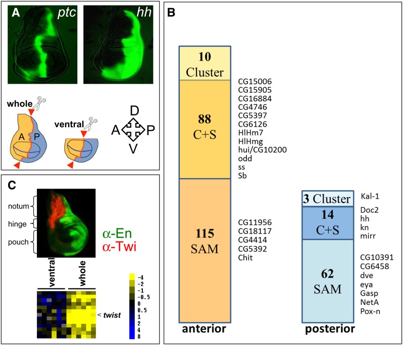
Figure 1.
Expression array analysis identifies genes with predominant expression in A or P cells and a dorsal subcluster. (A) GFP expression in the ptc and hh domains of wing imaginal discs was used to identify the compartment boundary for microdissection (genotypes: ptc-Gal4 or hh-Gal4 with UAS-GFP). Red arrowheads indicate locations of cuts that separated A and P cells of entire discs (whole) or discs without the dorsal part (ventral). The orientation of the discs is indicated: A, anterior; P, posterior; D, dorsal; V, ventral. (B) Bars indicate the numbers of transcripts identified with cluster analysis (Cluster), the significance analysis of microarrays algorithm (SAM), or both methods (S + C) to be enriched in anterior or posterior cells. The genes selected for in situ hybridization analysis as shown in Figure 2 are indicated. (C) The notum (dorsal) fragments identified a subcluster that includes twist. α-En (green) labels the P compartment and α-Twist (red) labels the adepithelial cells in the A region of the notum area. Heatmap legend shows log2-transformed ratios.
Results and Discussion
Comparison of transcript expression levels in wing disc A and P compartments
Because of the small size of the wing disc, obtaining sufficient material for microarray hybridization of defined cell populations, such as A and P cells, is challenging. We developed a method to analyze transcripts in A and P cells in single wing imaginal discs from third instar larvae. The method combines in vivo labeling, microdissection, linear RNA amplification, and microarray hybridization. We labeled the wing disc A/P compartment border in either of two ways, by expressing a GFP reporter transgene controlled by ptc or by hh (Figure 1A). Both reporter lines (ptc-Gal4 and hh-Gal4) are weak mutant alleles for the respective genes, but neither reporter line had visible phenotypes as a heterozygote. Nevertheless, to avoid bias, we performed half of the experiments with the ptc-Gal4 line and the other half with the hh-Gal4 line. Wing discs have several different cell types in addition to columnar cells, including peripodial cells, associated tracheal branches, and adepithelial mesodermal cells that are present in different proportions in A and P locations. To control for the contribution and influence by transcripts from these cell types, we removed the dorsal-most part of the discs that contains most of the tracheal and adepithelial cells in half of the experiments (Figure 1A). Wing disc A and P cells were manually dissected under a fluorescence microscope and were treated separately to amplify polyadenylated RNA (Klebes and Kornberg 2008). For both the ptc-Gal4 and hh-Gal4 genotypes, (1) the entire A compartment was compared to the entire P compartment, and (2) the ventral A compartment was compared with the ventral P compartment; three replicates of each experiment were performed, resulting in a total of twelve. To minimize variability, the pair-wise comparisons of A and P cells were of the same imaginal disc. Microarray hybridization was performed with custom-produced glass DNA microarrays that contained approximately 14,000 short PCR-generated cDNA fragments that represent approximately 75% of the currently annotated Drosophila genes as well as 72 spots representing GFP gene sequences.
We applied stringent filter settings (see Materials and Methods) that combine cluster analysis (Eisen et al. 1998) and a statistical algorithm, SAM (Tusher et al. 2001) to the hybridization data sets. Cluster analysis identified 98 A-enriched transcripts and 17 genes with preferential expression in P cells. The SAM analysis identified 203 A and 76 P transcripts. The 102 transcripts that were common to both methods (Figure 1B, Table S1) are considered as high confidence genes with preferential A (88) or P (14) expression. The following observations support the validity of this approach. GFP expression levels in each of 12 experiments were elevated more than 12-fold in A cells in probes made from the ptc-Gal4 line, and were elevated to similar levels in P cells for the hh-Gal4 line (Figure S1). The average ratios of hybridization signals for the genes known to be expressed in compartment-specific patterns were also consistent with the identity of the manually isolated cells. For the 12 experiments, the average A/P ratios of the A-expressed genes ci and dpp were 9.9 and 2.2, respectively. The average P/A ratios for the P-expressed genes en, hh, and inv were 9.6, 11.9, and 5.1, respectively (Table 1, Table S1). Expression levels of the anteriorly expressed ptc gene could not be analyzed due to technical problems with the spot representing the ptc sequence on the microarrays.
Table 1. Genes with more than twofold enrichment in transcript levels in A or P compartments.
| Gene Name | CG# | Annotated Function (www.flybase.org) | A#/P# (ratio) |
|---|---|---|---|
| Anterior | |||
| aristaless | CG3935 | Transcription factor | 20.3 |
| CG15611 | CG15611 | Regulation of Rho protein signal transduction | 11.6 |
| scute | CG3827 | Transcription factor | 11 |
| cubitus interruptus | CG2125 | Transcription factor | 9.9 |
| Drip | CG9023 | Water channel activity; cell homeostasis | 7.6 |
| Ect3 | CG3132 | Beta-galactosidase | 7.2 |
| achaete | CG3796 | Transcription factor | 7 |
| CG13044 | CG13044 | − | 6.1 |
| CG2663 | CG2663 | Transport, vitamin E binding | 5.3 |
| CG31705 | CG6528 | − | 4.9 |
| sister of odd and bowl | CG6993 | DNA binding | 4.9 |
| CG13023 | CG13023 | − | 4.8 |
| CG15714 | CG15714 | Protein folding | 4.6 |
| CG13574 | CG13574 | Learning or memory, olfactory learning | 4.2 |
| CG5966 | CG5966 | Lipid metabolic process, triglyceride lipase activity | 4.2 |
| Ecdysone-dependent gene 91 | CG7539 | Structural constituent of pupal cuticle | 4 |
| E(spl) region transcript 4 | CG6099 | Cell fate specification; sensory organ development | 4 |
| drumstick | CG10016 | Nucleic acid binding; zinc ion binding | 3.9 |
| Odorant-binding protein 56a | CG11797 | Odorant binding | 3.9 |
| CG3244 | CG3244 | Binding, C-type lectin 27kd | 3.7 |
| CG7090 | CG7090 | Oxidation-reduction process | 3.7 |
| Imaginal disc growth factor 4 | CG1780 | Imaginal disc growth factor, hydrolase activity | 3.6 |
| pxb | CG14874 | Learning and/or memory; olfactory learning; smoothened signaling pathway | 3.5 |
| CG5397 | CG5397 | Sterol O-acyltransferase activity | 3.4 |
| CG16884 | CG16884 | − | 3.3 |
| CG9338 | CG9338 | − | 3.3 |
| CG14598 | CG14598 | − | 3.2 |
| CG16885 | CG16885 | − | 3.2 |
| Imaginal disc growth factor 3 | CG4559 | NOT chitinase | 3.2 |
| Aldehyde dehydrogenase | CG3752 | Aldehyde dehydrogenase (NAD+) | 3.1 |
| Antennapedia | CG1028 | Transcription factor | 3.1 |
| CG9312 | CG9312 | − | 3.1 |
| opa | CG1133 | Transcription factor | 3.1 |
| Actin 57B | CG10067 | Structural constituent of cytoskeleton | 3 |
| CG10112 | CG10112 | Multicellular organism reproduction, structural constituent of chitin-based cuticle | 3 |
| CG8634 | CG8634 | Structural constituent of chitin-based cuticle, Cuticular protein 65Ec | 3 |
| Bearded | CG3096 | Calmodulin inhibitor | 2.9 |
| CG13060 | CG13060 | − | 2.9 |
| CG18634 | CG18634 | − | 2.9 |
| phyllopod | CG10108 | Protein binding; Ras protein signal transduction; peripheral nervous system development | 2.9 |
| CG12481 | CG12481 | − | 2.8 |
| CG15786 | CG15786 | − | 2.8 |
| CG6357 | CG6357 | Cysteine-type endopeptidase activity | 2.8 |
| CG8701 | CG8701 | − | 2.8 |
| Drop (msh) | CG1897 | Transcription factor | 2.8 |
| CG10625 | CG10625 | Structural constituent of cuticle | 2.7 |
| CG10962 | CG10962 | Oxidation-reduction process | 2.7 |
| E(spl) region transcript g | CG8333 | Transcription factor | 2.7 |
| Stubble | CG4316 | Serine-type endopeptidase | 2.7 |
| CG1674 | CG1674 | − | 2.6 |
| wunen-2 | CG8805 | Phosphatidate phosphatase, G-protein coupled receptor protein signaling pathway | 2.6 |
| CG3837 | CG3837 | Transmembrane receptor protein tyrosine kinase signaling pathway, protein phosphorylation | 2.5 |
| CG5391 | CG5391 | − | 2.5 |
| CG5888 | CG5888 | Transmembrane receptor activity | 2.5 |
| CG9336 | CG9336 | − | 2.5 |
| CG10311 | CG10311 | − | 2.4 |
| CG15006 | CG15006 | Structural constituent of chitin-based larval cuticle | 2.4 |
| CG18507 | CG18507 | − | 2.4 |
| CG7924 | CG7924 | − | 2.4 |
| Pherokine 3 | CG9358 | Protein serine/threonine kinase activity; carrier activity; Ras protein signal transduction | 2.4 |
| sob | CG3242 | RNA polymerase II transcription factor | 2.4 |
| Tetraspanin 42El | CG12840 | Receptor signaling protein activity | 2.4 |
| CG1368 | CG1368 | Structural constituent of chorion | 2.3 |
| CG4766 | CG4766 | − | 2.3 |
| CG8483 | CG8483 | − | 2.3 |
| odd skipped | CG3851 | Transcription factor | 2.3 |
| CG1572 | CG1572 | − | 2.2 |
| CG15785 | CG15785 | − | 2.2 |
| CG4382 | CG4382 | Carboxylesterase activity | 2.2 |
| decapentaplegic | CG9885 | Signal transducer, morphogen, growth factor | 2.2 |
| E(spl) region transcript 7 | CG8361 | Transcription factor | 2.2 |
| CG8502 | CG8502 | Structural constituent of chitin-based larval cuticle, Cuticular protein 49Ac | 2.1 |
| blown fuse | CG1363 | Mesoderm development; myoblast fusion | 2 |
| CG10200 | CG10200 | − | 2 |
| CG8216 | CG8216 | Regulation of transcription, DNA-dependent, DNA binding | 2 |
| CG9871 | CG9871 | Translation, structural constituent of ribosome, Ribosomal protein L22-like | 2 |
| Posterior | P#/A# (ratio) | ||
| hedgehog | CG4637 | Cysteine-type endopeptidase | 11.9 |
| engrailed | CG9015 | Transcription factor | 9.6 |
| invected | CG17835 | RNA polymerase II transcription factor | 5.1 |
| mirror | CG10601 | Transcription factor, smoothened signaling pathway | 3.5 |
| Inos | CG11143 | Enzyme, myo-inositol-1-phosphate synthase | 2.9 |
| CG10074 | CG30837 | − | 2.8 |
| Cytochrome P450-18a1 | CG6816 | Cytochrome P450 | 2.2 |
Genes are listed that show expression ratios ≥2 and that were identified by clustering and significance analysis (compare text and Table S1). Ratios were calculated with the average median intensities of the twelve arrays for A cells (A/P ratio) or P cells (P/A ratio). Ratios were rounded to one decimal place.
The microdissection strategy identifies sets of transcripts from A, P, and dorsal wing disc cells
Among the 102 genes in the high-stringency group, 88 segregated into an A group cluster, and 17 segregated into a P cluster (Figure 1B, Table 1, and Table S1). In addition to these A and P clusters, the separate probes that were prepared from wing discs that either included or lacked the dorsal-most cells (and retained or lacked adepithelial cells) identified a subcluster associated specifically with the presence of dorsal cells. twist (twi) encodes a transcriptional activator that regulates mesodermal development and is expressed specifically in mesoderm; its transcripts were elevated in preparations containing dorsal wing disc cells (Figure 1C). The twi-containing subcluster also detected 12 other genes with elevated transcript levels in preparations containing dorsal cells (Table S1). This group includes several that are known or are predicted to function in either tracheal (CG8748; Luschnig et al. 2006) or muscle and mesoderm development (CG15064/Him, CG6378/BM-40-SPARC; Furlong et al. 2001; Swan et al. 2004; Liotta et al. 2007), or have been identified in a previously published microarray screen comparing the dorsal and ventral parts of wing imaginal discs (eyg, Grip, CG9593, Him, CG11835; Butler et al. 2003).
Neuronal cells in the wing disc arise predominantly in the primordia for the notum and anterior wing margin, the regions that will produce most of the enervated sensory organs. Both the notum and anterior wing margin primordia are predominantly in the A compartment, and genes that are known to function in neuronal development (such as acheate and scute) are therefore expected to be expressed at high levels in A cells. Furthermore, because A cells at the A/P compartment border depend upon activation of the Notch pathway (Casso et al. 2011), Notch pathway components might also be expressed in greater levels in A cells. The A subcluster from the array analysis did include achaete and scute as well as the Notch pathway components Enhancer of spilt complex transcripts m7, m4, and γ.
In summary, these results indicate that microdissection effectively separated A and P cells, and that the expression array hybridization identified sets of genes that are expressed in subregions of the disc. Table 1 lists the genes in the A and P clusters that had expression ratios greater than 2. We presume that putative targets of en/Hh regulation will be represented in the A subcluster.
Expression patterns of candidate genes confirm hybridization array analysis
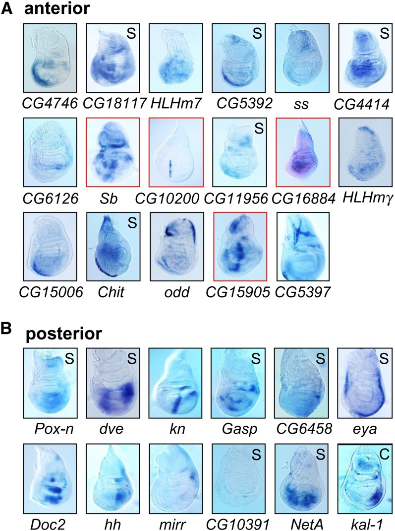
To further validate the expression array studies, we applied RNA in situ hybridization to whole wing discs for 29 genes. As illustrated in Figure 1B, we selected genes from each group for in situ analysis; 12 genes from the group of 88 that were identified by cluster and SAM analysis in the A group, five (of 115) that only SAM identified to be enriched in A cells, four of the high confidence group of 14 genes in P cells, seven (of 62) P transcripts that were identified by SAM only, and one of the three transcripts that only cluster analysis identified as P-enriched transcripts. The observed expression patterns confirmed the microarray predictions for 27 genes, revealing either predominant anterior (16 genes) or posterior (11 genes) expression (Figure 2). The only two exceptions that in situ hybridization did not confirm were HLHmγ, which in addition to the predicted A expression (by significance analysis and cluster) shows considerable expression in the P compartment, and NetA, which was predicted by significance analysis (but not by clustering analysis) to be predominantly in the P compartment. Note that knot (kn) is a Hh-target gene that is expressed in a stripe of A cells, but it is also expressed in a patch of cells in the P compartment (Vervoort et al. 1999; and Figure 2B). Because kn segregated with the P cluster, we assume that its expression in the P compartment was the greater influence on clustering. We conclude that the in situ patterns confirmed the array predictions for 27 of 29 genes. Transcripts of the A and P groups from cluster only, SAM only and the cluster and SAM overlap lists were confirmed indicating that both methods of data analysis produced list of candidate genes with a low rate of false-positive results (Table S1). The application of stringent filter settings that produced the list of 102 genes by combining cluster and significance analysis represents a high-confidence list. Based on predominantly A expression patterns, we selected four genes, Sb, CG16884, CG15905, and CG10200, for further analysis.
Figure 2.
In situ hybridization confirms the array analysis. In situ hybridization was carried out for 17 genes in the A cluster and twelve genes in the P cluster. Genes were identified by cluster and SAM analysis, only by SAM (S) or only by cluster analysis (C) as indicated in Figure 1. The full gene names are provided in Table S1. Discs were dissected from wandering third instar larvae. Dorsal is up and anterior to the left in all images. Genes that were analyzed in more detail are boxed in red.
Engrailed represses Stubble expression in posterior cells
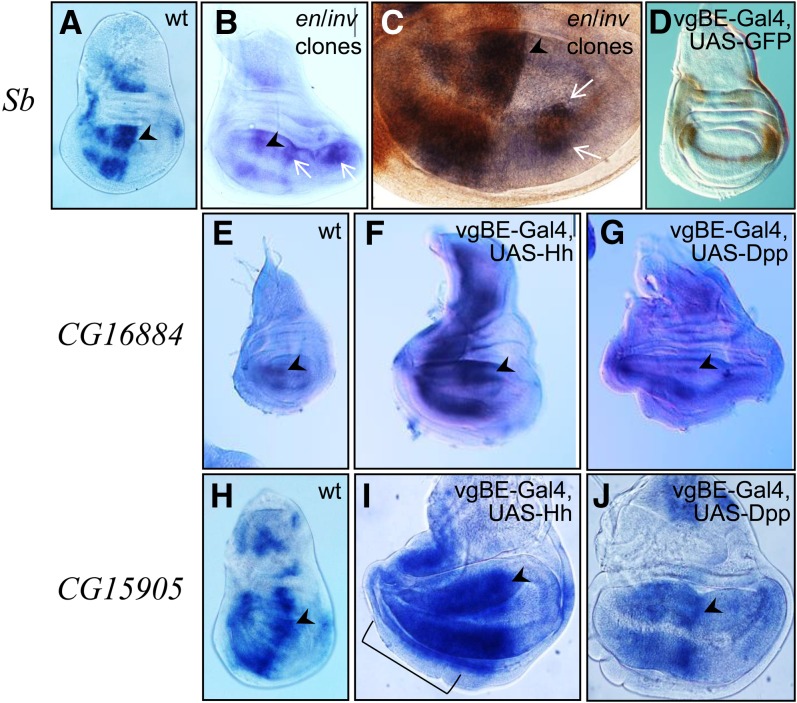
Stubble (Sb) encodes an endopeptidase that functions in cytoskeleton organization (Appel et al. 1993). Our microarray analysis indicated predominant anterior expression and in situ hybridization revealed that although many discrete areas in the wing disc expressed Sb, most were in the A compartment (Figure 3A). Because this expression pattern may indicate that Sb is repressed in P cells, we tested whether en has a role regulating Sb expression. We generated cell clones mutant for both en and inv, and in wing discs, P-cell clones lacking en/inv expressed Sb ectopically (Figure 3, B and C). The mutant cells also expressed ci, an A-specific gene that is normally repressed by en/inv. This finding indicates that En/Inv function is required to repress Sb in the P compartment either directly or indirectly.
Figure 3.
Sb is repressed by en; CG15905 and CG16884 respond to hh. in situ hybridization detected (A−C) Sb transcripts (blue) in a wild-type (wt) disc (A) and in discs with en/inv mutant clones (arrows, B, C). Mutant clones in the P compartment express both Sb (blue; in situ signal, arrows in B and C) and Ci (brown immunostaining in C). (D) Expression of GFP driven by the vestigial boundary enhancer (vgBE-Gal4, UAS-GFP) and immunostaining (HRP brown). Note the horseshoe-like pattern with a thin line along the dorsoventral compartment border. (E) CG16884 in situ signal in a wild-type disc is predominantly in the A compartment. (F, G) Overexpression of transgenic hh (F) or dpp (G) using vgBE-Gal4. Note that hh overexpression caused overproliferation in the A compartment and dpp caused overproliferation in both compartments. Signal was more intense in Hh-overexpressing discs compared with control or Dpp overexpressing discs (treated in parallel). (H−J) in situ hybridization detected CG15905 expression in wild-type (H), hh overexpressing (I), and dpp overexpressing (J) discs. Note the strong hh-induced up-regulation of CG15905 in the A compartment in (I), in contrast to the moderate expression levels in A cells apart from the stripe after dpp overexpression (J). The region with strong up-regulation is indicated by bracket in (I). Arrowheads point to the endogenous anterior stripe of expression at the A/P compartment borders; anterior, left and dorsal, up in all images. All discs are from wandering third instar larvae.
CG16884 and CG15905 expression is increased downstream of Hh signaling
In situ hybridization detected strong CG16884 expression in the central region of the wing pouch A compartment, as well as lower level expression in some P cells and other areas of the disc (Figure 2 and Figure 3E). Expression on the anterior side of the A/P compartment border is suggestive of an activating signal from P cells, such as Hh. To test this possibility, we overexpressed a hh transgene in A and P cells along the dorso/ventral compartment border in the pattern of the vestigial boundary enhancer (vgBE; Williams et al. 1994, Figure 3D). This ectopic expression caused extensive over-proliferation of A cells. In situ detection of CG16884 transcript revealed more intense signal in the A compartment compared to control discs (Figure 3, E and F). Because Hh up-regulates Dpp in A cells (but not in P cells), ectopic activation of CG16884 in A cells could be a consequence of Dpp-dependent activation. We therefore tested the response of CG16884 to ectopic expression of a dpp transgene by expressing Dpp under vgBE control. In these discs A and P cells over-proliferate due to the mitogenic activity of Dpp. However, CG16884 in situ hybridization did not reveal increased expression levels (Figure 3G).
In control discs CG15905 is expressed in numerous patches of wing disc A cells, including a prominent broad stripe along the compartment border (Figure 3H). Its expression in the A compartment increased in response to ectopic expression of Hh (Figure 3I) but was insensitive to Dpp (Figure 3J). These findings show that CG16884 and CG15905 expression can be activated by ectopic Hh.
CG10200-hase und igel is expressed in a dynamic pattern and responds to ectopic Hh
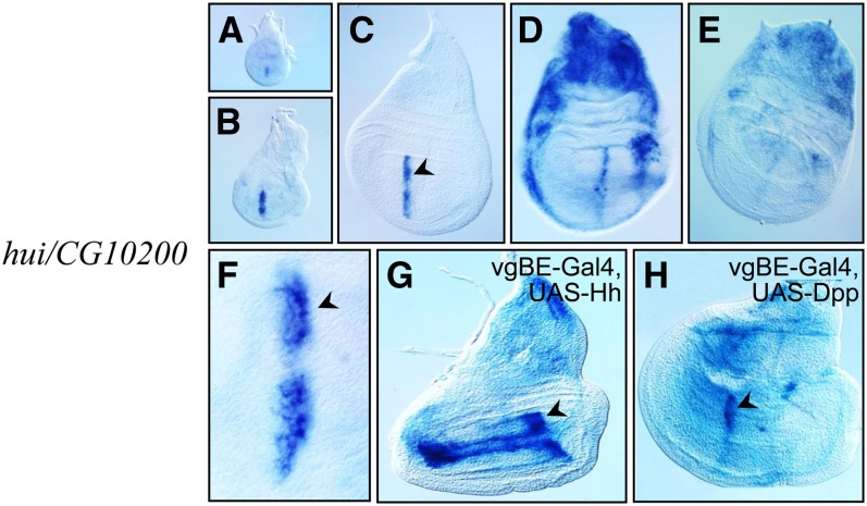
In situ hybridization detected CG10200 expression in wing discs in a narrow A-compartment stripe that abuts the A/P compartment of the wing pouch of early third instar discs (Figure 4, A−C). This stripe was four to five cells wide but was not uniformly intense. It had a gap in the area of the dorsal/ventral compartment border, and its intensity, which was greatest in the cells closest to the P compartment, decreased gradually with increasing distance from the border (Figure 4F). In late third instar discs, expression in the stripe diminished and increased both dorsally in the notum primordium and in the flanks of the disc (Figure 4D). Expression was at low levels in pre-pupal discs (Figure 4E). Monitoring the expression of CG10200 in discs that overexpressed hh in the vgBE domain revealed ectopic activation in response to the Hedgehog signal (Figure 4G). CG10200 expression was limited to two narrow stripes located at the dorsal and ventral sides of the D/V compartment border, an area that overlaps or is adjacent to the cells that expressed the hh transgene (compare Figure 4G with Figure 3D). In contrast to Hh overexpression, no ectopic activation of CG10200 was observed after Dpp overexpression (Figure 4H). This result suggests that Hh, but not Dpp, regulates expression of CG10200. We named CG10200 hase und igel (hare and hedgehog; hui).
Figure 4.
hui/CG10200 expression changes with time and responds to ectopic hh. (A−E) hui/CG10200 expression in wild-type young third instar (A, B), early wandering third instar (C), wandering third instar (D), and late third instar/early prepupa stage (E) discs. (F) Greater magnification view of the anterior stripe of expression from a disc comparable with (C). (G) Ectopic hh activates hui/CG10200 expression in two stripes adjacent to the D/V compartment border in A cells. (H) Dpp overexpression does not induce ectopic hui. Compare the ectopic expression of hui to the expression domain of the vgBE-Gal4 activator in Figure 3D.
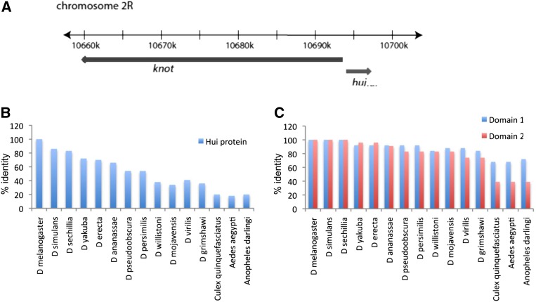
hui is one of three genes identified by our analysis that is expressed in a stripe along the wing disc A/P compartment boundary. The other two genes are CG15905 and kn (Figure 2). kn has two expression domains in third instar wing discs , a region with strong expression in the dorsal hinge primordium of the P compartment as well as the stripe in the A compartment that abuts the A/P border of the wing pouch primordium. The segregation of kn with the expression array posterior cluster (P/A = 1.7) indicates that the P compartment transcripts biased the cluster analysis, but the relevant and interesting issue for hui is that its transcription unit is immediately adjacent to kn (Figure 5A). Because the two genes are transcribed in opposite directions, it is possible that the 1.4-kbp intervening chromosomal region contains regulatory elements that control both genes.
Figure 5.
Genomic map of the knot/hui region and sequence conservation of the Hui protein (A) Drawing showing divergent orientations of the kn and hui genes at the indicated coordinate positions on chromosome 2R. (B) ClustalW (v1.4) comparison of the predicted D. melanogaster Hui sequence to Hui proteins of other Drosophilids and insects (C) and of the two most conserved regions (see Figure S3) designated arbitrarily as Domains 1 and 2. Numbers on the y-axes in B and C indicate identity in percentage.
The predicted Hui protein has 267 residues (Supporting Information, Table S2.) with no strong homology to known proteins or structures. It is highly conserved within the Drosophila genus (Figure 5B), but conservation outside the Drosophilids is low, and searching among genomes in the National Center for Biotechnology Information database (Basic Local Alignment Search Tool, i.e., BLAST) detected homologous sequences only in other insect genomes. Several regions of high conservation were noted after ClustalW alignment of insect sequences, the two largest of which are arbitrarily denoted as domains 1 and 2 (Figure S3); these several regions of strong conservation account for the limited homology to the apparent Hui proteins in non-Drosophilid insects (Figure 5C). In addition to these regions of high conservation, the Hui protein contains a putative signal peptide sequence at the amino-terminus that could mediate targeting to the endoplasmic reticulum for secretion or targeting to other organelles (Table S2). The fact that the Hui protein has two highly conserved domains (Figure 5) suggests that it is hui’s protein product that is under selective pressure.
RNAi knockdown indicates a requirement for Sb, CG16884, and hui during wing development
RNAi knockdown specificity:
To investigate how Sb, CG16884, CG15905, and hui function in wing development, we examined hypomorphic conditions for each gene by using RNAi to knock down expression in different regions of wing discs. Three lines expressing Gal4 were used: ptc-Gal4, which expresses in the A compartment with greatest expression levels in a stripe along the compartment border (Figure 1A); en-Gal4, which expresses in the P compartment (Figure 1C); and C765-Gal4, which expresses in most wing disc cells. We first monitored the specificity of the knockdown effects. The only extant mutant alleles of these genes are Sb mutants that are characterized by short, stubby bristles. By expressing RNAi directed against Sb RNA with C765-Gal4 throughout the wing disc, we selectively phenocopied this short, thick bristles phenotype in the notum and scutellum (not shown). This result is consistent with a previous report, which describes Sb-directed RNAi driven by Act5c-Gal4 (Dietzl et al. 2007). Importantly, head bristles, which are abnormal in Sb mutants, were normal in C765-Gal4 Sb-RNAi flies, indicative of the specific spatial targeting of the knock-down to the wing disc.
To estimate the efficiency of RNAi knock-down, we analyzed hui transcript levels by semiquantitative reverse transcription PCR. Transcript levels for hui were reduced relative to our reference Actin 42A in wing discs that activated expression of the RNAi construct in most cells of the wing imaginal disc (Figure S2). Three independent transgenic insertion lines for this construct showed comparable phenotypic effects (Please see RNAi knockdowns of Sb, CG16884, and hui/CG10200 cause wing malformations), indicating that the phenotypes were not caused by position effect of the transgenic insertions. However, because several putative off-targets were predicted for this RNAi construct (http://stockcenter.vdrc.at), additional tests were made of the specificity of the hui knockdown. First, we used three transgenic lines that carry different hui RNAi constructs and these independent transgenic lines produced comparable RNAi phenotypes (see next section). Second, we analyzed the transcript levels of one of the predicted off-targets, the BRDW3/CG31132 gene. We selected this gene because BRDW3/CG31132 is the most likely candidate to show effects on wing development, particularly on vein formation. RT-PCR analysis of BRDW3/CG31132 revealed no obvious reduction in levels of this transcript (Figure S2). We conclude that the effects of the hui RNAi-mediated knockdown are likely to be specific for the hui function.
RNAi knockdowns of Sb, CG16884, and hui/CG10200 cause wing malformations:
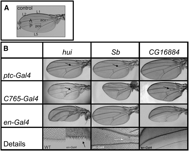
Knockdown of Sb expression reduced the size of the wing and caused vein malformations (Figure 6). RNAi expression in the ptc domain narrowed the L3-L4 intervein region and reduced the anterior crossvein (acv). Expression driven by C765-Gal4 reduced the size of both A and P compartments and appeared to primarily affect proximo-distal growth. Wings that developed in the C765-Gal4 Sb-RNAi genotype were 32% shorter than controls (t-test P-value <2.2E-8). In some cases wings were significantly smaller than the presented example and many were crumpled (not shown). In addition, vein bifurcations and ectopic veins, including a stretch positioned anterior to L4, were observed frequently. Induction of RNAi exclusively in the P compartment (en-Gal4 Sb-RNAi) had only mild effects. The overall wing size was not affected, and bifurcations of the posterior cross vein (pcv) that occurred were rare.
Figure 6.
RNAi knockdown reveals requirement for Sb, CG16884, and hui in wing development (A) Wild-type wing, with longitudinal veins L1-5, anterior (acv) and posterior crossveins (pcv) and the A/P compartment border indicated. (B) RNAi constructs directed against hui, Sb, and CG16884 (columns) were expressed along the A/P border (ptc-Gal4, top row), throughout the disc (C765-Gal4, middle row), or in the P compartment (en-Gal4, third row). hui-RNAi driven by ptc-Gal4 resulted in stubby wings with narrowed L3-L4 intervein region and missing acv (indicative of Hh signaling deficits, arrow). When driven by C765-Gal4 and en-Gal4, these wings have abnormal shape and venation. Phasing of the bristles at the posterior wing margin was disturbed following activation with en-Gal4 (arrow in detail view showing a comparison to a wild-type margin). Expression of Sb RNAi under ptc-Gal4 reduced or eliminated the acv (arrow in top left and detail view bottom row, compare to control in (A). Expression of Sb RNAi under C765-Gal4 reduced the size of both A and P compartments and caused some ectopic vein formation. Sb RNAi in P cells (en-Gal4) caused no apparent effect. CG16884-RNAi in the ptc domain caused narrowing of the L3-L4 intervein region. Knock-down with C765-Gal4 resulted in ectopic vein formation (arrow); RNAi in P cells caused size reduction of the P compartment and a fragile and less pigmented appearance of the cuticle (detail view). All images except for the detail views are to scale.
Whereas previous studies detected Sb only during metamorphosis (Appel et al. 1993), our results show that it is expressed earlier, in third instar discs. The importance of larval disc expression may be related to a more pleiotropic role for Sb. Various allelic combinations, such as Sb/stubbloid and Sb1/Sb63b, affect both leg and wing morphogenesis (Beaton et al. 1988). Interestingly, Sb1/Sb63b, wings are reduced in size and have an ectopic vein anterior to L4, abnormalities that are nearly indistinguishable from wings after C765-Gal4-mediated Sb knockdown (Figure 6). Consistent with its predominant anterior expression this function of Sb in wing development appears to be required in A cells, because Sb RNAi expression in the P compartment (with en-Gal4) revealed no or only very mild phenotypic defects. Additionally, the absence of an anterior crossvein in wings produced after ptc-Gal4−mediated knockdown suggests a role downstream of Hh in the L3-4 intervein region. It is interesting to note that in flies subjected to C765-Gal4-mediated Sb knock-down, both A- and P-wing compartments are abnormally small. We speculate that compensatory regulation between the two compartments may account for this effect.
Knockdown of CG16884 in the ptc pattern caused narrowing of the L3-L4 intervein region similar to the Sb and hui (see next section: hui RNAi and knot loss-of-function cause similar wing defects) RNAi phenotypes. In contrast to the other two genes, however, the acv was not ablated (Figure 6), although in a few wings the acv had small gaps (not shown). C756-Gal4−mediated RNAi resulted in slightly smaller wings with some ectopic vein formation, especially in the position of the anterior and posterior crossveins. A reduction in size was also observed after en-Gal4 activation. Similar to hui (see next section), the reduction affected the P compartment causing a posterior curvature of the wing. In addition to the growth disadvantage, en-Gal4−mediated RNAi resulted in defects in cuticle formation—the cuticle of the entire P compartment appeared fragile and less pigmented.
Expression of the CG15905 RNAi construct did not produce obvious defects with any Gal4 line that we tested (not shown). We do not know whether the absence of a morphological phenotype is a consequence of lack of requirement, unidentified genes with redundant function or insufficient knockdown.
hui RNAi and knot loss-of-function cause similar wing defects:
Phenotypes produced by all three of the hui-RNAi constructs we tested were indistinguishable; Figure 6 shows representative examples with characteristic defects that were observed. hui knockdown in the region between wing veins 3 and 4 with ptc-Gal4 ablated the acv and narrowed the L3-L4 intervein region, and in some wings, gaps in L3 were also observed (not shown). Expression throughout the wing using C765-Gal4 also caused L3-L4 malformation and occasional partial ablation of the acv (not shown). These phenotypes are characteristic of reduced Hh signaling (for example, see Casso et al. 2008), and of kn mutant phenotypes (Nestoras et al. 1997; Vervoort et al. 1999). Thus, hui and kn, whose expression at the A/P boundary is similarly responsive to Hh signaling, are both required for the intervein L3-L4 region.
Additional phenotypes induced by hui RNAi that are not observed for kn include a significant reduction in size of the wing and irregularly spaced bristles along the wing posterior margin (Figure 6). The small wing phenotype was observed after knockdown of hui throughout the wing disc (C765-Gal4; the entire wing was small) or specifically in the P compartment (en-Gal4; only the P compartment was small). In en-Gal4 wings, the L3-L4 intervein region was not reduced and the acv was present. Because the en-Gal4 induced phenotype did not affect the size or appearance of the L3-L4 intervein region, which is the most sensitive area of the wing to changes in Hh signaling, these effects of hui knockdown in the P compartment appear to be autonomous to the cells with reduced hui expression. This reasoning leads us to suggest that the targets of hui knock-down that are affected by en-Gal4 and C765-Gal4 knock-down are the hui-expressing cells along the flanks of the disc rather than the A stripe. If true, this would suggest that in addition to the kn-like function in A cells the growth of the wing primordium is dependent on hui expression in cells of the hinge primordium and/or the flanks of the wing pouch.
Anterior and posterior cells differ in expression of at least 102 genes:
In 1975, Garcia-Bellido proposed the term “selector gene” to designate the key regulatory genes that control the growth and differentiation of the groups of cells that populate developmental compartments (Garcia-Bellido 1975). His proposal was based on genetic studies of homeotic genes and of en, which had been shown to be specifically required in P-compartment cells of the wing disc (Garcia-Bellido et al. 1973; Morata and Lawrence 1975). Subsequent molecular studies have fully validated the selector gene hypothesis: the homeotic genes and en have expression patterns that correlate precisely with the cells that require their functions.
In the third instar wing disc, en is expressed in all P compartment cells (Kornberg et al.1985). Three other genes that are known to be expressed in all cells of either the A or P compartments are ci (A) and inv and hh (P). These genes were first isolated by either positional cloning based on the phenotype of insertional mutants (e.g., ci, Orenic et al. 1990; hh, Mohler and Vani 1992), by linkage and coregulation with en (Coleman et al. 1987), or based on their compartment-specific expression patterns that were revealed in enhancer trap lines (e.g., ci, Eaton and Kornberg 1990; and hh, Lee et al. 1992; Tabata et al. 1992). The compartment-specific expression of these four genes led in the ensuing years to numerous enhancer trap screens in search of other genes that express in a compartment-specific patterns, but these efforts identified no other genes that are expressed specifically in all A or P cells (T. Kornberg, unpublished data). It has not been apparent whether these negative results were due to the inadequacy of the insertional-based approaches used to screen for expression patterns or to the absence of other such genes.
The expression array screen described here was based on manual dissection and isolation of small numbers of cells from single imaginal discs. The cells were identified by patterns of GFP expression regulated either by ptc-Gal4 or hh-Gal4, and our results showed that the levels of GFP transcripts in these isolated cells correlated strongly with the domains that express GFP. The expression array analysis monitored approximately 75% of the currently annotated Drosophila genes and identified a list of 102 genes with preferential A or P expression. The three genes with the greatest P/A expression ratio were en, hh, and inv. The identification of the same set of genes by two dissimilar genome-wide scans (e.g., expression array hybridization and enhancer trap screening) suggests that en, hh and ci may be unique among Drosophila genes in their compartment-specific expression. Thus, whereas en, hh, and ci had the most robust differences in expression levels, the other genes in our list produced more moderate differences in signal intensities (Table 1 and Table S1). Most of these genes appear to be expressed by subsets of cells in both compartments with only moderate enrichment in one or the other (Figure 2). Nevertheless, several expression patterns are suggestive of regulation downstream of en or hh, and we analyzed four of these genes: Sb, CG16884, CG15905, and CG10200/hui, of which three are involved in wing development. The data we present indicate that Sb is a regulatory target of en/inv and that hui, CG16884, and CG15905 are inducible by the en/inv-regulated gene hh. It is interesting to note that the Sb protein is thought to be a transmembrane serine protease that could process as yet unidentified signaling proteins (Bayer et al. 2003), and that hui, CG15905, and CG16884 encode short proteins with amino-terminal putative endoplasmic reticulum signal peptides Table S2. These observations raise the possibility that all three proteins could be secreted or could process secreted factors that act on cells beyond their expression domains.
Supplementary Material
Acknowledgment
Supported by National Institutes of Health grant GM030637 (to T.B.K.) and FK funds of the Freie Universität Berlin (to A.K.) supported this work.
Footnotes
Communicating editor: T. R. Hughes
Literature Cited
- Amin A., Li Y., Finkelstein R., 1999. Hedgehog activates the EGF receptor pathway during Drosophila head development. Development 126: 2623–2630 [DOI] [PubMed] [Google Scholar]
- Appel L. F., Prout M., Abu-Shumays R., Hammonds A., Garbe J. C., et al. , 1993. The Drosophila Stubble-stubbloid gene encodes an apparent transmembrane serine protease required for epithelial morphogenesis. Proc. Natl. Acad. Sci. USA 90: 4937–4941 [DOI] [PMC free article] [PubMed] [Google Scholar]
- Basler K., Struhl G., 1994. Compartment boundaries and the control of Drosophila limb pattern by the Hedgehog protein. Nature 368: 208–215 [DOI] [PubMed] [Google Scholar]
- Bayer C. A., Halsell S. R., Fristrom J. W., Kiehart D. P., von Kalm L., 2003. Genetic interactions between the RhoA and Stubble-stubbloid loci suggest a role for a type II transmembrane serine protease in intracellular signaling during Drosophila imaginal disc morphogenesis. Genetics 165: 1417–1432 [DOI] [PMC free article] [PubMed] [Google Scholar]
- Beaton A. H., Kiss I., Fristrom D., Fristrom J. W., 1988. Interaction of the Stubble-stubbloid locus and the Broad-complex of Drosophila melanogaster. Genetics 120: 453–464 [DOI] [PMC free article] [PubMed] [Google Scholar]
- Butler M. J., Jacobsen T. L., Cain D. M., Jarman M. G., Hubank M., et al. , 2003. Discovery of genes with highly restricted expression patterns in the Drosophila wing disc using DNA oligonucleotide microarrays. Development 130: 659–670 [DOI] [PubMed] [Google Scholar]
- Casso D. J., Liu S., Iwaki D. D., Ogden S. K., Kornberg T. B., 2008. A screen for modifiers of hedgehog signaling in Drosophila melanogaster identifies swm and mts. Genetics 178: 1399–1413 [DOI] [PMC free article] [PubMed] [Google Scholar]
- Casso D. J., Biehs B., Kornberg T. B., 2011. A novel interaction between hedgehog and Notch promotes proliferation at the anterior-posterior organizer of the Drosophila wing. Genetics 187: 485–499 [DOI] [PMC free article] [PubMed] [Google Scholar]
- Coleman K. G., Poole S. J., Weir M. P., Soeller W. C., Kornberg T. B., 1987b The invected gene of Drosophila: sequence analysis and expression studies reveal a close kinship to the engrailed gene. Genes Dev. 1: 19–28 [DOI] [PubMed] [Google Scholar]
- Dietzl G., Chen D., Schnorrer F., Su K. C., Barinova Y., et al. , 2007. A genome-wide transgenic RNAi library for conditional gene inactivation in Drosophila. Nature 448: 151–156 [DOI] [PubMed] [Google Scholar]
- Eaton S., Kornberg T. B., 1990. Repression of ci-D in posterior compartments of Drosophila by engrailed. Genes Dev. 4: 1068–1077 [DOI] [PubMed] [Google Scholar]
- Eisen M. B., Spellman P. T., Brown P. O., Botstein D., 1998. Cluster analysis and display of genome-wide expression patterns. Proc. Natl. Acad. Sci. USA 95: 14863–14868 [DOI] [PMC free article] [PubMed] [Google Scholar]
- Furlong E. E., Andersen E. C., Null B., White K. P., Scott M. P., 2001. Patterns of gene expression during Drosophila mesoderm development. Science 293: 1629–1633 [DOI] [PubMed] [Google Scholar]
- Garcia-Bellido A., 1975. Genetic control of wing disc development in Drosophila. Ciba Found. Symp. 0: 161–182 [DOI] [PubMed] [Google Scholar]
- Garcia-Bellido A., Ripoll P., Morata G., 1973. Developmental compartmentalization of the wing disc of Drosophila. Nat. New Biol. 245: 251–253 [DOI] [PubMed] [Google Scholar]
- Gustavson E., Goldsborough A. S., Ali Z., Kornberg T. B., 1996. The Drosophila engrailed and invected genes: partners in regulation, expression and function. Genetics 906: 893–906 [DOI] [PMC free article] [PubMed] [Google Scholar]
- Ingham P. W., Fietz M. J., 1995. Quantitative effects of hedgehog and decapentaplegic activity on the patterning of the Drosophila wing. Curr. Biol. 5: 432–440 [DOI] [PubMed] [Google Scholar]
- Klebes A., Kornberg T. B., 2008. Linear RNA amplification for the production of microarray hybridization probes. Methods Mol. Biol. 420: 303–317 [DOI] [PubMed] [Google Scholar]
- Klebes A., Biehs B., Cifuentes F., Kornberg T. B., 2002. Expression profiling of Drosophila imaginal discs. Genome Biol. 3: 1–16 [DOI] [PMC free article] [PubMed] [Google Scholar]
- Klebes A., Sustar A., Kechris K., Li H., Schubiger G., et al. , 2005. Regulation of cellular plasticity in Drosophila imaginal disc cells by the Polycomb group, trithorax group and lama genes. Development 132: 3753–3765 [DOI] [PubMed] [Google Scholar]
- Kornberg T., Siden I., O’Farrell P., Simon M., 1985. The engrailed locus of Drosophila: in situ localization of transcripts reveals compartment-specific expression. Cell 40: 45–53 [DOI] [PubMed] [Google Scholar]
- Lawrence P. A., Struhl G., 1996. Morphogens, compartments, and pattern: lessons from Drosophila? Cell 85: 951–961 [DOI] [PubMed] [Google Scholar]
- Lee J. J., von Kessler D. P., Parks S., Beachy P. A., 1992. Secretion and localized transcription suggest a role in positional signaling for products of the segmentation gene hedgehog. Cell 71: 33–50 [DOI] [PubMed] [Google Scholar]
- Liotta D., Han J., Elgar S., Garvey C., Han Z., et al. , 2007. The Him gene reveals a balance of inputs controlling muscle differentiation in Drosophila. Curr. Biol. 17: 1409–1413 [DOI] [PMC free article] [PubMed] [Google Scholar]
- Luschnig S., Batz T., Armbruster K., Krasnow M. A., 2006. serpentine and vermiform encode matrix proteins with chitin binding and deacetylation domains that limit tracheal tube length in Drosophila. Curr. Biol. 16: 186–194 [DOI] [PubMed] [Google Scholar]
- Mohler J., Vani K., 1992. Molecular organization and embryonic expression of the hedgehog gene involved in cell-cell communication in segmental patterning of Drosophila. Development 115: 957–971 [DOI] [PubMed] [Google Scholar]
- Morata G., Lawrence P. A., 1975. Control of compartment development by the engrailed gene of Drosophila. Nature 255: 614–618 [DOI] [PubMed] [Google Scholar]
- Motzny C. K., Holmgren R., 1995. The Drosophila Cubitus interruptus protein and its role in the wingless and hedgehog signal transduction pathways. Mech. Dev. 52: 137–150 [DOI] [PubMed] [Google Scholar]
- Nellen D., Burke R., Struhl G., Basler K., 1996. Direct and long-range action of a DPP morphogen gradient. Cell 85: 357–368 [DOI] [PubMed] [Google Scholar]
- Nestoras K., Lee H., Mohler J., 1997. Role of knot (kn) in wing patterning in Drosophila. Genetics 147: 1203–1212 [DOI] [PMC free article] [PubMed] [Google Scholar]
- Orenic T. V., Slusarski D. C., Kroll K. L., Holmgren R. A., 1990. Cloning and characterization of the segment polarity gene cubitus interruptus Dominant of Drosophila. Genes Dev. 4: 1053–1067 [DOI] [PubMed] [Google Scholar]
- Phillips R. G., Roberts I. J., Ingham P. W., Whittle J. R., 1990. The Drosophila segment polarity gene patched is involved in a position-signalling mechanism in imaginal discs. Development 110: 105–114 [DOI] [PubMed] [Google Scholar]
- Schnepp B., Grumbling G., Donaldson T., Simcox A., 1996. Vein is a novel component in the Drosophila epidermal growth factor receptor pathway with similarity to the neuregulins. Genes Dev. 10: 2302–2313 [DOI] [PubMed] [Google Scholar]
- Speicher S., Thomas U., Hinz U., Knust E., 1994. The Serrate locus of Drosophila and its role in morphogenesis of the wing imaginal discs: control of cell proliferation. Development 120: 535–544 [DOI] [PubMed] [Google Scholar]
- Sullivan W., Ashburner M., Hawley R. S., 2000. Drosophila Protocols. Cold Spring Harbor Laboratory Press, New York [Google Scholar]
- Swan L. E., Wichmann C., Prange U., Schmid A., Schmidt M., et al. , 2004. A glutamate receptor-interacting protein homolog organizes muscle guidance in Drosophila. Genes Dev. 18: 223–237 [DOI] [PMC free article] [PubMed] [Google Scholar]
- Tabata T., Kornberg T. B., 1994. Hedgehog is a signaling protein with a key role in patterning Drosophila imaginal discs. Cell 76: 89–102 [DOI] [PubMed] [Google Scholar]
- Tabata T., Eaton S., Kornberg T. B., 1992. The Drosophila hedgehog gene is expressed specifically in posterior compartment cells and is a target of engrailed regulation. Genes Dev. 6: 2635–2645 [DOI] [PubMed] [Google Scholar]
- Tabata T., Schwartz C., Gustavson E., Ali Z., Kornberg T. B., 1995. Creating a Drosophila wing de novo, the role of engrailed, and the compartment border hypothesis. Development 121: 3359–3369 [DOI] [PubMed] [Google Scholar]
- Tanimoto H., Itoh S., ten Dijke P., Tabata T., 2000. Hedgehog creates a gradient of DPP activity in Drosophila wing imaginal discs. Mol. Cell 5: 59–71 [DOI] [PubMed] [Google Scholar]
- Thisse B., Stoetzel C., Gorostiza-Thisse C., Perrin-Schmitt F., 1988. Sequence of the twist gene and nuclear localization of its protein in endomesodermal cells of early Drosophila embryos. EMBO J. 7: 2175–2183 [DOI] [PMC free article] [PubMed] [Google Scholar]
- Tusher V. G., Tibshirani R., Chu G., 2001. Significance analysis of microarrays applied to the ionizing radiation response. Proc. Natl. Acad. Sci. USA 98: 5116–5121 [DOI] [PMC free article] [PubMed] [Google Scholar]
- Vervoort M., Crozatier M., Valle D., Vincent A., 1999. The COE transcription factor Collier is a mediator of short-range Hedgehog-induced patterning of the Drosophila wing. Curr. Biol. 9: 632–639 [DOI] [PubMed] [Google Scholar]
- Williams J. A., Paddock S. W., Vorwerk K., Carroll S. B., 1994. Organization of wing formation and induction of a wing-patterning gene at the dorsal/ventral compartment boundary. Nature 368: 299–305 [DOI] [PubMed] [Google Scholar]
Associated Data
This section collects any data citations, data availability statements, or supplementary materials included in this article.


