

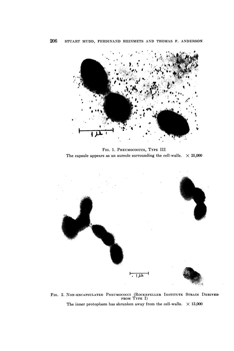
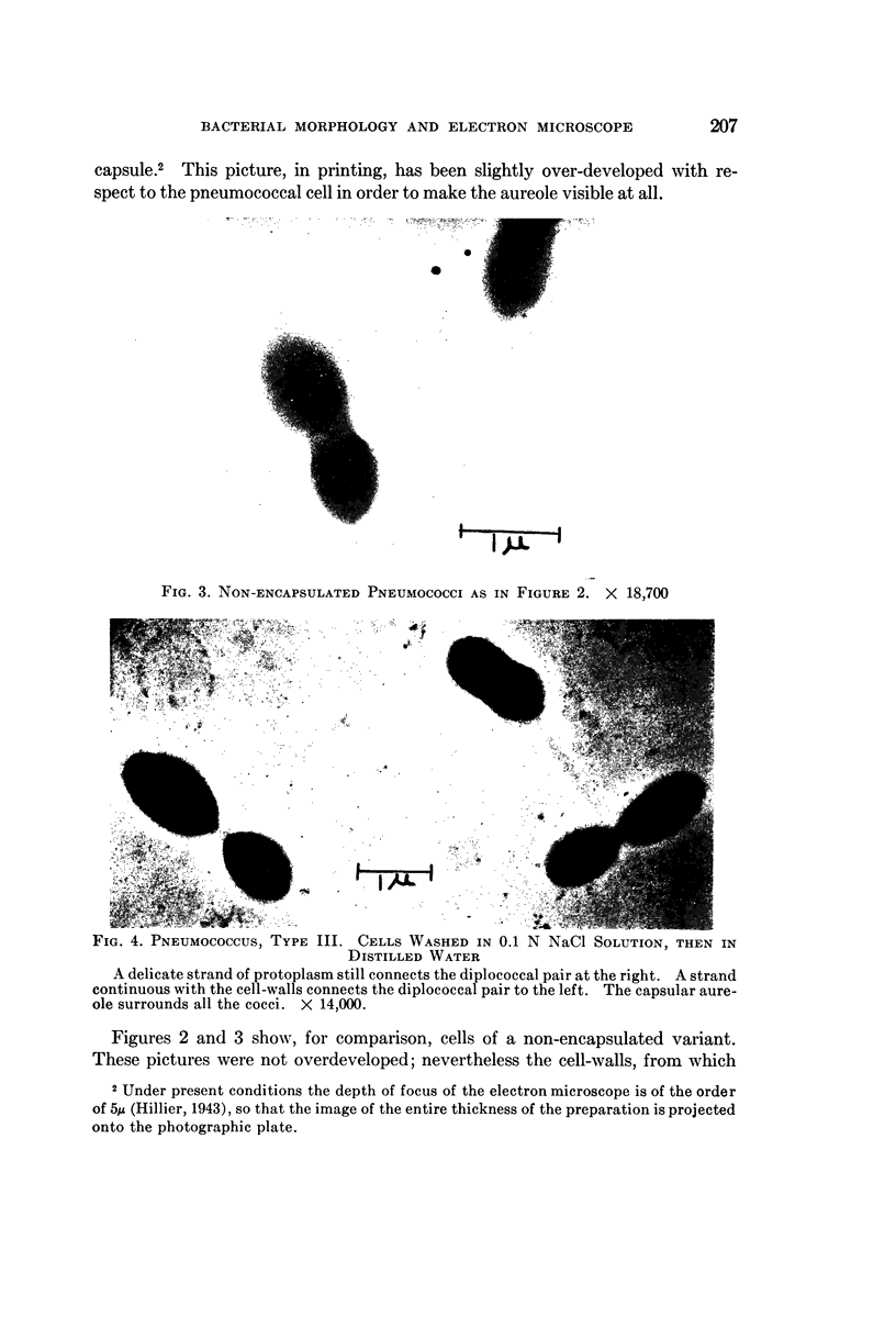
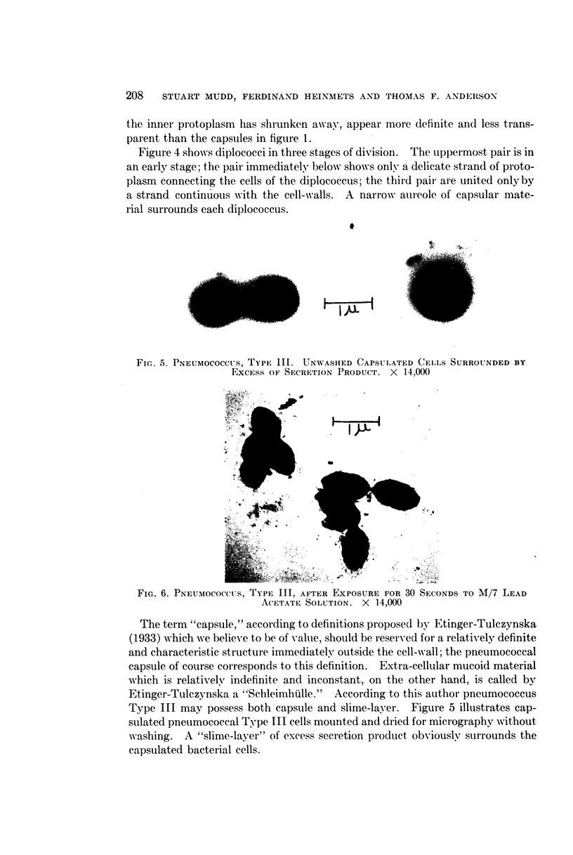
Full text
PDF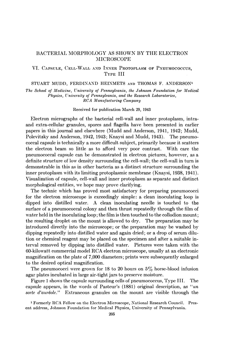


Images in this article


Selected References
These references are in PubMed. This may not be the complete list of references from this article.
- Knaysi G., Mudd S. The Internal Structure of Certain Bacteria as Revealed by the Electron Microscope-A Contribution to the Study of the Bacterial Nucleus. J Bacteriol. 1943 Apr;45(4):349–359. doi: 10.1128/jb.45.4.349-359.1943. [DOI] [PMC free article] [PubMed] [Google Scholar]
- Knaysi G. Observations on the Cell Division of Some Yeasts and Bacteria. J Bacteriol. 1941 Feb;41(2):141–153. doi: 10.1128/jb.41.2.141-153.1941. [DOI] [PMC free article] [PubMed] [Google Scholar]
- Mudd S., Polevitzky K., Anderson T. F. Bacterial Morphology as shown by the Electron Microscope: V. Treponema pallidum, T. macrodentium and T. microdentium. J Bacteriol. 1943 Jul;46(1):15–24. doi: 10.1128/jb.46.1.15-24.1943. [DOI] [PMC free article] [PubMed] [Google Scholar]