Abstract
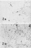
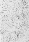
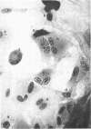
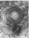
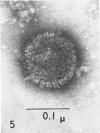
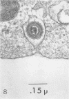
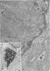
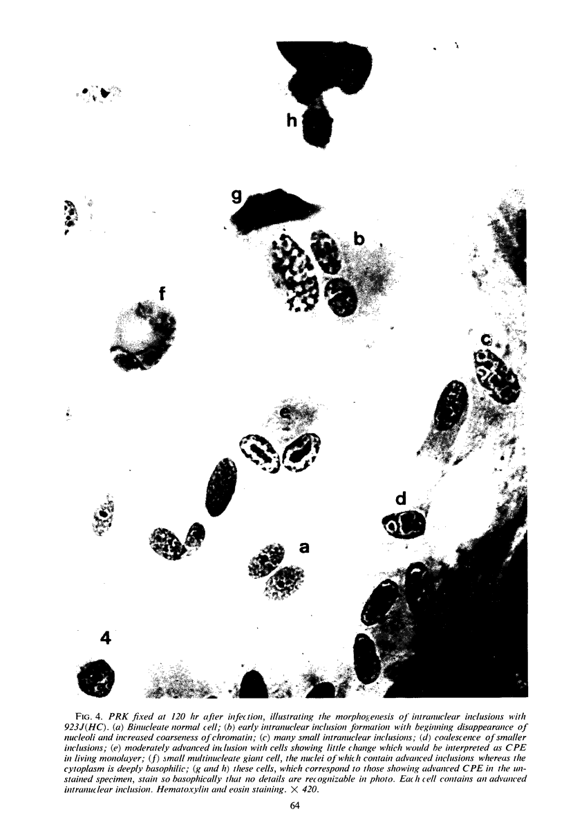
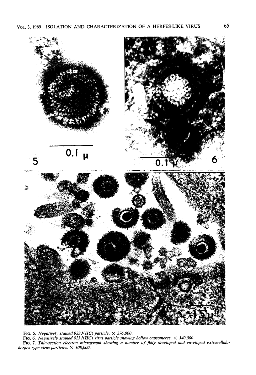
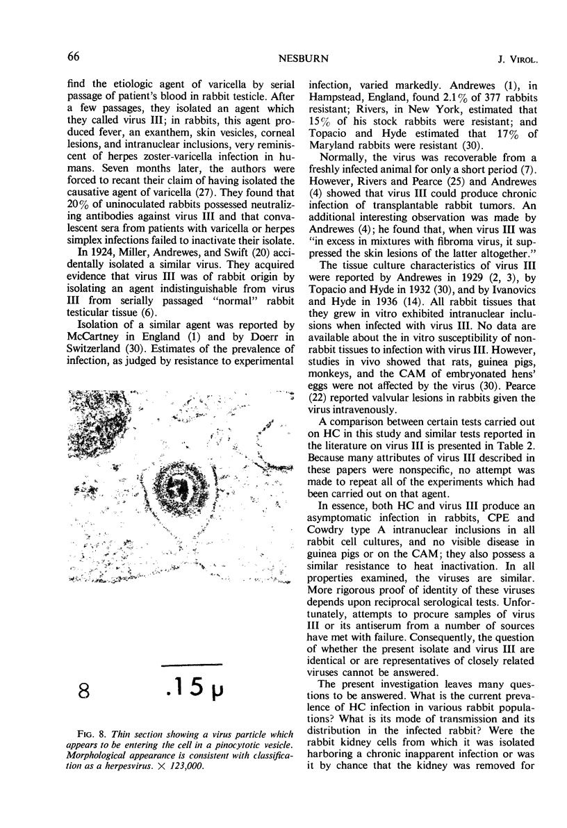
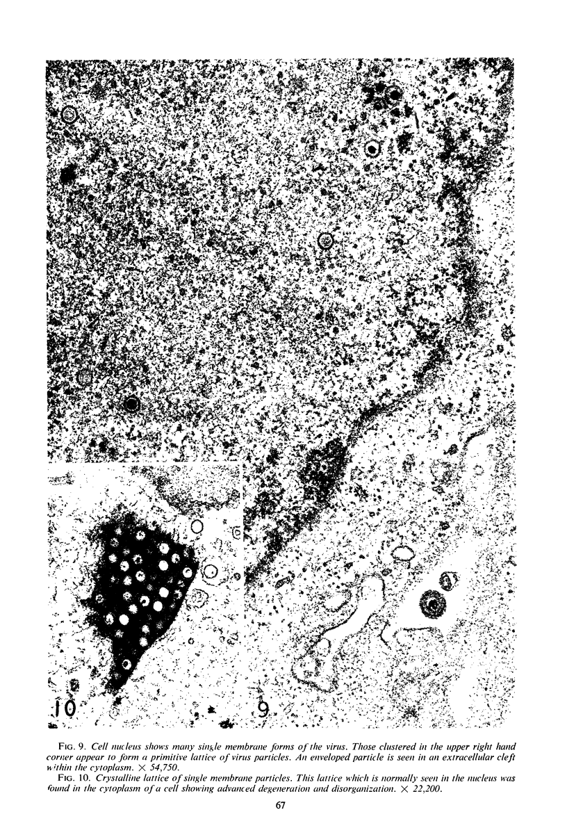
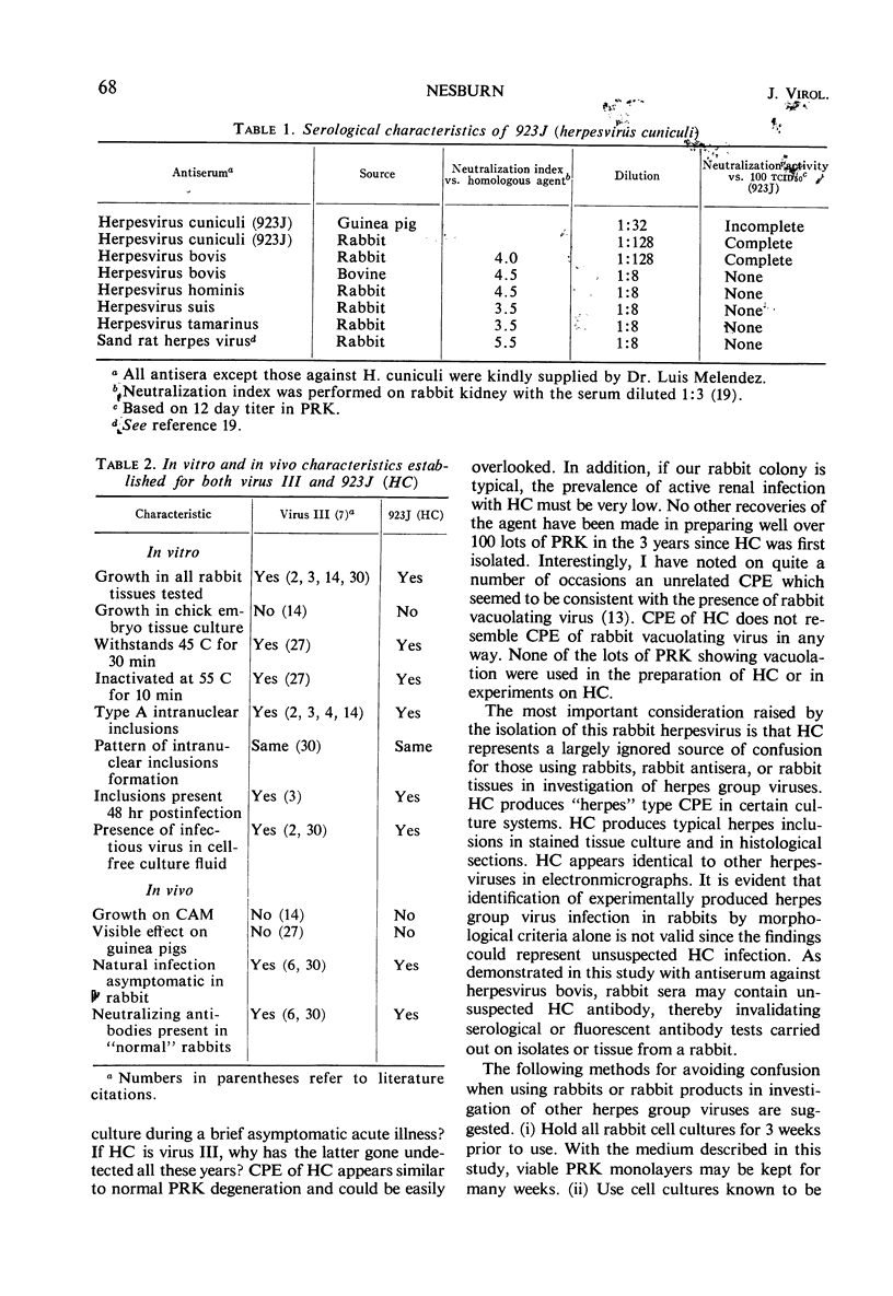
An agent which possesses the physical, chemical, cytopathic, histological, and electron microscopic attributes of a herpes group virus was isolated from an uninoculated batch of primary rabbit kidney cell cultures. Preliminary evidence indicates that antibodies against the agent are found in some sera of other “normal” New Zealand albino rabbits. In cell cultures, the virus grew best and almost exclusively in cells of rabbit origin. On the basis of these facts, the name herpesvirus cuniculi (HC) is suggested for the isolate. A batch of anti-herpesvirus bovis antiserum prepared in rabbits was found to be “contaminated” with unsuspected neutralizing antibodies against HC. Caution is mandatory when using rabbits, rabbit tissues, or rabbit sera for work with any herpes group virus unless precautions are taken to rule out unsuspected infection with or antibodies against HC. This agent may well represent a reisolation of virus III, a rabbit herpes virus, described by Rivers in 1923; the isolation of this virus has not been reported since 1940. It is important to reemphasize the existence of this agent in an animal which is commonly used for laboratory investigation of herpes group viruses.
Full text
PDF
Images in this article
Selected References
These references are in PubMed. This may not be the complete list of references from this article.
- DART L. H., Jr, TURNER T. R. Fluorescence microscopy in exfoliative cytology. Report of acridine orange examination of 5491 cases, with comparison by the Papanicolaou technic. Lab Invest. 1959 Nov-Dec;8:1513–1522. [PubMed] [Google Scholar]
- ENDERS J. F. Bovine amniotic fluid as tissue culture medium in cultivation of poliomyelitis and other viruses. Proc Soc Exp Biol Med. 1953 Jan;82(1):100–105. doi: 10.3181/00379727-82-20035. [DOI] [PubMed] [Google Scholar]
- ENDERS J. F., PEEBLES T. C. Propagation in tissue cultures of cytopathogenic agents from patients with measles. Proc Soc Exp Biol Med. 1954 Jun;86(2):277–286. doi: 10.3181/00379727-86-21073. [DOI] [PubMed] [Google Scholar]
- GUENALP A. GROWTH AND CYTOPATHIC EFFECT OF RUBELLA VIRUS IN A LINE OF GREEN MONKEY KIDNEY CELLS. Proc Soc Exp Biol Med. 1965 Jan;118:85–90. [PubMed] [Google Scholar]
- HARTLEY J. W., ROWE W. P. NEW PAPOVAVIRUS CONTAMINATING SHOPE PAPILLOMATA. Science. 1964 Jan 17;143(3603):258–260. doi: 10.1126/science.143.3603.258. [DOI] [PubMed] [Google Scholar]
- MCGAVRAN M. H., SMITH M. G. ULTRASTRUCTURAL, CYTOCHEMICAL, AND MICROCHEMICAL OBSERVATIONS ON CYTOMEGALOVIRUS (SALIVARY GLAND VIRUS) INFECTION OF HUMAN CELLS IN TISSUE CULTURE. Exp Mol Pathol. 1965 Feb;76:1–10. doi: 10.1016/0014-4800(65)90019-5. [DOI] [PubMed] [Google Scholar]
- MILOVANOVIC M. V., ENDERS J. F., MITUS A. Cultivation of measles virus in human amnion cells and in developing chick embryo. Proc Soc Exp Biol Med. 1957 May;95(1):120–127. doi: 10.3181/00379727-95-23140. [DOI] [PubMed] [Google Scholar]
- Meléndez L. V., Hunt R. D., King N. W., Garcia F. G., Like A. A., Mike E. A herpes virus from sand rats (Psammomys obesus). Lab Anim Care. 1967 Jun;17(3):302–309. [PubMed] [Google Scholar]
- PEARCE J. M. Heart disease and filtrable viruses. Circulation. 1960 Mar;21:448–455. doi: 10.1161/01.cir.21.3.448. [DOI] [PubMed] [Google Scholar]
- PLUMMER G., WATERSON A. P. Equine herpes viruses. Virology. 1963 Mar;19:412–416. doi: 10.1016/0042-6822(63)90083-7. [DOI] [PubMed] [Google Scholar]
- YOUNGNER J. S. Monolayer tissue cultures. I. Preparation and standardization of suspensions of trypsin-dispersed monkey kidney cells. Proc Soc Exp Biol Med. 1954 Feb;85(2):202–205. doi: 10.3181/00379727-85-20830. [DOI] [PubMed] [Google Scholar]


