Abstract
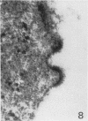
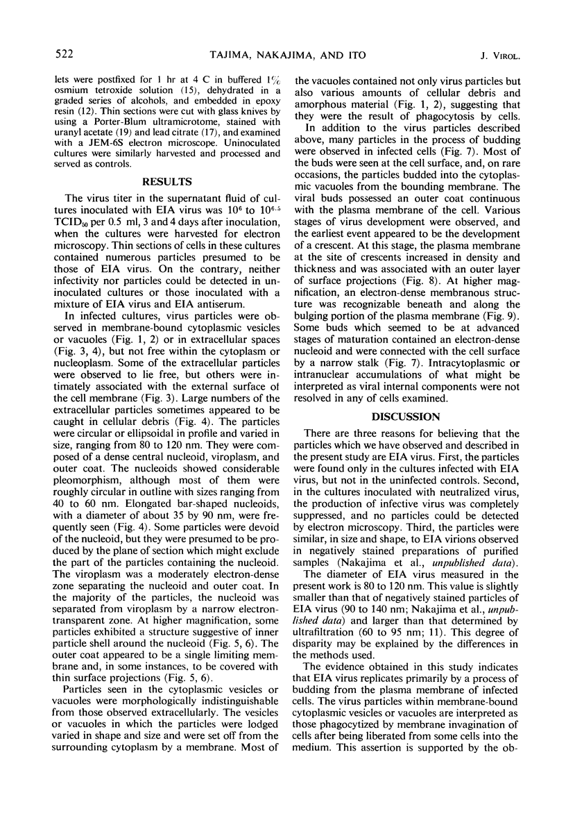
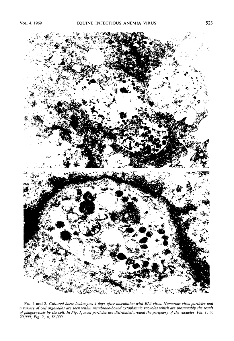
Equine infectious anemia (EIA) virus was observed in thin sections of infected cultured horse leukocytes by electron microscopy. The virus particles had a spherical shape and were between 80 and 120 nm in diameter. Most of them contained an electron-dense nucleoid 40 to 60 nm in diameter. They were observed to form by a process of budding from the plasma membrane and appeared to have thin surface projections. The particles described were not detected in uninfected cultured cells, and their appearance could be prevented by adding EIA immune serum to the inoculum. The implications of these findings in the classification of EIA virus are discussed.
Full text
PDF
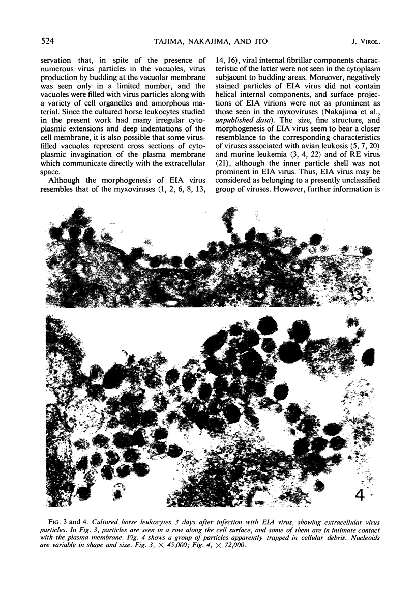
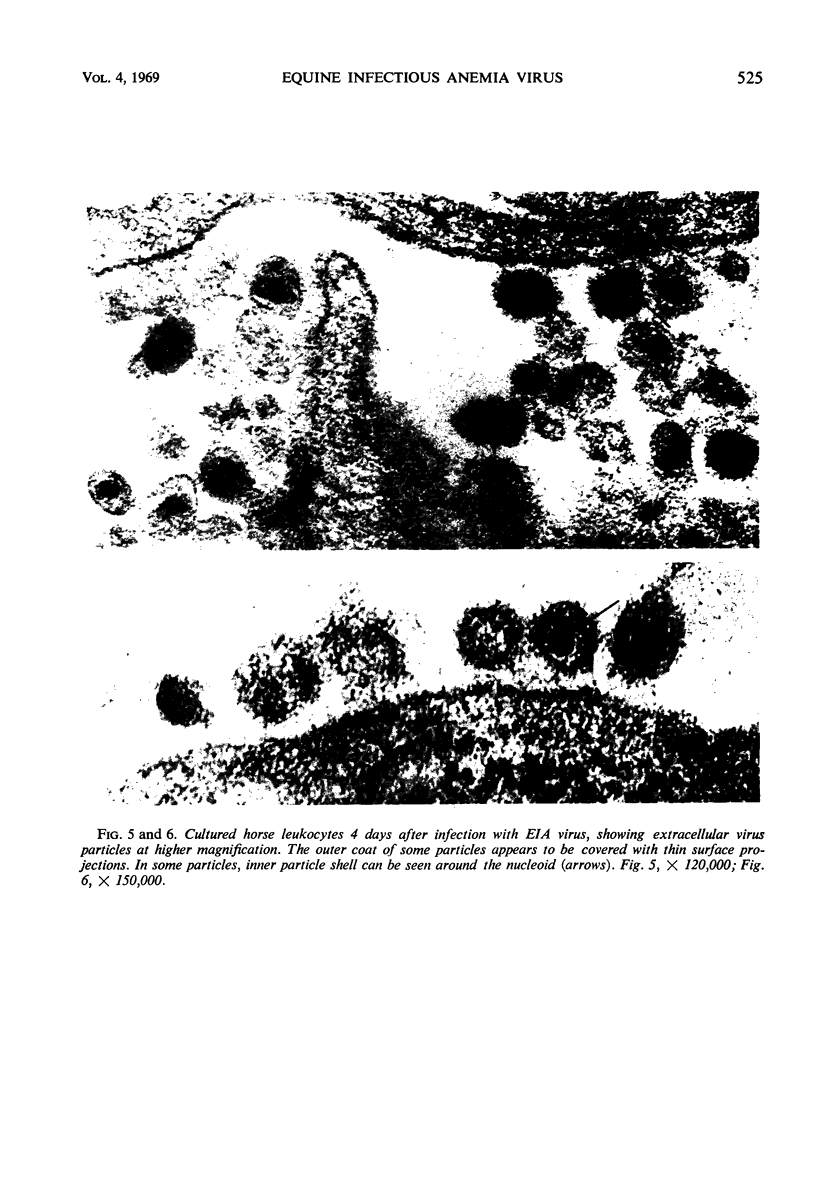
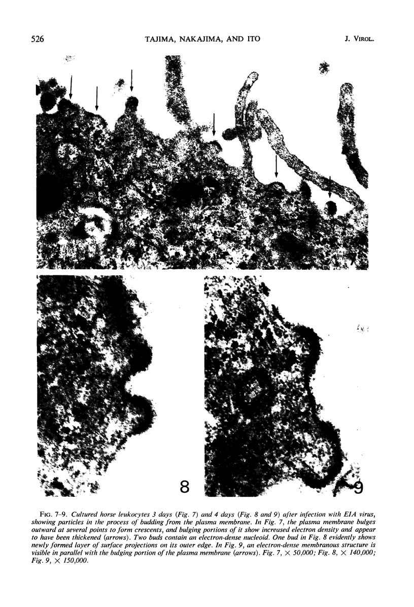
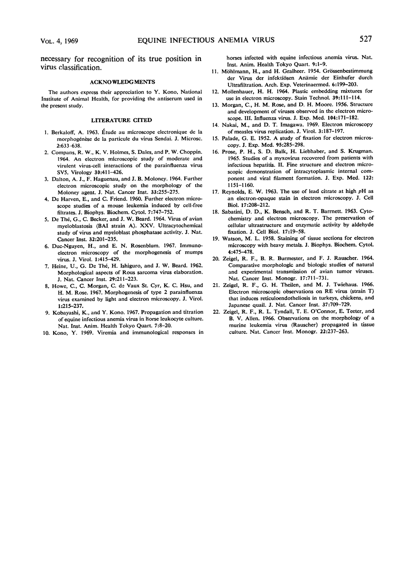
Images in this article
Selected References
These references are in PubMed. This may not be the complete list of references from this article.
- Compans R. W., Holmes K. V., Dales S., Choppin P. W. An electron microscopic study of moderate and virulent virus-cell interactions of the parainfluenza virus SV5. Virology. 1966 Nov;30(3):411–426. doi: 10.1016/0042-6822(66)90119-x. [DOI] [PubMed] [Google Scholar]
- DALTON A. J., HAGUENAU F., MOLONEY J. B. FURTHER ELECTRON MICROSCOPIC STUDIES ON THE MORPHOLOGY OF THE MOLONEY AGENT. J Natl Cancer Inst. 1964 Aug;33:255–275. [PubMed] [Google Scholar]
- DE HARVEN E., FRIEND C. Further electron microscope studies of a mouse leukemia induced by cell-free filtrates. J Biophys Biochem Cytol. 1960 Jul;7:747–752. doi: 10.1083/jcb.7.4.747. [DOI] [PMC free article] [PubMed] [Google Scholar]
- Duc-Nguyen H., Rosenblum E. N. Immuno-electron microscopy of the morphogenesis of mumps virus. J Virol. 1967 Apr;1(2):415–429. doi: 10.1128/jvi.1.2.415-429.1967. [DOI] [PMC free article] [PubMed] [Google Scholar]
- HEINE U., DE THE G., ISHIGURO H., BEARD J. W. Morphologic aspects of Rous sarcoma virus elaboration. J Natl Cancer Inst. 1962 Jul;29:211–223. [PubMed] [Google Scholar]
- Howe C., Morgan C., de Vaux St Cyr C., Hsu K. C., Rose H. M. Morphogenesis of type 2 parainfluenza virus examined by light and electron microscopy. J Virol. 1967 Feb;1(1):215–237. doi: 10.1128/jvi.1.1.215-237.1967. [DOI] [PMC free article] [PubMed] [Google Scholar]
- Kobayashi K., Kono Y. Propagation and titration of equine infectious anemia virus in horse leukocyte culture. Natl Inst Anim Health Q (Tokyo) 1967 Spring;7(1):8–20. [PubMed] [Google Scholar]
- Kono Y. Viremia and immunological responses in horses infected with equine infectious anemia virus. Natl Inst Anim Health Q (Tokyo) 1969 Spring;9(1):1–9. [PubMed] [Google Scholar]
- MOLLENHAUER H. H. PLASTIC EMBEDDING MIXTURES FOR USE IN ELECTRON MICROSCOPY. Stain Technol. 1964 Mar;39:111–114. [PubMed] [Google Scholar]
- MORGAN C., ROSE H. M., MOORE D. H. Structure and development of viruses observed in the electron microscope. III. Influenza virus. J Exp Med. 1956 Aug 1;104(2):171–182. doi: 10.1084/jem.104.2.171. [DOI] [PMC free article] [PubMed] [Google Scholar]
- Nakai M., Imagawa D. T. Electron microscopy of measles virus replication. J Virol. 1969 Feb;3(2):187–197. doi: 10.1128/jvi.3.2.187-197.1969. [DOI] [PMC free article] [PubMed] [Google Scholar]
- PALADE G. E. A study of fixation for electron microscopy. J Exp Med. 1952 Mar;95(3):285–298. doi: 10.1084/jem.95.3.285. [DOI] [PMC free article] [PubMed] [Google Scholar]
- Prose P. H., Balk S. D., Liebhaber H., Krugman S. Studies of a myxovirus recovered from patients with infectious hepatitis. II. Fine structure and electron microscopic demonstration of intracytoplasmic internal component and viral filament formation. J Exp Med. 1965 Dec 1;122(6):1151–1160. doi: 10.1084/jem.122.6.1151. [DOI] [PMC free article] [PubMed] [Google Scholar]
- REYNOLDS E. S. The use of lead citrate at high pH as an electron-opaque stain in electron microscopy. J Cell Biol. 1963 Apr;17:208–212. doi: 10.1083/jcb.17.1.208. [DOI] [PMC free article] [PubMed] [Google Scholar]
- SABATINI D. D., BENSCH K., BARRNETT R. J. Cytochemistry and electron microscopy. The preservation of cellular ultrastructure and enzymatic activity by aldehyde fixation. J Cell Biol. 1963 Apr;17:19–58. doi: 10.1083/jcb.17.1.19. [DOI] [PMC free article] [PubMed] [Google Scholar]
- THEG D. E., BECKER C., BEARD J. W. VIRUS OF AVIAN MYELOBLASTOSIS (BAI STRAIN A). XXV. ULTRACYTOCHEMICAL STUDY OF VIRUS AND MYELOBLAST PHOSPHATASE ACTIVITY. J Natl Cancer Inst. 1964 Jan;32:201–235. [PubMed] [Google Scholar]
- WATSON M. L. Staining of tissue sections for electron microscopy with heavy metals. J Biophys Biochem Cytol. 1958 Jul 25;4(4):475–478. doi: 10.1083/jcb.4.4.475. [DOI] [PMC free article] [PubMed] [Google Scholar]
- Zeigel R. F., Theilen G. H., Twiehaus M. J. Electron microscopic observations on RE virus (strain T) that induces reticuloendotheliosis in turkeys, chickens, and Japanese quail. J Natl Cancer Inst. 1966 Dec;37(6):709–729. [PubMed] [Google Scholar]
- Zeigel R. F., Tyndall R. L., O'Connor T. E., Teeter E., Allen B. V. Observations on the morphology of a murine leukemia virus (Rauscher) propagated in tissue culture. Natl Cancer Inst Monogr. 1966 Sep;22:237–263. [PubMed] [Google Scholar]


