Abstract
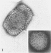
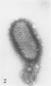
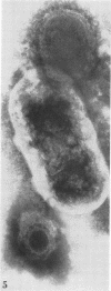
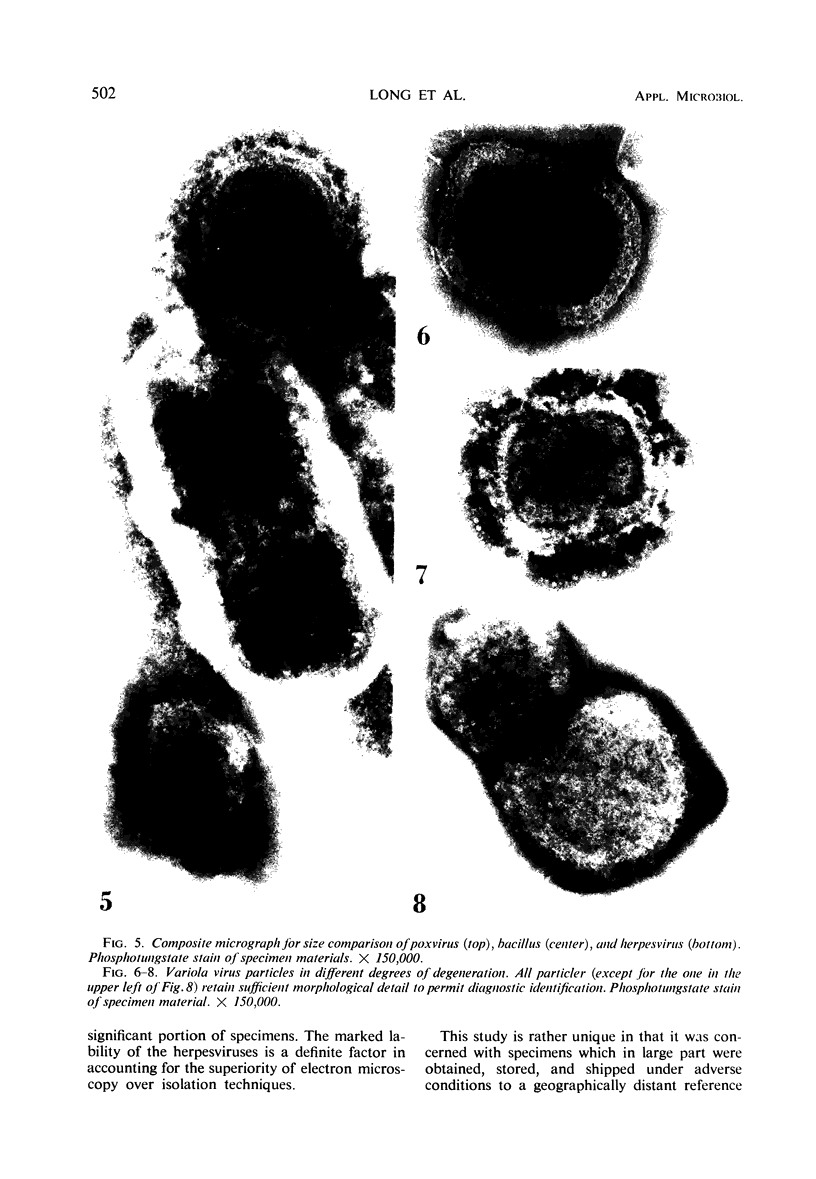
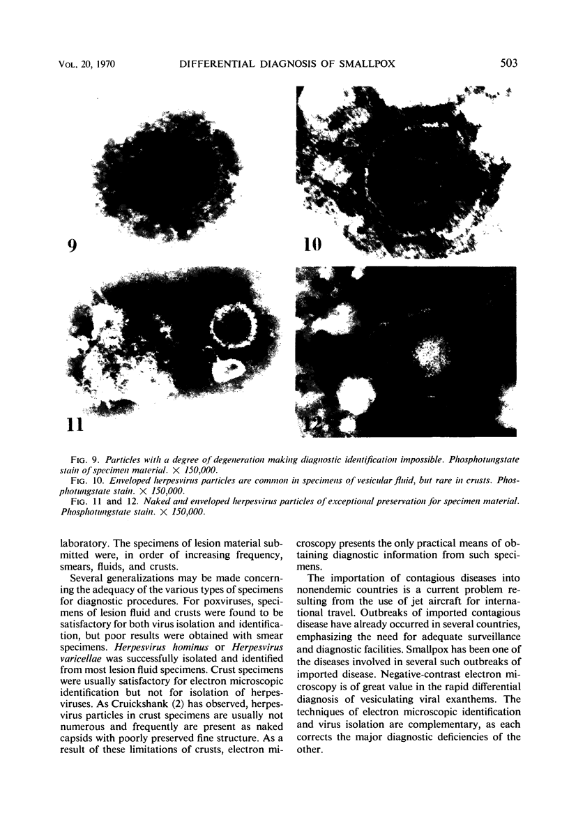
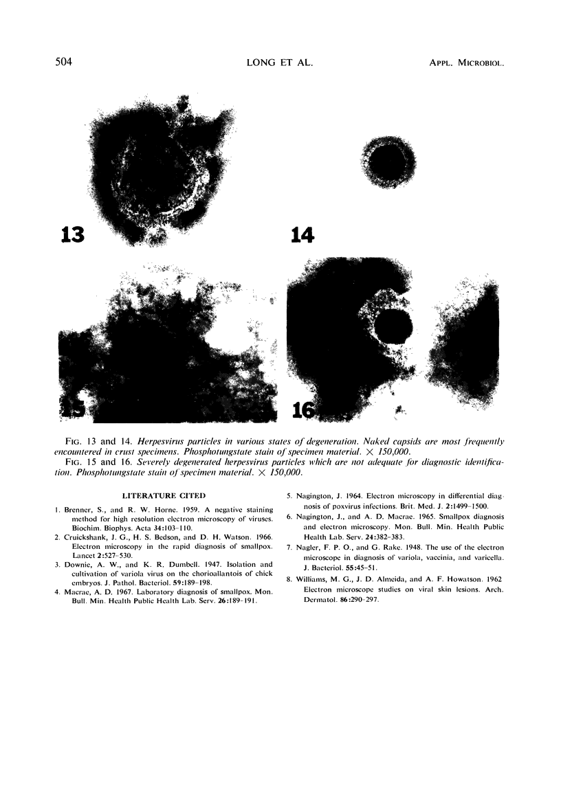
The usefulness of negative-contrast electron microscopy in the rapid differential diagnosis of poxvirus and herpesvirus exanthems is described in this study of 301 specimens from patients with vesicular exanthematous diseases. Specimens from patients with smallpox, various forms of vaccination complications, varicella, zoster (shingles), and herpes simplex are included in this evaluation. Electron microscopy, when applied to the study of lesion material, was found to be more sensitive than the classical techniques of virus isolation in the diagnosis of both poxvirus and herpes/varicella virus infections. However, since specific identification of a virus within a group cannot be made morphologically by electron microscopy, it is recommended that both electron microscopy and virus isolation methods be employed for the routine differential diagnosis of vesicular exanthematous diseases in the reference diagnostic laboratory.
Full text
PDF
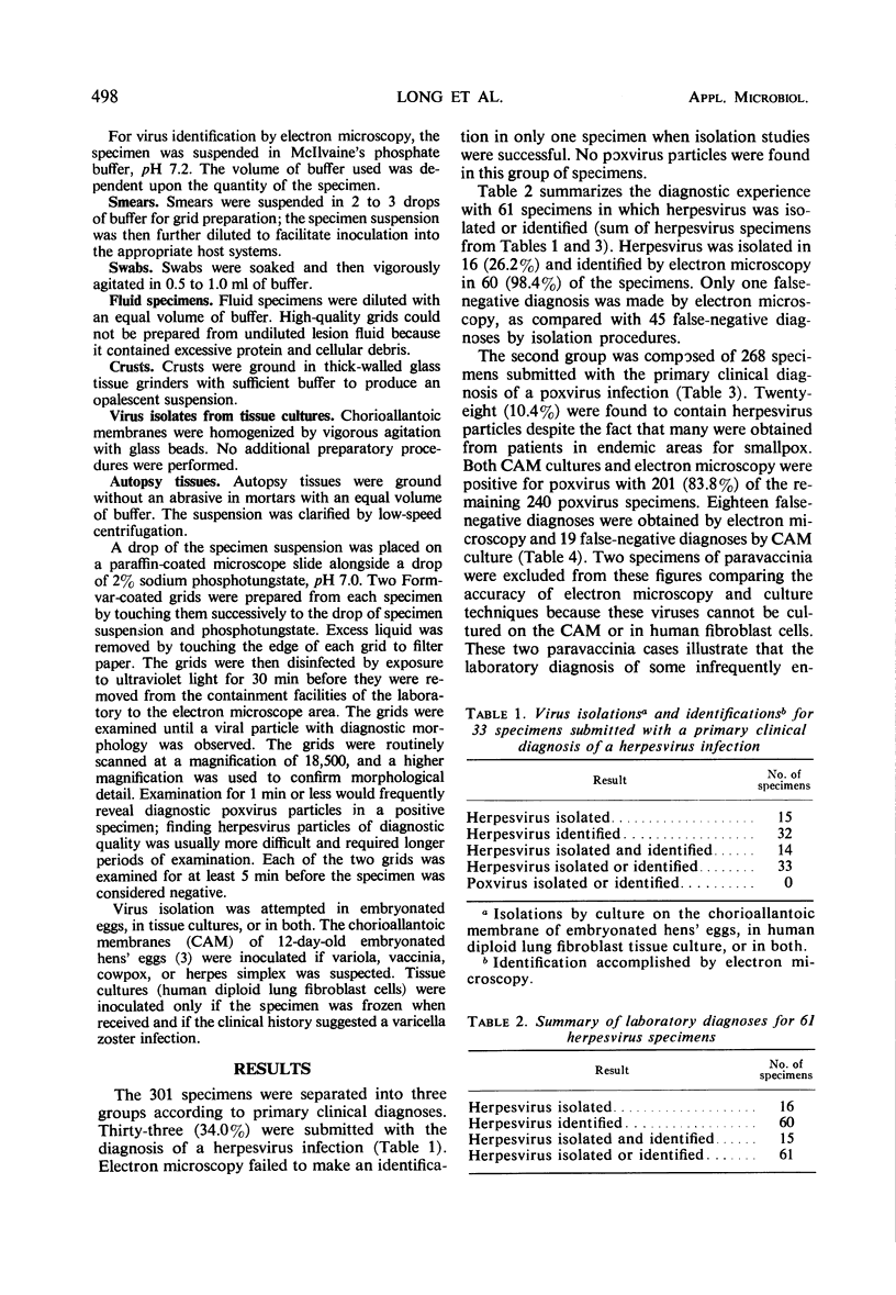
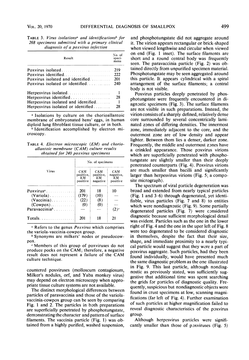
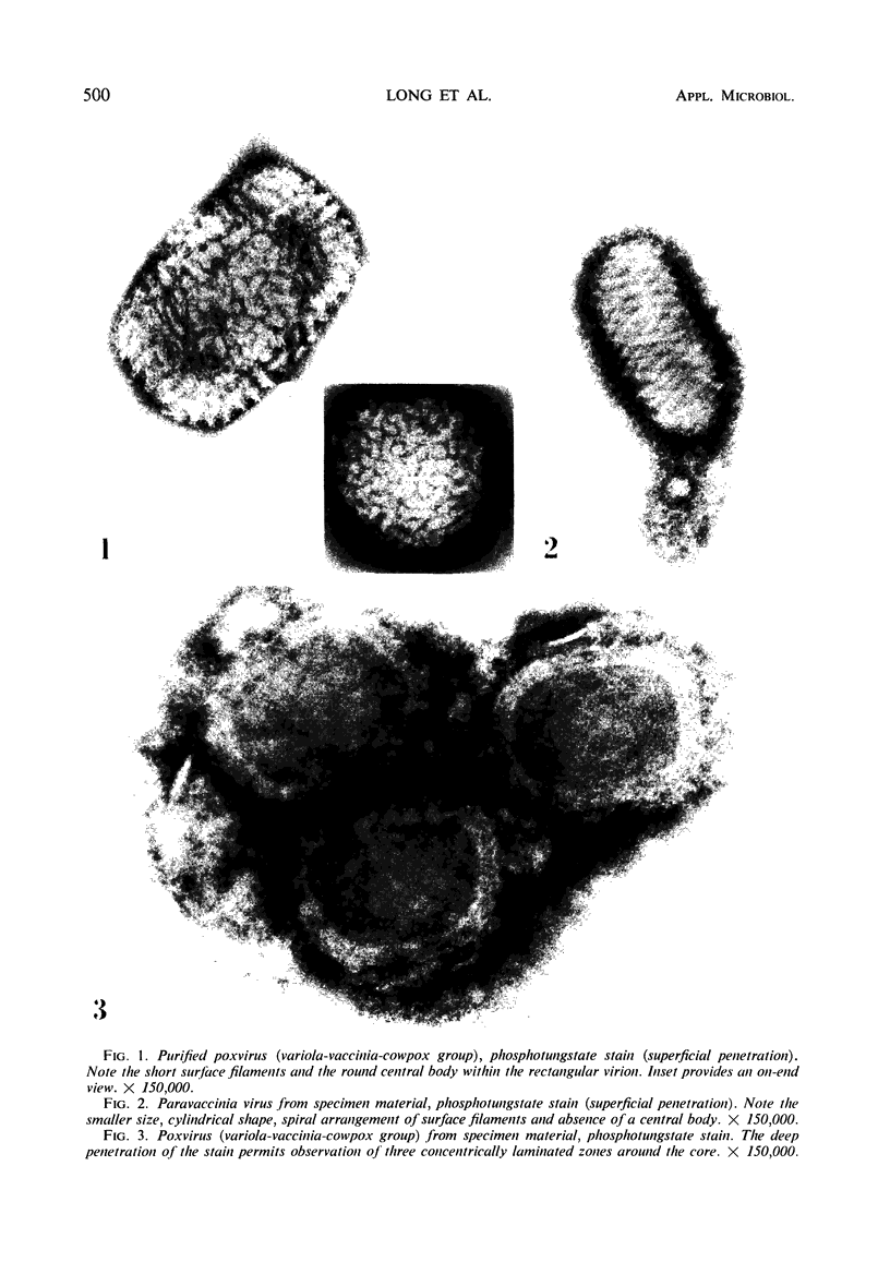
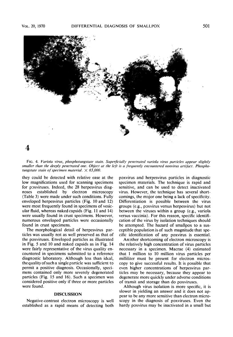
Images in this article
Selected References
These references are in PubMed. This may not be the complete list of references from this article.
- BRENNER S., HORNE R. W. A negative staining method for high resolution electron microscopy of viruses. Biochim Biophys Acta. 1959 Jul;34:103–110. doi: 10.1016/0006-3002(59)90237-9. [DOI] [PubMed] [Google Scholar]
- Cruickshank J. G., Bedson H. S., Watson D. H. Electron microscopy in the rapid diagnosis of smallpox. Lancet. 1966 Sep 3;2(7462):527–530. doi: 10.1016/s0140-6736(66)92882-0. [DOI] [PubMed] [Google Scholar]
- Macrae A. D. Laboratory diagnosis of smallpox. Mon Bull Minist Health Public Health Lab Serv. 1967 Oct;26:189–191. [PubMed] [Google Scholar]
- NAGINGTON J. ELECTRON MICROSCOPY IN DIFFERENTIAL DIAGNOSIS OF POXVIRUS INFECTIONS. Br Med J. 1964 Dec 12;2(5423):1499–1500. doi: 10.1136/bmj.2.5423.1499. [DOI] [PMC free article] [PubMed] [Google Scholar]
- Nagington J., Macrae A. D. Smallpox diagnosis and electron microscopy. Mon Bull Minist Health Public Health Lab Serv. 1965 Nov;24:382–384. [PubMed] [Google Scholar]
- Nagler F. P., Rake G. The Use of the Electron Microscope in Diagnosis of Variola, Vaccinia, and Varicella. J Bacteriol. 1948 Jan;55(1):45–51. doi: 10.1128/jb.55.1.45-51.1948. [DOI] [PMC free article] [PubMed] [Google Scholar]
- WILLIAMS M. G., ALMEIDA J. D., HOWATSON A. F. Electron microscope studies on viral skin lesions. A simple and rapid method of identifying virus particles. Arch Dermatol. 1962 Sep;86:290–297. doi: 10.1001/archderm.1962.01590090032010. [DOI] [PubMed] [Google Scholar]


