Abstract
The Tetraodontidae are an Acantomorpha fish family with circumglobal distribution composed of 189 species grouped in 19 genera, occurring in seas, estuaries, and rivers between the tropical and temperate regions. Of these, the genus Colomesus is confined to South America, with what have been up to now considered only two species. C. asellus is spread over the entire Amazon, Tocantins-Araguaia drainages, and coastal environments from the Amazon mouth to Venezuela, and is the only freshwater puffers on that continent. C. psittacus is found in coastal marine and brackish water environments from Cuba to the northern coast of South America as far south as to Sergipe in Brazil. In the present contribution we used morphological data along with molecular systematics techniques to investigate the phylogeny and phylogeography of the freshwater pufferfishes of the genus Colomesus. The molecular part is based on a cytochrome C oxidase subunit I dataset constructed from both previously published and newly determined sequences, obtained from specimens collected from three distinct localities in South America. Our results from both molecular and morphological approaches enable us to identify and describe a new Colomesus species from the Tocantins River. We also discuss aspects of the historical biogeography and phylogeography of the South American freshwater pufferfishes, suggesting that it could be more recent than previously expected.
Introduction
The Tetraodontidae is an Acantomorpha fish family with circumglobal distribution composed of 189 species in 19 genera, occurring in seas, estuaries, and rivers between the tropical and temperate regions [1]. They are mainly characterized by their typical four large dental plates; the ability to inflate their body in stressful situations; the presence of the neurotoxin Tetrodotoxin/Saxitoxin in its tissues, being responsible for numerous cases of fatal poisoning in many countries, including Brazil; and by having the smallest genome among vertebrates, therefore being considered as a model for the genome evolution of the group.
Among the Amazonian taxa exploited by the ornamental fish industry in South America are those of Colomesus [2], a genus confined to South America, with what is presently considered two species, C. asellus and C. psittacus. C. asellus [3] is spread in the entire Amazon, Tocantins-Araguaia drainages, and coastal environments from the Amazon mouth to Venezuela, being the only freshwater puffers on that continent. C. psittacus [4] is found in coastal marine and brackish water environments from Cuba and the northern coast of South America to Sergipe in Brazil.
Mainly located in tropical and subtropical regions all around the world, including the Amazon region, the ornamental fish industry is one of the largest transporters of live animals and plants with an annual trade volume estimated at U$15–25 billion [5]–[7], in a scenario where species identification problems, mainly related to border biosecurity are not rare.
The DNA barcode is a widely accepted tool for species determination mainly due to its enhanced attention on standardization and data validation [8], being a rapid and low cost method of identification [9]. The use of DNA barcoding techniques has been utilized in many taxa, including bacteria, birds, bivalves, butterflies, fishes, flies, macroalgae, mammals, spiders, sprigtails, and also for plants [10]–[27].
The DNA barcode technique for Metazoans uses a short (∼650 bp) and standardized gene region from the mitochondrial 5′ region of the cytochrome C oxidase subunit I (COI) for a rapid and cost-effective animal identification. This has been demonstrated to be an effective fish identification tool in numerous situations, including consumer protection [28]–[30], fisheries management/conservation [31], border biosecurity in the ornamental fish trade [5], and in the identification of overlooked or cryptic species [32].
Here we used both morphological and molecular methodologies in an integrative taxonomical approach to investigate the diversity of the Amazonian freshwater pufferfishes of the genus Colomesus based on specimens collected from three distinct populations from both Brazil and Peru. Additionally, we describe a new Colomesus species from the Upper Tocantins drainage based on both morphological and molecular data.
Methods
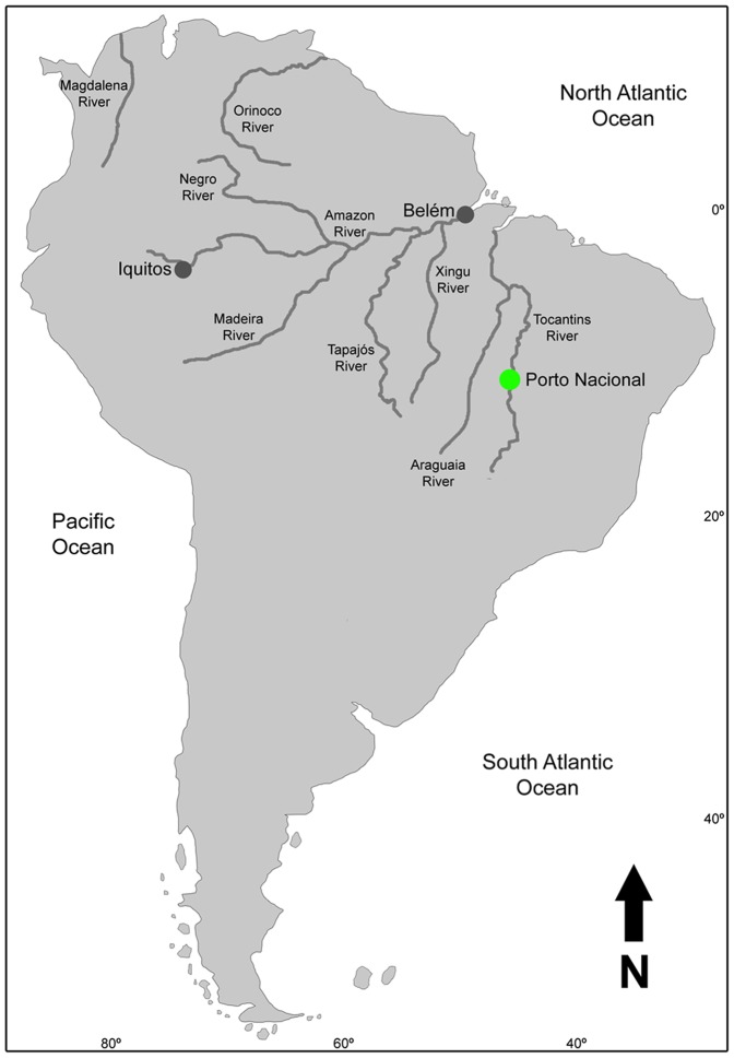
Specimens of Colomesus asellus were collected from three distinct populations with about 2200 km of mean distance separating them. The collection localities were Ilha do Mosqueiro, Belém, Brazil; Upper Tocantins River - Porto Nacional, Tocantins, Brazil; and Nanay River - Iquitos, Peru (Figure 1).
Figure 1. Map of South America showing the northern hydrology and the localities where the specimens were collected (grey and green marks).
Ethics Statement
No statement from an ethics committee was necessary, and the manuscript did not involve any endangered or protect species. All samples were extracted from dead specimens collected with appropriate permissions under authorization number 22512 issued by SISBIO/Instituto Chico Mendes de Conservação da Biodiversidade. We used the ice-slurry method for killing following [33] as they are tropical warm water species and the collected specimens are all smaller than 5 cm SL. All specimens were preserved in alcohol. The reported localities do not include protected areas.
Nomenclatural Acts
The electronic version of this document does not represent a published work according to the International Code of Zoological Nomenclature (ICZN), and hence the nomenclatural acts contained in the electronic version are not available under that Code from the electronic edition. Therefore, a separate edition of this document was produced by a method that assures numerous identical and durable copies, and those copies were simultaneously obtainable (from the publication date noted on the first page of this article) for the purpose of providing a public and permanent scientific record, in accordance with Article 8.1 of the Code. The separate print-only edition is available on request from PLoS by sending a request to PLoS ONE, 1160 Battery Street, Suite 100, San Francisco, CA 94111, USA along with a check for $10 (to cover printing and postage) payable to “Public Library of Science”.
In addition, this published work and the nomenclatural acts it contains have been registered in ZooBank, the proposed online registration system for the ICZN. The ZooBank LSIDs (Life Science Identifiers) can be resolved and the associated information viewed through any standard web browser by appending the LSID to the prefix “http://zoobank.org/”. The LSID for this publication is: urn:lsid:zoobank.org:pub:033A323A-18F7-4788-8405-32D78BF65B13.
Morphological Analyses
Specimens from all three localities were cleared and stained following the methodology of [34].
Molecular Analyses
The molecular systematic analyses used newly determined sequences obtained from the mitochondrial barcode marker COI as well as previously published sequences obtained from the NCBI and BOLD databases.
For the newly determined sequences, a fragment of epaxial musculature was submitted to the standard protocol for DNA extraction and purification from the Qiagen QIAamp DNA FFPE Tissue kit. The fragments were amplified and sequenced using the primers VF2_t1 and FishR2_t1 [35]–[37]. All primers were appended with M13 tails on sequencing reactions. The PCR profile consisted of 2 min at 95°C, 35 cycles of 30 sec at 94°, 40 sec at 52°C, and 1 min at 72°C, with a final extension step for 10 min at 72°C. Sequencing reactions were performed with the use of the BigDye® Terminator v.3.1 Cycle Sequencing kit (Applied Biosystems, Inc.), with 25 cycles of 10 sec at 95°C, 5 sec at 50°C and 4 min at 60°C. Sequencing products were processed in an ABI 3500 capillary system (Applied Biosystems, Inc.).
The chromatograms were checked and aligned using the BioEdit 7.053 [38] software with its built-in ClustalW routine [39]. The alignment was visually inspected for accuracy and to minimize missing data. All the newly determined sequences are available at the BOLD database (http://www.barcodinglife.com) under the project acronym PUFER. The GenBank accession numbers for all newly determined and previously published sequences used in the present manuscript are summarized in Table 1. The dataset consisted of a 651 bp COI matrix, and we used the MEGA 5.06 software [40] to determinate the TN93+G+I as the most appropriate model of sequence evolution based on the Akaike criterion (AIC) [41].
Table 1. Taxonomic sampling and accession numbers.
| Taxon | Accession No. | Taxon | Accession No. |
| Família Triodontidae | JQ841396 | ||
| Triodon macropterus | AP009170 | JQ840304 | |
| Família Diodontidae | GU225449 | ||
| Diodon holocanthus | AP009177 | Sphoeroides annulatus | GU440524 |
| Chilomycterus reticulatus | AP009188 | Sphoeroides testudineus | KC959927* |
| Família Tetraodontidae | KC959928* | ||
| Lagocephalus laevigatus | AP011934 | GU225665 | |
| JQ365394 | GU225453 | ||
| JQ365392 | GU225450 | ||
| JQ365395 | JQ842706 | ||
| JQ365393 | JQ843064 | ||
| KC959926* | JQ840306 | ||
| Lagocephalus inermis | FJ434549 | GU225664 | |
| GU804920 | JQ365576 | ||
| Lagocephalus lunaris | DSFSG9111 | JQ365575 | |
| Lagocephalus lagocephalus | AP011933 | JQ365574 | |
| Lagocephalus guentheri | HQ149858 | GU440524 | |
| JF493722 | Colomesus psittacus | KC959923* | |
| JF493724 | KC959924* | ||
| Lagocephalus wheeleri | JF952772 | KC959925* | |
| AP009538 | Colomesus asellus | KC959904* | |
| FJ434551 | KC959907* | ||
| Lagocephalus spadiceus | EU595163 | KC959908* | |
| EU595161 | KC959909* | ||
| Takifugu ocellatus | AP009536 | KC959910* | |
| Takifugu poecilonotus | AP009539 | KC959911* | |
| Takifugu snyderi | AP009531 | KC959913* | |
| Takifugu oblongus | AP009535 | KC959914* | |
| Takifugu pardalis | AP009528 | KC959915* | |
| Takifugu niphobles | AP009526 | KC959916* | |
| Takifugu porphyreus | AP009529 | Colomesus tocantinensis | KC959905* |
| Tetraodon biocellatus | KC959929* | KC959906* | |
| Tetraodon nigroviridis | KC959930* | KC959912* | |
| Sphoeroides pachygaster | EU074597 | KC959917* | |
| EU074598 | KC959918* | ||
| EU869843 | KC959919* | ||
| CSFOM07310 | KC959920* | ||
| JF494544 | KC959921* | ||
| JF494541 | KC959922* | ||
| EU869842 | |||
| EU869839 | |||
| AP006745 | |||
| Sphoeroides greeleyi | JQ365572 | ||
| Sphoeroides nephelus | JQ842698 | ||
| JQ842695 | |||
| JQ842699 | |||
| Sphoeroides spengleri | JQ842704 | ||
| JQ842695 | |||
| JQ842701 | |||
| JQ841395 |
(*)Sequences newly determined in this study.
The neighbor-joining (NJ) and maximum-likelihood (ML) trees that encompass the genera Triodon, Diodon, Chilomycterus, Lagocephalus, Tetraodon, Takifugu, Sphoeroides, and Colomesus, were constructed using the MEGA 5.06 software [40].
The neighbor-joining sequence divergences were calculated based on the Kimura Two Parameter (K2P) distance model [42] on BOLD workbench and MEGA 5.06 software [40]. The haplotype determination was carried with the use of the server FaBox (http://birc.au.dk/software/fabox/).
Results and Discussion
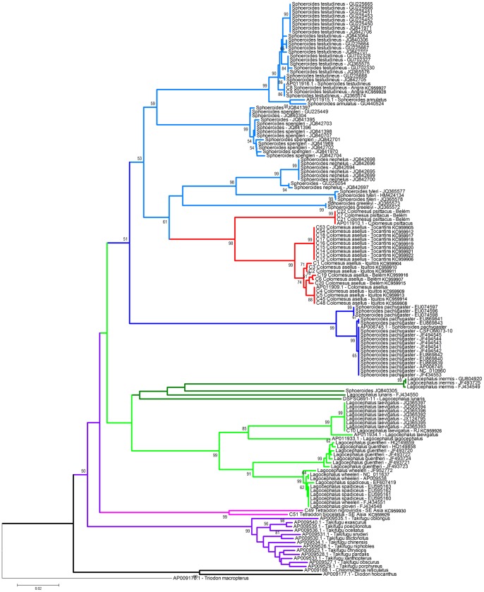
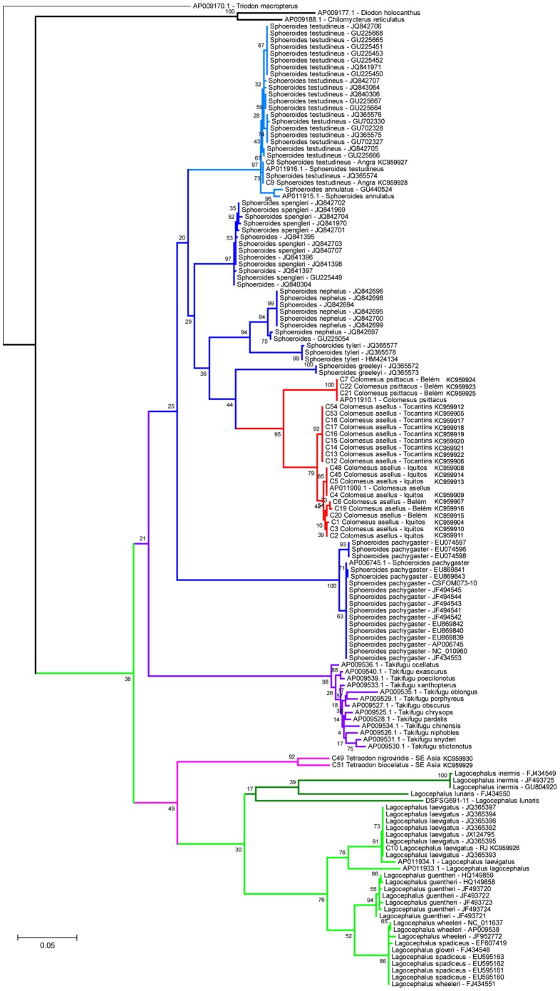
The neighbor-joining (NJ) and maximum-likelihood (ML) result trees are presented in Figures 2 and 3, respectively. The genus Colomesus was recovered as monophyletic inside the group formed by the sampled Sphoeroides species, in except for Sphoeroides pachygaster. Lagocephalus was recovered in a basal phylogenetic position in relation to Sphoeroides and Colomesus, therefore corroborating recent results such as those presented by [43]–[45].
Figure 2. Neighbor-Joining tree based on the barcode region of the COI.
The numbers near the branches represent bootstrap probabilities higher than 50%.
Figure 3. Maximum-likelihood phylogeny based on the barcode region of the COI marker.
The numbers near the branches represent the bootstrap probabilities.
Colomesus was recovered deep inside the group formed by the remaining Sphoeroides species, therefore suggesting Sphoeroides as paraphyletic, with S. pachygaster being recovered as basal in relation to all the remaining Sphoeroides species in all the analyses. Additionally, Colomesus was also recovered as the sister-taxa of the group formed by the species Sphoeroides nephelus, S. tyleri, and S. greeleyi in the NJ result, although it was recovered as the sister-taxa of S. greeleyi in the ML results.
In the same way, Colomesus is clearly distinguishable from the group formed by all the Sphoeroides species mainly by the banded pigmentation pattern present in all the Colomesus species; the presence of two lateral lines, with the ventral line running the full length of the caudal peduncle; and the absence of an upraised horizontal ridge of skin ventrolaterally along the caudal peduncle. The color pattern was used by [46], along with pectoral fin ray counts, the presence of a dark bar underside of caudal peduncle, and the presence of dermal flaps across the chin, to distinguish between what at that time were considered to be the only two species of the genus, the marine/estuarine C. psittacus, and the freshwater C. asellus. The presence of a dark bar on the underside of the caudal peduncle is a prominent feature for specimens of C. asellus from Iquitos, but this bar is present or not in specimens from both Belém and Tocantins. Dermal flaps were observed in all specimens from both Iquitos and Belém, but such flaps were not observed in any of the examined specimens from the Tocantins drainage.
DNA Barcode and Deep Sequence Divergence
COI amplicons were obtained from all the specimens included in the analyses. The obtained sequences clearly identified both previous accepted Colomesus species (C. asellus and C. psittacus), therefore being in accordance with the previous morphological diagnose presented by [46].
The K2P divergence distances between congeneric species ranged from 5.557% to 12.394% with a mean distance of 8.546%, while the uncorrected K2P distance ranged from 0 to 4.472% within species. The mean K2P distance within the analyzed populations was 0.657% and the mean normalized distance within species is 1.079% (Figure 4).
Figure 4. Distribution of K2P distances (%) for COI: A) within species; B) within genera; C, normalized distribution of K2P distance (%) within species.

The analyses included the following taxa: Tetraodon nigroviridis, Tetraodon biocellatus, Sphoeroides testudineus, Lagocephalus laevigatus, Colomesus asellus, Colomesus psittacus, and the freshwater Colomesus from the Tocantins drainage.
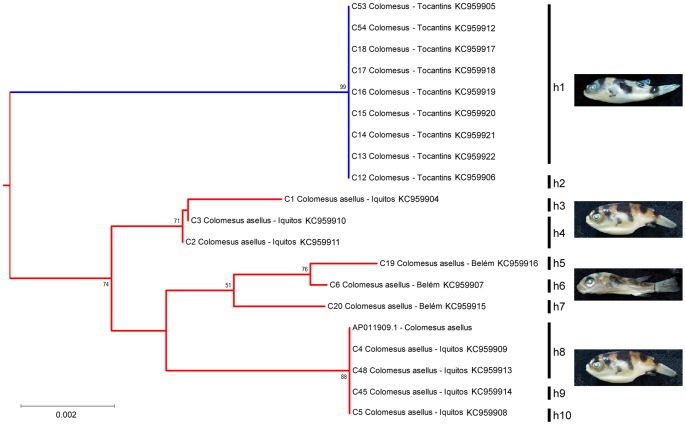
Deep sequence divergence was observed regarding the freshwater Colomesus from the Tocantins drainage (Figure 5). The mean sequence divergence of the specimens from both Belém and Iquitos was estimated at 1.079%, while the Tocantins distances ranged from 1.955% to 3.063%, with a mean distance of 2.166%. The observed sequence divergence values together with the congruence observed from both molecular and morphological phylogenetic approaches used here suggest the existence of an overlooked species within the genus Colomesus.
Figure 5. Neighbor-Joining phylogeny of the freshwater Colomesus and haplotype determination.
A new Colomesus species from the Tocantins River, Brazil
Systematics
Tetraodontiformes sensu Tyler, 1980 [47]
Tetraodontidae sensu Santini & Tyler, 2003 [45]
Colomesus Gill, 1885 [2]
Colomesus tocantinensis nov. sp. urn:lsid:zoobank.org:act:9B8ACCB5-FF55-4514-901B-6366FB6EA307
Derivation of name
The specific epithet tocantinensis refers to the type locality, Porto Nacional, State of Tocantins, Brazil.
Holotype
PNT.UERJ.405 (Figure 6).
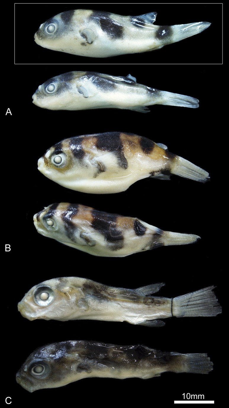
Figure 6. External morphology of the genus Colomesus.
A) Colomesus tocantinensis nov. sp. – Tocantins (holotype PNT.UERJ.405 highlighted in white); B) Colomesus asellus – Iquitos; C) Colomesus asellus – Belém.
Paratypes
PNT.UERJ.396, PNT.UERJ.397, PNT.UERJ.398, PNT.UERJ.399, PNT.UERJ.400, PNT.UERJ.401, PNT.UERJ.402, PNT.UERJ.403, PNT.UERJ.404.
Type-locality
The specimens are from the Tocantins River near Porto Nacional, State of Tocantins, Brazil.
Diagnosis
Colomesus species diagnosed by six to seven basal pterygiophores and nine rays in the anal fin (contra ten to eleven in both C. asellus and C. psittacus); ten basal pterygiophores and rays in the dorsal fin (contra eleven for both C. asellus and C. psittacus); the absence of dermal flaps across the chin (contra its presence uniquely in C. asellus); a caudal peduncle with eight vertebrae; and an opercle with a posterior ventral border subdivided in a ventral and a posterior region, the herein called “inverted V” shape (contra the triangular opercle exhibited by both C. asellus and C. psittacus).
Description
The holotype (PNT.UERJ.405) is 29,62 mm SL (Figure 6), with 10,37 mm HL; the entire type-series ranges from 27.02 mm to 34.9 mm SL. The meristic and morphometric data of the type series is presented in Table 2. The extent of the dorsal and ventral lateral lines is similar to those found in C. asellus. The prickles extend along the dorsal, lateral, and ventral surfaces of the body, from the level of the eye to the origin of the dorsal fin.
Table 2. Morphometric and meristic data of the type series of Colomesus tocantinensis nov. sp.
| Register | SL | HL | PR | DR | AR | CR | IOL |
| PNT.403 | 30.84 | 10.75 | 15 | 10 | 9 | 11 | 5.8 |
| PNT.404 | 34.9 | 11.83 | 15 | 10 | 9 | 11 | 5.9 |
| PNT.405* | 29.62 | 10.37 | 16 | 10 | 9 | 11 | 5.47 |
| PNT.395 | 30.35 | 11.25 | 15 | 10 | 9 | 11 | 6.35 |
| PNT.396 | 29.28 | 11.1 | 16 | 9 | 9 | 11 | 6.78 |
| PNT.397 | 29.59 | 10.79 | 16 | 10 | 9 | 11 | 5.48 |
| PNT.398 | 29.46 | 10.6 | 15 | 10 | 9 | 11 | 5.77 |
| PNT.399 | 30.66 | 11.99 | 16 | 10 | 9 | 11 | 5.55 |
| PNT.400 | 32.92 | 11.75 | 15 | 10 | 9 | 11 | 6.03 |
| PNT.401 | 27.02 | 9.61 | 16 | 10 | 9 | 11 | 5.12 |
SL, standard length; HL, head length; PR, pectoral fin rays; DR, dorsal fin rays; AR, anal fin rays; CR, caudal fin rays; IOL, interorbital length.
(*)Holotype.
The color pattern of Colomesus tocantinensis nov. sp. is essentially the same as that of Colomesus asellus, with five transverse dark bars across the dorsal region of the body. A dark blotch on the underside of the caudal peduncle, which is a state used by [46] to diagnose Colomesus asellus, is present or absent, being vestigial to unobservable or absent in several specimens. The interspaces between the dark bars are light yellow, with gradually decreasing pigmentation and becoming white in the ventral region (Figure 6). However, the light yellow to pale pattern presented by C. tocantinensis nov. sp. clearly contrasts with the gold-yellow pattern present in specimens from Iquitos and Belém.
The nasal sac is higher than that presented in the specimens of C. asellus. Two large lateral and anteromedial nostrils are present. They are similar to those found on C. psittacus, rather than the two small nostrils exhibited by C. asellus. The anterior surface of the nasal sac is smooth while the posterior surface of it is folded as in C. psittacus, exhibiting a “T-shaped” ridge with a relatively small dorsal flap. This flap seems much smaller than the one found on C. asellus, although more flexible when compared to C. psittacus.
The presence of dermal flaps across the chin is another character used by [46] to distinguish C. asellus from C. psittacus. No dermal flaps could be seen in the examined specimens from the Tocantins River, although they are always present in examined specimens from Iquitos and Belém.
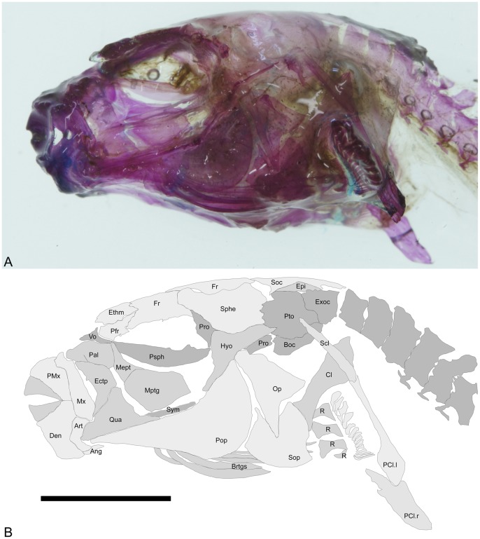
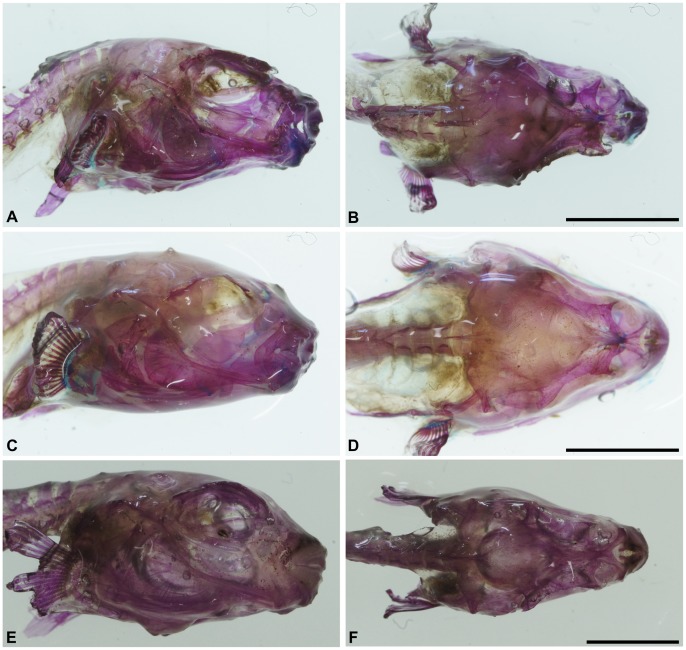
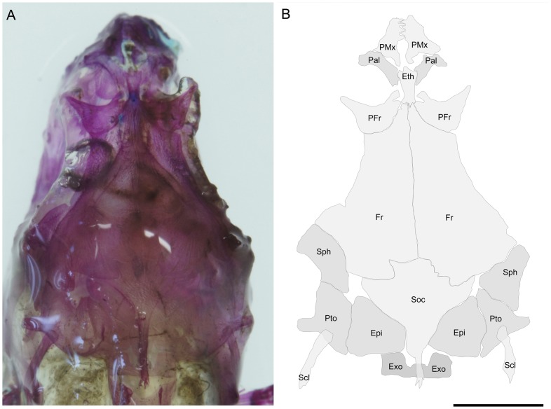
The skull is partially similar to those found in Colomesus asellus described and figured by [46], although the frontals exhibit a wide posterior border and prominently participate in the orbital margin (Figures 7–9). The prefrontals are triangular and articulate medially with the ethmoid, which posteriorly articulates with the frontals and anteriorly with the palatines (Figure 8). The supraoccipital is roughly triangular and well developed, with an elongate posterior process which covers the first vertebrae (Figure 8). The sphenotics articulate postero-laterally with the frontals and, in the examined specimens, they neither contact nor closely approach the prefrontals. The lateral wing of the sphenotics is only partially developed (Figure 8). Posterior to the sphenotics, the pterotics (Figures 7 and 8) articulates posteriorly with the slender supracleithrum and medially with the epiotics, which articulates medially with the supraoccipital (Figure 9).
Figure 7. Colomesus tocantinensis nov. sp. (PNT.UERJ.398).
A) left photograph of the head; B) anatomical interpretations. Abbreviations: Ang, angular; Art, articular; Boc, basioccipital; Brstgs, branchiostegals; Cl, cleithrum; Den, dentary; Epi, epiotic; Ethm, ethmoid; Exo, exoccipital; Fr, frontal; Hyo, hyomandibula; Ecptg, ectopterygoid; Mept, mesopterygoid; Mtptg, metapterygoid; Mx, Maxilla; Op, opercle; Pal, palatine; PCl.l/r, ventral post-cleithrum left and right; Pfr, prefrontal; PMx, premaxilla; Pop, preopercle; Pro, prootic; Psph, parasphenoid; Pto, pterotic; Qua, quadrate; R, radials; Scl, supracleithrum; Soc, supraocciptal; Sop, subopercle; Sphe, sphenotic; Sym, sympletic; Vo, vomer. Scale bar equals 5 mm.
Figure 9. Right and top photographs from cleared-and-stained specimens of: A–B) Colomesus tocantinensis nov. sp. – Tocantins (PNT.UERJ.398); C–D) Colomesus asellus – Iquitos (PNT.UERJ.470); E–F) Colomesus asellus – Belém (PNT.UERJ.386).
Figure 8. Colomesus tocantinensis nov. sp. (PNT.UERJ.398).
A) top photograph of the head; B) anatomical interpretations. Abbreviations: Epi, epiotic; Ethm, ethmoid; Exo, exoccipital; Fr, frontal; Pal, palatine; Pfr, prefrontal; PMx, premaxilla; Pto, pterotic; Scl, supracleithrum; Soc, supraocciptal; Sphe, sphenotic. Scale bar equals 5 mm.
In lateral view, the skull is characterized by the wide preopercle with about 110 degrees between both horizontal and vertical rami (Figure 7), with the preopercular canal running along its anterior border, and by the opercle which is divided in two distinct regions, having ventral and posterior wings, the herein called “inverted V” shape, distinct from the condition found in all other examined specimens of Colomesus (Figure 10). The subopercle is sturdy, with two small dorsal processes.
Figure 10. Isolated opercles from: A) Colomesus tocantinensis nov. sp. – Tocantins (PNT.UERJ.398); B) Colomesus asellus – Iquitos (PNT.UERJ.470); C) Colomesus asellus – Belém (PNT.UERJ.386); D) Colomesus psittacus – Belém (PNT.UERJ.387).

Scale bar equals 1
The parasphenoid is elongate and does not exhibit any developed dorsal flange (Figures 7 and 9). The hyomandibula is roughly triangular and has a slender ventral region; its wide head articulates dorsally with the sphenotics, and its upper posterior edge with the anterior end of the opercle (Figure 7).
The palatine is wide and somewhat triangular, with a robust anterior process for the maxilla (Figure 7). The maxilla is robust, with an anterodorsal region articulating with the premaxilla and a posterior expanded region, medially concave for muscle insertion. The ectopterygoid articulates dorsally with the palatine and ventrally with the anterodorsal border of the quadrate. The metapterygoid is wide and composes almost the entire ventral orbital region (Figure 7). It articulates anteriorly with the mesopterygoid (Figure 7), and with the posterior end of the large and triangular quadrate (Figure 7). The quadrate exhibits a well-developed posteroventral spine articulating posteriorly with the slender symplectic (Figure 7), and anteriorly with the articular. The articular is “L” shaped and articulates anteriorly with the robust dentary and ventrally with the small angular (Figure 7).
Five branchiostegal rays (Figure 7) are present and the branchial apparatus is strikingly similar in all the examined specimens (Figure 11).
Figure 11. Isolated branchial apparatus from: A) Colomesus tocantinensis nov. sp. – Tocantins (PNT.UERJ.404); B) Colomesus asellus – Iquitos (PNT.UERJ.470); C) Colomesus asellus – Belém (PNT.UERJ.386).
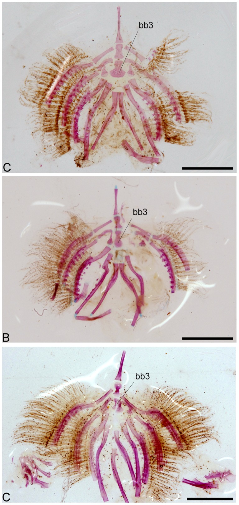
Scale bar equals 2.5
The pectoral girdle is robust and formed by a wide cleithrum, somewhat triangular and posteriorly expanded, articulating dorsally with the slender supracleithrum. The supracleithrum articulates ventrally with the two postcleithra; a slender dorsal postcleithrum, followed by the posteriorly expanded ventral postcleithrum (Figure 7). There are four radials and sixteen pectoral fin rays (Figure 7).
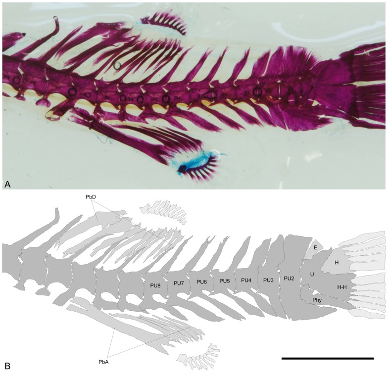
The axial skeleton has 19 vertebrae. The dorsal fin originates between vertebrae 7–8 and has ten basal pterygiophores and ten fin rays (Figures 12–14). The anal fin is located beneath the 9th vertebra and has six basal pterygiophores and nine fin rays.
Figure 12. Colomesus tocantinensis nov. sp. (PNT.UERJ.403).
A) left photograph of the unpaired fins and caudal endoskeleton; B) anatomical interpretations. Abbreviations: E, epural; H, dorsal hypural plate; H-H, ventral hypural plate fused with the ural centrum; Phy, parhypural; PU, pre-ural vertebrae; PbD, dorsal-fin basal pterigiophores; PbA, anal-fin basal pterigiophores. Scale bar equals 5 mm.
Figure 14. High-definition x-ray images of: A–D, Colomesus psittacus USNM.393077; E–G, Colomesus asellus USNM.191569.
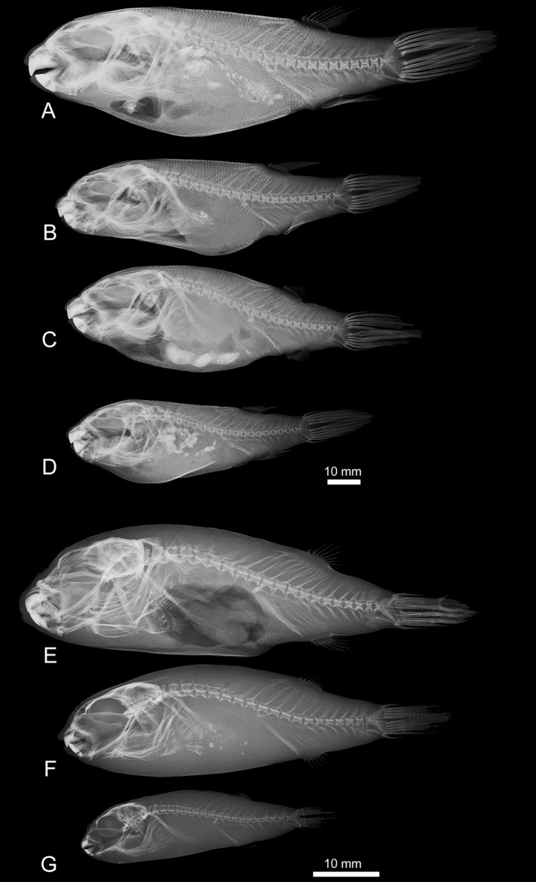
Scale bar equals 10
Figure 13. Left view photographs from cleared-and-stained specimens of: A) Colomesus tocantinensis nov. sp. (PNT.UERJ.403); B) Colomesus asellus – Iquitos (PNT.UERJ.470); C) Colomesus asellus – Belém (PNT.UERJ.386).
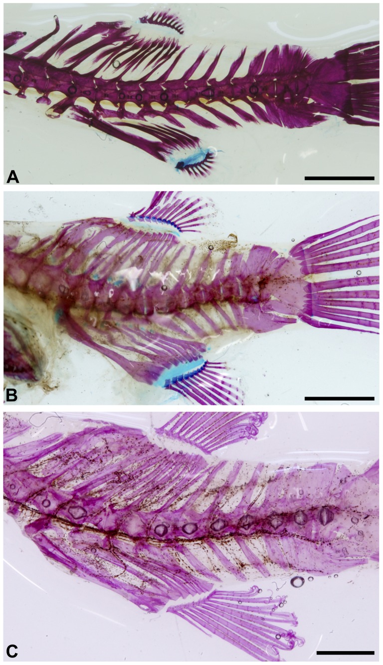
Scale bar equals 5
The caudal skeleton (Figure 12) has a wide ural centrum formed by the preural centrum 1, the ural centrum, the ventral hypural plate, and the postero-dorsal expansion which articulates anteriorly with the almost triangular epural, and posteriorly with the dorsal hypural plate (Figure 12). Eleven caudal fin rays, five dorsal and six ventral, are present in all of the specimens, both the uppermost and the two lowermost rays are unbranched.
Phylogeography of the South American Freshwater Pufferfishes
Although the influence of marine incursions after the Miocene is still under debate, the Caribbean (or Miocene) marine incursion, via the Llanos Basin (Colombia-Venezuela), is well accepted based on both geological and paleontological evidence, suggesting that these incursions may have isolated marine taxa within the western South America freshwater environments [48]–[52]. This might be the case for the freshwater tetraodontids. As pointed by [53], this scenario predicts that the distribution of the marine sister groups of marine lineages should be related with the Caribbean or western Atlantic, the age of freshwater taxa should be coincident with marine incursions, and the biogeographic congruence should be observed among multiple unrelated taxa, conditions only partially filled by the genus Colomesus.
The timing of divergence between the brackish/marine C. psittacus and the freshwater C. asellus was recently discussed [45], based on a multiple loci approach including both nuclear and mitochondrial markers. The authors dated the split between 2,5-7My, therefore postdating the Miocene marine incursions usually used to explain the presence of several marine groups within the western Amazon. In this sense, as observed by [45], the colonization carried by the tetraodontids in South America could be presumably related to the Pliocene global climate oscillations. Additionally, the basal split of the Colomesus from Tocantins and from Iquitos/Belém agrees with the general area cladogram of neotropical fishes presented by [54] in which the Xingu/Tocantins-Araguaia group was recovered in a basal position in relation to the groups from the lowlands of Western and Eastern Amazon.
It was recently proposed [55], based on the distribution of characiforms, that recent marine incursions would have isolated fish populations in upland terrains or refuges, where lineage divergence is maximized, followed by dispersal episodes back to the lowlands. The “museum hypothesis” predicts that lowlands exhibit a higher number of species, but lower levels of endemism, than highlands, and that the upland refuges would represent areas of high endemism.
Looking on the molecular phylogeny of the serrasalmids Pygocentrus and Serrasalmus, [56] proposed a phylogenetic test which predicts that basal lineages in a phylogeny of widespread fishes would occur in highland areas, and lowland lineages would have originated only during the last 5 Ma. Additionally, [57] studying the genetics of Symphysodon cichlids, indicated the effects that marine incursions would have in population structure, stating that populations in upland terrains or refuges would exhibit reduced genetic variation, while populations in lowlands would represent multiple upland sources, therefore exhibiting a high level of genetic variation, and that populations in lowlands would show a demographic pattern of expansion.
Our results recovered the Upper Tocantins lineages in a basal position in relation to all the remaining specimens, with the sequences being collapsed in uniquely two haplotypes (Figure 5), the first one (h1), represented by eight sequences, and the second haplotype (h2), represented by a unique sequence. This suggests low genetic variation, at least among the studied sampling, and a history initially related with the eastern Amazon, followed by a subsequently expansion to the western South America.
The Tocantins-Araguaia Ichthyofauna
The Tocantins-Araguaia drainage is the fourth largest Brazilian drainage, draining part of the northern end of the Brazilian shield directly to the eastern end of the Amazon Basin. It exhibits a recent geomorphological history, within a still tectonically active sedimentary basin with recent subsidence episodes, which are related with the high load of sediments observed within the basin, leading the development of the Bananal Plain, in the lower part of the drainage, mainly during the Quaternary [58]–[59].
The Tocantins drainage, specially the Upper Tocantins River, is constantly regarded as an area of high endemism, with several fish taxa restricted to this area having been described, such as Leporinus taeniofasciatus (Anostomidae), Sternarchorhynchus mesensis (Apteronotidae), Aspidoras albater, A. eurycephalus (Callichthyidae), Acestrocephalus maculosus, Astyanax unitaeniatus, Astyanacinus goyanensis, Creagrutus atrisignum, C. britskii, C. mucipu, C. saxatilis, Hyphessobrycon hamatus, Moenkhausia tergimaculata, Cetopsis caiapo, C. sarcodes (Cetopsidae), Characidium stigmosum (Crenuchidae), Pimelodella spelaea (Heptapteridae), Ancistrus aguaboensis, A. jataiensis, A. minutus, A. reisi, Corumbataia veadeiros, Hemiancistrus micrommatos, Hypostomus ericae, Gymnotocinclus anosteos, Lamontichthys avacanoeiro (Loricariidae), Apareiodon argenteus and A. cavalcante (Parodontidae), Cynolebias griseus, C. notatus, Rivulus planaltinus, and Simpsonichthys marginatus (Rivulidae), the herein described Colomesus tocantinensis nov. sp. (Tetraodontidae), and Ituglanis bambui and I. mambai (Trichomycteridae).
In the same way, [60]–[62] pointed that the Tocantins River is the only river system related with the Amazon Basin, and draining shield areas which exhibit a considerable number of fish taxa known from the lowlands of the central and western Amazon. The faunal similarity between both basins is constantly related with the evolution of the Bananal Plain as a selective barrier for the lowland fauna and the upper part of the drainage, and the list of typical lowland ichthyofauna shared by the Tocantins-Araguaia and the Amazon drainages includes, as presented by [60], Leporinus trifasciatus (Anostomidae), Arapaima gigas (Arapaimatidae), Auchenipterichthys coracoideus (Auchenipteridae), Mylossoma spp., Pygocentrus nattereri (Characidae), Cetopsis candiru, C. coecutiens (Cetopsidae), Cichla monoculus, C. pleiozona, C. kelberi (Cichlidae), Psectrogaster amazonica (Curimatidae), Thorachocharax stellatus (Gasteropelecidae), Osteoglossum bicirrhosum (Osteoglossidae), Pellona castelnaeana, Pristigaster cayana (Pristigasteridae), and Colomesus asellus (Tetraodontidae). In this sense, even still under debate, it seems clear that the Tocantins-Araguaia drainage has a composite nature including both lowland and upland ichthyofauna, as argued by [62].
Conclusions
Based on a comprehensive analysis including both morphological and molecular methodologies using the cytochrome C oxidase I gene, we were able to discuss aspects of the phylogeny and phylogeography of the South American freshwater pufferfishes of the genus Colomesus.
Our molecular results based on the COI marker agrees with the recent results such as [43]–[45], and suggest that the genus Sphoeroides should be revised, mainly regarding the phylogenetic position recovered for the genus Colomesus, deeply nested within the Sphoeroides tree, and the basal position recovered for S. pachygaster. We plan further investigations along these lines to reconcile any conflicts between these molecular hypotheses presented herein and morphologically based interpretations [47] of the phylogeny of the taxa of Colomesus, Sphoeroides, and Lagocephalus.
The use of molecular systematic techniques together with morphological methodologies confirmed the identification of a new cryptic pufferfish species from the Upper Tocantins drainage, Colomesus tocantinensis nov. sp. Morphological features such as the color pattern, the absence of dermal flaps across the chin, the distinct ‘inverted V’ opercle shape, and the caudal peduncle morphology, all support the description of Colomesus tocantinensis nov. sp., as a new pufferfish species from the South American freshwater drainages.
The timing of divergence between the marine/brackish species Colomesus psittacus and the freshwater group formed by C. asellus and C. tocantinensis nov. sp., as recovered by [45], postdates the Miocene marine incursions usually used to explain the presence of tetraodontids within the Amazon freshwater environments. Therefore, it suggests that the freshwater colonization in South America, at least for the tetraodontids, could be more recent than previously expected. Additionally, together with the observed distribution of haplotypes, our results suggest that the history of tetraodontids into the Amazonian freshwater environments could be presumably related to the Pliocene global climate oscillations and its effects inside the eastern Amazon and subsequently to the western South America.
Finally, our results reinforce the Upper Tocantins drainage as an area of high endemism within the Tocantins-Araguaia drainage, although the composite nature of the entire drainage is unquestionable.
Acknowledgments
We would like to thank Dr. James C. Tyler (Smithsonian Institution, Washington) for his support and helpfull comments on the manuscript. We are also grateful to Dr. Francesco Santini (Universitá degli Studi di Torino), Dr. Dorothée Huchon (Tel Aviv University), and an anonymous reviewer for the valuable suggestions during the review of the manuscript, Dr. Leonor Gusmão and Dr. Antonio Amorim (Universidade do Porto) for the comments during the initial discussion of the results, Yuri Modesto (Universidade do Estado do Rio de Janeiro) for the specimens from the Tocantins drainage, Lúcio Paulo Machado and Diogo de Mayrink (Universidade do Estado do Rio de Janeiro) for the specimens from Iquitos; Ms. Sandra Raredon (Smithsonian Institution, Washington) for the x-rays of tetraodontids, Dr. Richard Pyle (Hawaii Biological Survey) for the LSID numbers, and Kleyton M. C. Severiano and Anna Carolina Chaves (Universidade do Estado do Rio de Janeiro) for the technical assistance.
Funding Statement
This study was supported by the Brazilian National Counsel of Technological and Scientific Development and Fundação Carlos Chagas Filho de Amparo à Pesquisa do Estado do Rio de Janeiro. The funders had no role in study design, data collection and analysis, decision to publish, or preparation of the manuscript.
References
- 1.Froese R, Pauly D (2012) FishBase World Wide Web electronic publication. www.fishbase.org, 2012, version (02/2012).
- 2. Gill TN (1885) Synopsis of the Plectognath fishes. Proc United States Natl Mus 7: 411–427. [Google Scholar]
- 3.Müller J, Troschel FH (1849) Fische. In: Reisen in Britisch-Guiana in den Jahren, 1840–1844. Im Auftrag Sr. Mäjestat des Königs von Preussen ausgeführt von Richard Schomburgk. [Versuch einer Fauna und Flora von Britisch-Guiana.] 3. Berlin.
- 4.Bloch ME, Schneider JG (1801) Systema Ichthyologiae iconibus cx illustratum. Post obitum auctoris opus inchoatum absolvit, correxit, interpolavit Jo. Gottlob Schneider, Saxo. Berolini. Sumtibus Auctoris Impressum et Bibliopolio Sanderiano Commissum. M. E. Blochii, Systema Ichthyologiae.: i-lx +1–584, Pls. 1–110.
- 5. Collins RA, Armstrong KF, Meier R, Yi Y, Brown SDJ, et al. (2012) Barcoding and border Biosecurity: Identifying Cyprinid Fishes in the Aquarium Trade. PLoS ONE 7(1): e28381 doi:10.1371/journal.pone.0028381 [DOI] [PMC free article] [PubMed] [Google Scholar]
- 6. Padilla DK, Williams SL (2004) Beyond ballast water: aquarium and ornamental trades as sources of invasive species in aquatic ecosystems. Front Ecol Environ 2: 131–138. [Google Scholar]
- 7.Ploeg A, Bassleer G, Hensen R (2009) Biosecurity in the Ornamental Aquatic Industry. Maarssen: Orn Fish Intl. 148 p. [Google Scholar]
- 8. Mabragaña E, Díaz de Astarloa JM, Hanner R, Zhang J, González Castro M (2011) DNA Barcoding Identifies Argentine Fishes from Marine and Brackish Waters. PLoS ONE 6(12): e28655 doi:10.1371/journal.pone.0028655 [DOI] [PMC free article] [PubMed] [Google Scholar]
- 9.Golding GB, Hanner R, Hebert PDN (2009) Preface. Molecular Ecology Resources 9(Suppl. 1): iv–vi. [DOI] [PubMed]
- 10. Hogg ID, Hebert PDN (2004) Biological identification of springtails (Collembola: Hexapoda) from the Canadian Arctic, using mitochondrial DNA barcodes. Can J Zoo 82: 749–754. [Google Scholar]
- 11. Barrett RDH, Hebert PDN (2005) Identifying spiders through DNA barcodes. Can J of Zoo 83: 481–491. [Google Scholar]
- 12. Hebert PDN, Cywinska A, Ball SL, deWaard JR (2003a) Biological identification through DNA barcodes. Proc Roy Soc London, Series B: Biol Sci 270: 313–321. [DOI] [PMC free article] [PubMed] [Google Scholar]
- 13. Hebert PDN, Ratnasingham S, deWaard JR (2003b) Barcoding animal life: cytochrome c oxidase subunit 1 divergences among closely related species. Proc Roy Soc London, Series B: Biol Sci (Suppl 1) 270: 96–99. [DOI] [PMC free article] [PubMed] [Google Scholar]
- 14. Janzen DH, Hajibabaei M, Burns JM (2005) Wedding biodiversity inventory of a large and complex Lepidoptera fauna with DNA barcoding. Phil Trans Roy Soc London, Series B, Biol Sci 1462: 1835–1846. [DOI] [PMC free article] [PubMed] [Google Scholar]
- 15. Hajibabaei M, Janzen DH, Burns JM, Hallwachs W, Hebert PDN (2006) DNA barcodes distinguish species of tropical Lepidoptera. Proc Natl Acad Sci U S A 103: 968–971. [DOI] [PMC free article] [PubMed] [Google Scholar]
- 16. Lukhtanov VA, Sourakov A, Zakharov EV, Hebert PDN (2009) DNA barcoding Central Asian butterflies: increasing geographical dimension does not significantly reduce the success of species identification. Mol Ecol Res 9: 1302–1310. [DOI] [PubMed] [Google Scholar]
- 17. Smith MA, Wood DM, Janzen DH, Hallwachs W, Hebert PDN (2007) DNA barcodes affirm that 16 species of apparently generalist tropical parasitoid flies (Diptera, Tachinidae) are not all generalists. Proc Natl Acad Sci U S A 104: 4967–4972. [DOI] [PMC free article] [PubMed] [Google Scholar]
- 18. Järnegren J, Schander C, Sneli JA, Ronningen V, Young CM (2007) Four genes, morphology and ecology: distinguishing a new species of Acesta (Mollusca; Bivalvia) from the Gulf of Mexico. Mar Biol 152: 43–55. [Google Scholar]
- 19. Ward RD, Zemlak TS, Innes BH, Last PR, Hebert PDN (2005) DNA barcoding Australia’s fish species. Phil Trans Roy Soc London, Series B, Biol Sci 360: 1847–1857. [DOI] [PMC free article] [PubMed] [Google Scholar]
- 20. Hebert PDN, Stoeckle MY, Zemlak TS, Francis CM (2004) Identification of birds through DNA barcodes. PLoS Biol 2: 1657–1663. [DOI] [PMC free article] [PubMed] [Google Scholar]
- 21. Kerr KCR, Lijtmaer DA, Barreira AS, Hebert PDN, Tubaro PL (2009) Probing evolutionary patterns in neotropical birds through DNA barcodes. PloS ONE 4: e4379. [DOI] [PMC free article] [PubMed] [Google Scholar]
- 22. Clare EL, Lim BK, Engstrom MD, Eger JL, Hebert PDN (2007) DNA barcoding of Neotropical bats: species identification and discovery within Guyana. Mol Ecol Notes 7: 184–190. [Google Scholar]
- 23. Amaral AR, Sequeira M, Coelho MM (2007) A first approach to the usefulness of cytochrome c oxidase I barcodes in the identification of closely related delphinid cetacean species. Mar Fresh Res 58: 505–510. [Google Scholar]
- 24. Borisenko AB, Lim BK, Ivanova NV, Hanner RH, Hebert PDN (2008) DNA barcoding in surveys of small mammal communities: a field study in Suriname. Mol Ecol Res 8: 471–479. [DOI] [PubMed] [Google Scholar]
- 25. Hollingsworth PM, Forrest LL, Spouge JL, Hajibabaei M, Ratnasingham S, et al. (2009) A DNA barcode for land plants. Proc Natl Acad Sci U S A 106: 12794–12797. [DOI] [PMC free article] [PubMed] [Google Scholar]
- 26. Saunders GW (2005) Applying DNA barcoding to red macroalgae: a preliminary appraisal holds promise for future applications. Phil Trans Roy Soc London, Series B, Biol Sci 360: 1879–1888. [DOI] [PMC free article] [PubMed] [Google Scholar]
- 27. Sogin ML, Morrison HG, Huber JA, Welch DM, Huse SM, et al. (2006) Microbial diversity in the deep sea and the underexplored ‘rare biosphere’. Proc Natl Acad Sci U S A 103: 12115–12120. [DOI] [PMC free article] [PubMed] [Google Scholar]
- 28. Lowenstein JH, Amato G, Kolokotronis SO (2009) The real maccoyii: identifying tuna sushi with DNA barcodes - contrasting characteristic attributes and genetic distances. PLoS ONE 4: e7866. [DOI] [PMC free article] [PubMed] [Google Scholar]
- 29. Lowenstein JH, Burger J, Jeitner CW, Amato G, Kolokotronis SO, et al. (2010) DNA barcodes reveal species-specific mercury levels in tuna sushi that pose a health risk to consumers. Biol Lett 6: 692–695. [DOI] [PMC free article] [PubMed] [Google Scholar]
- 30. Cohen NJ, Deeds JR, Wong ES, Hanner RH, Yancy HF, et al. (2009) Public health response to puffer fish (tetrodotoxin) poisoning from mislabeled product. J Food Prot 72: 810–817. [DOI] [PubMed] [Google Scholar]
- 31. Holmes BH, Steinke D, Ward RD (2009) Identification of shark and ray fins using DNA barcoding. Fish Res 95: 280–288. [Google Scholar]
- 32. Steinke D, Zemlak TS, Hebert PDN (2009) Barcoding Nemo: DNA-Based identifications for the Ornamental Fish Trade. PLoS ONE 4(7): e6300 doi:10.1371/journal.pone.0006300 [DOI] [PMC free article] [PubMed] [Google Scholar]
- 33. Blessing JJ, Marshall JC, Balcombe SR (2010) Humane killing of fishes for scientific research: a comparison of two methods. J Fish Biol. 76(10): 2571–2577 doi: –10.1111/j.1095–8649.2010.02633.x [DOI] [PubMed] [Google Scholar]
- 34. Song J, Parenti L (1995) Clearing and Staining Whole Fish Specimens for Simultaneous Demonstration of Bone, Cartilage, and Nerves. Copeia 1995(1): 114–118. [Google Scholar]
- 35.Palumbi SR (1996) Nucleic acids II: the polymerase chain reaction. In: Molecular Systematics, Hillis DM, Moritz C, Mable BK (Eds), Sinauer & Associates Inc., Sunderland, Massachusetts: 205–247.
- 36. Ward RD, Zemlak TS, Innes BH, Last PR, Hebert PDN (2005) DNA barcoding Australia’s fish species. Phil Trans R Soc London. Series B, Biol Sci 360: 1847–1857. [DOI] [PMC free article] [PubMed] [Google Scholar]
- 37. Ivanova NV, Zemlak TS, Hanner RH, Hebert PDN (2007) Universal primer cocktails for fish DNA barcoding. Mol Ecol Notes 7: 544–548 doi: –10.1111/j.1471–8286.2007.01748.x [Google Scholar]
- 38. Hall TA (1999) BioEdit: a user-friendly biological sequence alignment editor and analysis program for Windows 95/98/NT. Nucleic Acids Symposium Series 41: 95–98. [Google Scholar]
- 39. Thompson JD, Gibson TJ, Plewniak F, Jeanmougin F, Higgins DG (1994) The Clustal X windows interface: Flexible strategies for multiple sequence alignment aided by quality analysis tools. Nuc Acid Res 24: 4876–4882. [DOI] [PMC free article] [PubMed] [Google Scholar]
- 40. Tamura K, Peterson D, Peterson N, Stecher G, Nei M, et al. (2011) MEGA5: Molecular Evolutionary Genetics Analysis using Maximum Likelihood, Evolutionary Distance, and Maximum Parsimony Methods. Mol Biol Evol 28: 2731–2739. [DOI] [PMC free article] [PubMed] [Google Scholar]
- 41.Akaike H (1973). Information theory and an extension of the maximum likelihood principle. Proc. 2nd Inter. Symposium on Information Theory, Budapest: 267–281.
- 42. Kimura M (1980) A simple method for estimating evolutionary rate of base substitutions through comparative studies of nucleotide sequences. J Mol Evol 16: 111–120. [DOI] [PubMed] [Google Scholar]
- 43. Yamanoue Y, Miya M, Doi H, Mabuchi K, Sakai H, et al. (2011) Multiple Invasions into Freshwater by Pufferfishes (Teleostei: Tetraodontidae): A Mitogenomic Perspective. PLoS ONE 6(2): e17410 doi:10.1371/journal.pone.0017410 [DOI] [PMC free article] [PubMed] [Google Scholar]
- 44. Santini F, Tyler JC (2003) A phylogeny of the families of fossil and extant tetraodontiform fishes (Acanthomorpha, Tetraodontiformes), Upper Cretaceous to recent. Zoo J Linn Soc 139: 565–617. [Google Scholar]
- 45.Santini F, Nguyen MTT, Sorenson L, Waltzek TB, Lynch Alfaro JW, et al.. (2013) Do habitat shifts drive diversification in teleost fishes? An example from the pufferfishes (Tetraodontidae). J Evol Biol: 1–16. [DOI] [PubMed]
- 46. Tyler JC (1964) A diagnosis of the two species of South American puffer fishes (Tetraodontidae, Plectognathi) of the genus Colomesus . Proc Acad Natl Sci Philad 116: 119–148. [Google Scholar]
- 47. Tyler JC (1980) Osteology, phylogeny, and higher classification of the fishes of the order Plectognathi (Tetraodontiformes). National Oceanic and Atmospheric Administration technical reports, Natl Mar Fish Serv 434: 1–422. [Google Scholar]
- 48. Nuttall CP (1990) A review of the Tertiary non-marine molluscan faunas of the Pebasian and other inland basins of north-western South America. Bull Brit Mus Natl Hist, Geol 45: 165–371. [Google Scholar]
- 49. Webb SD (1995) Biological implications of the Middle Miocene Amazon seaway. Science 269: 361–362. [DOI] [PubMed] [Google Scholar]
- 50. Lovejoy NR (1997) Stingrays, parasites, and Neotropical biogeography: A closer look at Brooks, et al’s hypotheses concerning the origins of Neotropical freshwater rays (Potamotrygonidae). Sys Biol 46: 218–230. [Google Scholar]
- 51. Lovejoy NR, Bermingham E, Martin AP (1998) Marine incursion into South America. Nature 396: 421–422. [Google Scholar]
- 52. Lovejoy NR, Albert JS, Crampton WGR (2006) Miocene marine incursions and marine/freshwater transitions: evidence from Neotropical fishes. J Sou Am Ear Sci 21: 5–13. [Google Scholar]
- 53.Bloom DD, Lovejoy NR (2011) The Biogeography of Marine Incursions in South America. In: Albert JS, Reis RE (eds). Historical biogeography of neotropical freshwater fishes. 406p.
- 54.Albert JS, Carvalho TP (2011) Neogene Assembly of Modern Faunas. In: Albert JS, Reis RE (eds). Historical biogeography of neotropical freshwater fishes. 406p.
- 55. Hubert N, Renno J-F (2006) Historical biogeography of South American freshwater fishes. J Biog 33: 1414–1436. [Google Scholar]
- 56. Hubert N, Duponchelle F, Nuñez J, Garcia-Davila C, Paugy D, et al. (2007) Phylogeography of the piranha genera Serrasalmus and Pygocentrus: Implications for the diversification of the Neotropical ichthyofauna. Mol Ecol 16: 2115–2136. [DOI] [PubMed] [Google Scholar]
- 57. Farias IP, Hrbek T (2008) Patterns of diversification in the discus fishes (Symphysodon spp. Cichlidae) of the Amazon basin. Mol Phyl Evol 49: 32–43. [DOI] [PubMed] [Google Scholar]
- 58. Saadi A (1993) Neotectônica da plataforma brasileira: Esboço e interpretação preliminares. Geonomos 1: 1–15. [Google Scholar]
- 59.Saadi A, Bezerra FHR, Costa RD, Igreja HLS, Franzinelli E (2005) Neotectônica da Plataforma Brasileira. In: Quaternário do Brasil, edited by Souza CRG, Suguio K, Oliveira AMS, Oliveira PE, 211–234. Ribeirão Preto: Holos Editora.
- 60. Jégu M, Keith P (1999) Lower Oyapock River as northern limit for the Western Amazon fish fauna or only a stage in its northward progression. Compt Rend Biol Acad Sci 322: 1133–1143. [Google Scholar]
- 61. Hrbek T, Seckinger J, Meyer A (2007) A phylogenetic and biogeographic perspective on the evolution of poeciliid fishes. Mol Phyl Evol 43: 986–998. [DOI] [PubMed] [Google Scholar]
- 62.Lima FCT, Ribeiro AC (2011) Continental-Scale Tectonic Controls of Biogeography and Ecology. In: Albert JS, Reis RE (eds). Historical biogeography of neotropical freshwater fishes. 406p.


