Abstract
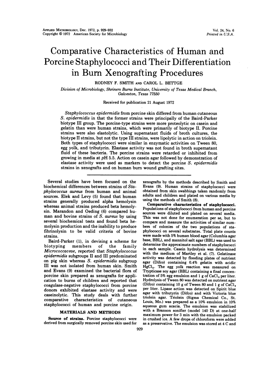
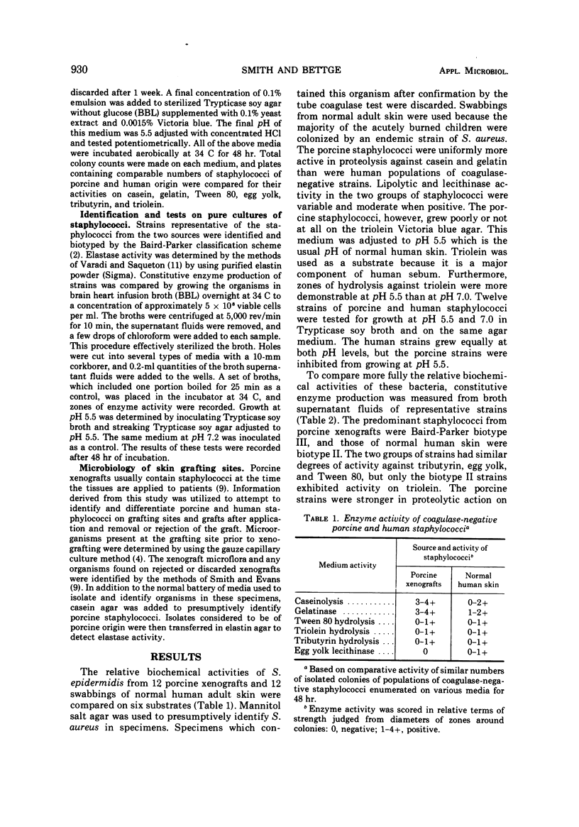
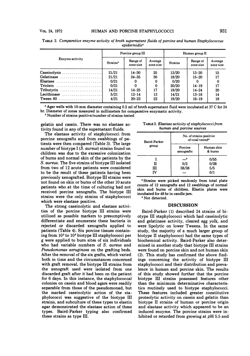
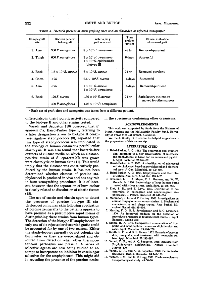
Staphylococcus epidermidis from porcine skin differed from human cutaneous S. epidermidis in that the former strains were principally of the Baird-Parker biotype III group. The porcine-type strains were more proteolytic on casein and gelatin than were human strains, which were primarily of biotype II. Porcine strains were also elastolytic. Using supernatant fluids of broth cultures, the biotype II strains, but not the type III strains, were lipolytic in action on triolein. Both types of staphylococci were similar in enzymatic activities on Tween 80, egg yolk, and tributyrin. Elastase activity was not found in broth supernatant fluid of these bacteria. The porcine strains were retarded or inhibited from growing in media at pH 5.5. Action on casein agar followed by demonstration of elastase activity were used as markers to detect the porcine S. epidermidis strains in xenografts and on human burn wound grafting sites.
Full text
PDF


Selected References
These references are in PubMed. This may not be the complete list of references from this article.
- BAIRD-PARKER A. C. A classification of micrococci and staphylococci based on physiological and biochemical tests. J Gen Microbiol. 1963 Mar;30:409–427. doi: 10.1099/00221287-30-3-409. [DOI] [PubMed] [Google Scholar]
- Baird-Parker A. C. Staphylococci and their classification. Ann N Y Acad Sci. 1965 Jul 23;128(1):4–25. doi: 10.1111/j.1749-6632.1965.tb11626.x. [DOI] [PubMed] [Google Scholar]
- Brentano L., Gravens D. L., Moyer C. A., Monafo W. W., Jr Bacteriology of large human burns treated with silver nitrate. Arch Surg. 1966 Sep;93(3):456–466. doi: 10.1001/archsurg.1966.01330030086019. [DOI] [PubMed] [Google Scholar]
- ELEK S. D., LEVY E. Distribution of haemolysins in pathogenic and non-pathogenic staphylococci. J Pathol Bacteriol. 1950 Oct;62(4):541–554. doi: 10.1002/path.1700620405. [DOI] [PubMed] [Google Scholar]
- Marandon J. L., Oeding P. Investigations on animal Staphylococcus aureus strains. 1. Biochemical characteristics and phage typing. Acta Pathol Microbiol Scand. 1966;67(1):149–156. doi: 10.1111/apm.1966.67.1.149. [DOI] [PubMed] [Google Scholar]
- Martley F. G., Jayashankar S. R., Lawrence R. C. An improved agar medium for the detection of proteolytic organisms in total bacterial counts. J Appl Bacteriol. 1970 Jun;33(2):363–370. doi: 10.1111/j.1365-2672.1970.tb02208.x. [DOI] [PubMed] [Google Scholar]
- Smith R. F. Comparative enumeration of lipophilic and nonlipophilic cutaneous diphtheroids and cocci. Appl Microbiol. 1970 Feb;19(2):254–258. doi: 10.1128/am.19.2.254-258.1970. [DOI] [PMC free article] [PubMed] [Google Scholar]
- Smith R. F., Evans B. L. Bacteria of porcine skin, xenografts, and treatment with neomycin sulfate. Appl Microbiol. 1972 Feb;23(2):293–297. doi: 10.1128/am.23.2.293-297.1972. [DOI] [PMC free article] [PubMed] [Google Scholar]
- Varadi D. P., Saqueton A. C. Elastase from Staphylococcus epidermidis. Nature. 1968 May 4;218(5140):468–470. doi: 10.1038/218468a0. [DOI] [PubMed] [Google Scholar]
- Varadi D. P., Saqueton A. C. Perifollicular elastolysis. Br J Dermatol. 1970 Jul;83(1):143–150. doi: 10.1111/j.1365-2133.1970.tb12875.x. [DOI] [PubMed] [Google Scholar]
- Vistnes L. M., Hogg G. R. The burn eschar. A histopathological study. Plast Reconstr Surg. 1971 Jul;48(1):56–60. doi: 10.1097/00006534-197107000-00012. [DOI] [PubMed] [Google Scholar]


