

Abstract
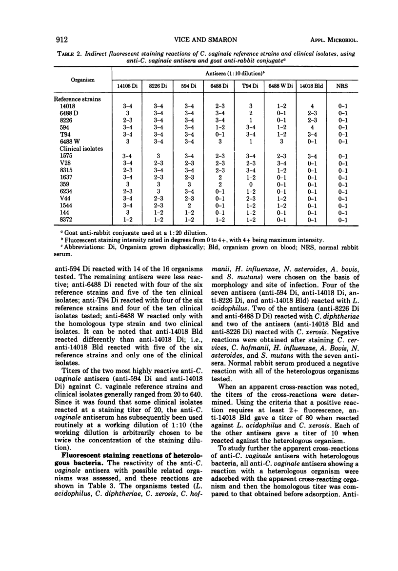
The indirect fluorescent-antibody technique was employed in an attempt to develop a rapid method of identification of Corynebacterium vaginale. Six reference strains and ten clinical isolates selected on the basis of morphology and conventional biochemical tests were compared. Antisera were prepared in rabbits against the six reference strains. The most satisfactory antiserum was that prepared using strain 14018 grown diphasically (14018 Di) as the antigen. Certain of the antisera did exhibit a cross-reacting titer when reacted against Corynebacterium diptheriae, Corynebacterium xerosis, or Lactobacillus acidophilus. However, antisera adsorbed with these bacteria did not exhibit a significant decrease in titer when reacted against the homologous strain. Various other species of Corynebacterium as well as species of Nocardia, Actinomyces, Hemophilus, and Streptococcus did not fluoresce with the antisera. A specific antiserum was prepared by adsorbing anti-14018 Di with L. acidophilus. The adsorption removed the cross-reacting antibody but did not affect the staining reaction with C. vaginale strains. All reference strains and clinical isolates characterized as C. vaginale gave a definite positive reaction with the adsorbed anti-14018 Di. The specificity of the reactions was assessed by adsorbing the antiserum with the homologous strain. The data suggest that the indirect staining method will be of value in the rapid presumptive identification of C. vaginale.
Full text
PDF
Images in this article
Selected References
These references are in PubMed. This may not be the complete list of references from this article.
- DUDMAN W. F. IMMUNE DIFFUSION ANALYSIS OF THE EXTRACELLULAR SOLUBLE ANTIGENS OF TWO STRAINS OF RHIZOBIUM MELILOTI. J Bacteriol. 1964 Sep;88:782–794. doi: 10.1128/jb.88.3.782-794.1964. [DOI] [PMC free article] [PubMed] [Google Scholar]
- Dunkelberg W. E., Jr, McVeigh I. Growth requirements of Haemophilus vaginalis. Antonie Van Leeuwenhoek. 1969;35(2):129–145. doi: 10.1007/BF02219124. [DOI] [PubMed] [Google Scholar]
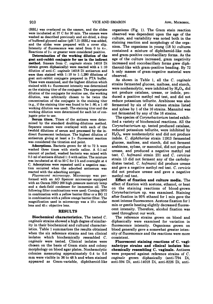
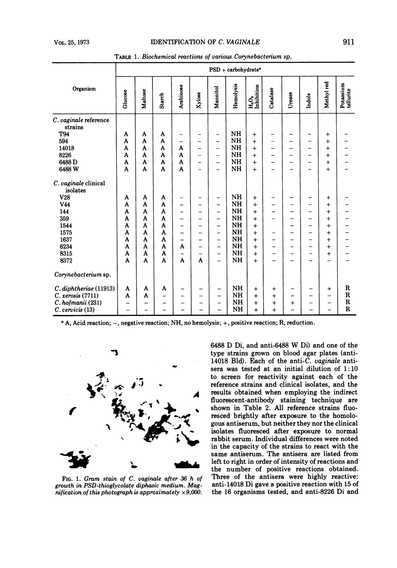
- Dunkelberg W. E., Jr, Skaggs R., Kellogg D. S., Jr A study and new description of Corynebacterium vaginale (Haemophilus vaginalis). Am J Clin Pathol. 1970 Mar;53(3):370–377. doi: 10.1093/ajcp/53.3.370. [DOI] [PubMed] [Google Scholar]
- Dunkelberg W. E., Jr, Skaggs R., Kellogg D. S., Jr Method for isolation and identification of Corynebacterium vaginale (Haemophilus vaginalis). Appl Microbiol. 1970 Jan;19(1):47–52. doi: 10.1128/am.19.1.47-52.1970. [DOI] [PMC free article] [PubMed] [Google Scholar]
- EDMUNDS P. N. The biochemical, serological and haemagglutinating reactions of "Haemophilus vaginalis". J Pathol Bacteriol. 1962 Apr;83:411–422. doi: 10.1002/path.1700830211. [DOI] [PubMed] [Google Scholar]
- GARDNER H. L., DUKES C. D. Haemophilus vaginalis vaginitis: a newly defined specific infection previously classified non-specific vaginitis. Am J Obstet Gynecol. 1955 May;69(5):962–976. [PubMed] [Google Scholar]
- LAPAGE S. P. Haemophilus vaginalis and its role in vaginitis. Acta Pathol Microbiol Scand. 1961;52:34–54. doi: 10.1111/j.1699-0463.1961.tb01547.x. [DOI] [PubMed] [Google Scholar]
- LEOPOLD S. Heretofore undescribed organism isolated from the genitourinary system. U S Armed Forces Med J. 1953 Feb;4(2):263–266. [PubMed] [Google Scholar]
- MOODY M. D., JONES W. L. IDENTIFICATION OF CORYNEBACTERIUM DIPHTHERIAE WITH FLUORESCENT ANTIBACTERIAL REAGENTS. J Bacteriol. 1963 Aug;86:285–293. doi: 10.1128/jb.86.2.285-293.1963. [DOI] [PMC free article] [PubMed] [Google Scholar]
- Pavlova M. T., Beauvais E., Brezenski F. T., Litsky W. Fluorescent-antibody techniques for the identification of group D streptococci: direct staining method. Appl Microbiol. 1972 Mar;23(3):571–577. doi: 10.1128/am.23.3.571-577.1972. [DOI] [PMC free article] [PubMed] [Google Scholar]
- Pease P. The antigenic structure of PPLO (Mycoplasma hominis) and related bacteria. J Gen Microbiol. 1965 Dec;41(3):299–308. doi: 10.1099/00221287-41-3-299. [DOI] [PubMed] [Google Scholar]
- REDMOND D. L., KOTCHER E. COMPARISON OF CULTURAL AND IMMUNOFLUORESCENT PROCEDURES IN THE IDENTIFICATION OF HAEMOPHILUS VAGINALIS. J Gen Microbiol. 1963 Oct;33:89–94. doi: 10.1099/00221287-33-1-89. [DOI] [PubMed] [Google Scholar]
- Reyn A., Birch-Andersen A., Lapage S. P. An electron microscope study of thin sections of Haemophilus vaginalis (Gardner and Dukes) and some possibly related species. Can J Microbiol. 1966 Dec;12(6):1125–1136. doi: 10.1139/m66-154. [DOI] [PubMed] [Google Scholar]
- Vickerstaff J. M., Cole B. C. Characterization of Haemophilus vaginalis, Corynebacterium cervicis, and related bacteria. Can J Microbiol. 1969 Jun;15(6):587–594. doi: 10.1139/m69-100. [DOI] [PubMed] [Google Scholar]